Abstract
This work evaluates sperm head morphometric characteristics in adolescents from 12 to 18 years of age, and the effect of varicocele. Volunteers between 150 and 224 months of age (mean 191, n = 87), who had reached oigarche by 12 years old, were recruited in the area of Barranquilla, Colombia. Morphometric analysis of sperm heads was performed with principal component (PC) and discriminant analysis. Combining seminal fluid and sperm parameters provided five PCs: two related to sperm morphometry, one to sperm motility, and two to seminal fluid components. Discriminant analysis on the morphometric results of varicocele and nonvaricocele groups did not provide a useful classification matrix. Of the semen-related PCs, the most explanatory (40%) was related to sperm motility. Two PCs, including sperm head elongation and size, were sufficient to evaluate sperm morphometric characteristics. Most of the morphometric variables were correlated with age, with an increase in size and decrease in the elongation of the sperm head. For head size, the entire sperm population could be divided into two morphometric subpopulations, SP1 and SP2, which did not change during adolescence. In general, for varicocele individuals, SP1 had larger and more elongated sperm heads than SP2, which had smaller and more elongated heads than in nonvaricocele men. In summary, sperm head morphometry assessed by CASA-Morph and multivariate cluster analysis provides a better comprehension of the ejaculate structure and possibly sperm function. Morphometric analysis provides much more information than data obtained from conventional semen analysis.
Keywords: adolescence, CASA-Morph system, seminal quality, sperm head morphometry, spermiogram, subpopulation
INTRODUCTION
Genetic, environmental, and nutritional factors have a great influence on the time to reach puberty. Therefore, it is difficult to predict the age at which a particular individual will achieve his reproductive maturity, i.e., the sperm production corresponding to that of an adult. In addition, biomedical information about the age of first conscious ejaculation (oigarche1), the presence or absence of spermatozoa in seminal fluid, the biological characteristics of spermatozoa during this maturational process, and the time required to reach adult spermatogenesis, is currently scarce and controversial. The few studies reported on this topic correspond to adolescents under the age of 18 years and are related to different pathologies such as testicular cancer.2,3,4
The age at which men start producing spermatozoa, whether spermatogenesis and accessory gland secretions start on one particular day, and the time required for stabilizing the production, maturation, and the release of spermatozoa are not known exactly. One of the problems is the method used to obtain the sample for a semen analysis. The requirement that adolescents have to masturbate may bring some conflict with the moral and ethical values of a part of society although it has been pointed out that masturbation has no effect on health.5 On the other hand, varicocele usually increases around adolescence and is rarely reported to appear later on in adult men. Patients are commonly referred to the urologist either after detection of a scrotal mass, classically described as a “bag of worms,” or after detection of a difference in testicular size during child or a sports-related physical examination. Most varicoceles are asymptomatic. However, testicular pain or the presence of a scrotal mass may be the presenting symptom.6
The main objective of this study was to investigate sperm features in adolescents between 12 and 18 years of age. These data are part of a social project carried out in Barranquilla, Colombia, directed to develop a sexual educational program because of the high teenage pregnancy rate in this area. Therefore, it is necessary to know the mean age at the onset of sperm production in adolescents from this area to provide the best counseling. In addition, we analyzed the effect of varicocele and used morphometric data analysis to assess sperm subpopulations in human semen samples from these adolescents.
MATERIALS AND METHODS
Participants
Semen samples were provided by 87 adolescents with ages ranging from 150 to 224 months (mean of 190.69 ± 18.4 months) from the area of Barranquilla, Colombia. All the participants referred that had their oigarche and previous ejaculations by self-stimulation. Ejaculatory abstinence interval ranged between 1 and 16 days (mean of 4.31 ± 2.43 days).
Ethical approval
The study was approved by the Committee of Medical Ethics of Universidad del Norte and the Program of Sexual Education of teh School (Barranquilla, Colombia). The purpose of the study was explained to the participants, and they had to provide a semen sample. Written informed consent was obtained from all participants and their parents or tutors.
Collection and analysis of semen samples
A single semen sample was produced by masturbation from each participant, and conventional semen analysis was performed according to the World Health Organization (WHO, 1999) recommendations.7 The semen samples were collected at the laboratory of Centro Médico del Hombre and analyzed within 1 h of collection.
The conventional semen analysis included the following semen parameters: semen volume (ml), total sperm count (millions per ejaculate), sperm concentration (million ml−1), total motility (progressive + nonprogressive, %), progressive motility (%), vitality (live spermatozoa after staining with eosin-nigrosin, %), sperm morphology (normal forms, after staining with Papanicolau, %), pH and fructose levels (μmol per ejaculate).
In addition, 64 samples from the total population were analyzed by the ISAS® v1 (Proiser R+D S.L., Paterna, Valencia, Spain) CASA-Morph system to obtain the sperm head morphometric characteristics. For the morphology assessment, air-dried fixed semen smears were stained with Diff-Quik (Medion Diagnostics, Düdingen, Switzerland) following the kit instructions, washed free of excess colorant water, air dried, and mounted with Eukitt (Sigma-Aldrich, St. Louis, MO, USA).
All analyses were performed by the same technician (PC) from the camera Proiser 782M attached to a microscope UB203 (UOP/Proiser, Paterna, Valencia, Spain) with an eyepiece 100 × oil immersion bright feld objective. The final resolution of the images was 746 × 578 pixels. Depending on the quality of the staining, between 100 and 220 images were randomly captured from the smears. The ISAS® v1 system outcomes include head descriptors: length (μm), width (μm), area (μm2), perimeter (μm), Ellipticity (L/W), Elongation ([L − W]/[L + W]), Regularity (πLW/4A), Rugosity (4πA/P2) (shape factors are dimensionless), and percentage of head covered by the acrosome (%).
Statistical analysis
Discriminant analysis was performed on the effect of the absence or presence of varicocele to obtain a classification matrix. This analysis was performed by considering the standard semen parameters and morphometric characteristics independently, using the linear stepwise procedure to identify those parameters that were most useful in classifying individuals into one of the two categories. In both cases, variables were first added to the discriminant functions one by one until the addition of extra variables did not result in a significantly better discrimination. In both cases, it was found that all the variables were useful for discrimination. The classification matrix obtained after this discriminant analysis was applied to the whole population to establish the proportion of cases in each category.
Principal component analysis (PCA) for semen parameters and for the CASA-Morph morphometric characteristics was as follows: the database was obtained from 87 donors and for the morphometric database, a total number of 11 358 spermatozoa were analyzed from 64 donors. An additional database was produced by combining the seminal data with the sperm morphometric data of each volunteer. To select which components should be used in the next round of analysis, the criterion used for selecting the number of principal components (PCs) with an eigenvalue (variance extracted for that particular PC) >1 (Kaiser criterion) was followed. The second step was to perform a two-step cluster procedure with the sperm-derived indices obtained after the PCA. Varimax rotation with the Kaiser normalization method was used for the definition of the PCs.
The correlation between the different parameters and PC with age was assessed from linear Pearson's correlation coefficients. Because of the high number of correlations made with morphometric parameter variables, the Bonferroni correction was used to establish the significance level.
Finally, we performed an additional study directed to analyze the morphometric subpopulations in the sperm samples, for which a nonhierarchical analysis with the k-means model that uses Euclidean distances to calculate the center of the clusters was developed. Spermatozoa were then assigned to one of these clusters according to morphometric descriptors.
Morphometric data obtained from the subpopulations belonging to the nonvaricocele and varicocele groups were first tested for normality and homoscedasticity using Shapiro–Wilks and Kolmogorov–Smirnov tests, respectively. Because no parameters satisfied both criteria, nonparametric analyses were performed using a Kruskal–Wallis test.
Multivariate analysis of variance (MANOVA) was based on Wilk's lambda criterion and cluster analysis on the average linkage method. The multivariate linear model: Y=Xβ+E, where X is the design matrix, β the vector of regression parameters and E the matrix of random deviations. E~N(0,σ2). When the Bonferroni correction was not needed, the level of significance was set to P < 0.05. All statistical analyses were performed with SPSS software version 20.0 for Windows software (SPSS Inc., Chicago, IL, USA).
RESULTS
Oigarche
The results of the questionnaire used before sampling indicated that the mean age for the appearance of the first conscious ejaculation was of 12.8 ± 1.0 years (range of 10–15 years of age).
Discriminant analysis
From the seminal variables of the participants with no varicocele and those with varicocele, 72.6% of the original clusters of cases were correctly assigned to the correspondent class (Table 1). Although this value is not informative enough for the classification matrix to be a good predictor of varicocele presence, it can explain the presence of mathematical differences in the data distribution. This indicates that the individuals cannot be considered as comprising a homogeneous population, so it is necessary to consider them as two populations for subsequent analysis.
Table 1.
Prediction ability of discriminant analysis to classify individuals with and without varicocele by taking into account the seminogram and morphometric data
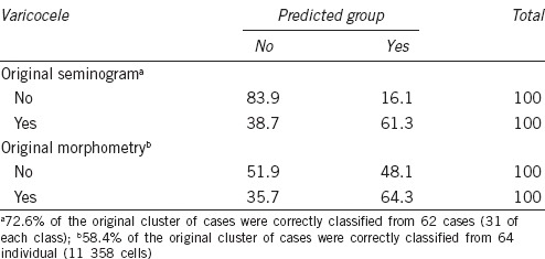
Adolescents with no varicocele were better recognized (83.9%) than those presenting varicocele (61.3%) (Table 1). The morphometric data were the least informative, identifying only 58.4% of the cells from a given class (Table 1). None of these results gave values informative enough to be considered as good predictors of the classification matrix. Therefore, they cannot be used for predicting if a given individual has or has not a varicocele from the whole set of data, including the semen analysis and the morphometric characteristics.
Semen parameter PCA
Three PCs were generated from the whole population and also when varicocele and nonvaricocele populations were considered independently (Table 2). For the whole population, PC1 (40.03% of the variance) was positively correlated with sperm motility and vitality; PC2 (15.81%) was positively correlated with seminal volume and fructose, and PC3 (13.59%) with seminal pH and negatively correlated with sperm concentration. Adolescents with no varicocele showed similar PC values except for PC3, where no negative correlation with sperm concentration was found, providing a considerable significance for morphology. Larger differences were observed in spermatozoa from adolescents with varicoceles where PC2 was positively correlated with sperm concentration and negatively correlated with pH, and PC3 was correlated with semen volume and fructose levels. The percentage of variance explained by PC2 and PC3 was very similar in all cases (Table 2).
Table 2.
Principal components (PC1-PC3) from the total population, divided into no varicocele and varicocele populations, as obtained from seminogram data

Head sperm morphometric parameter PCA
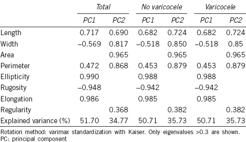
Two PCs were required to explain these population data. For both, the whole population and the subpopulations in semen in adolescents with or without varicocele, PC1 was correlated with shape parameters (positively correlated with ellipticity and elongation, and negatively correlated with rugosity) and PC2 with size. In all cases, a total variance explanation gave similar values (Table 3).
Table 3.
Principal components (PC1, PC2) from the total population, divided into no varicocele and varicocele populations, obtained from the morphometric data
Joint seminal and morphometric parameter PCA
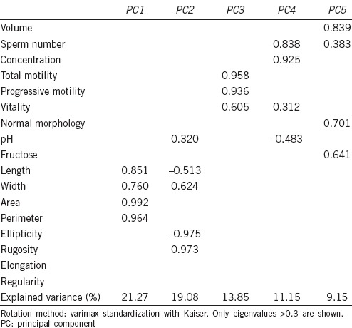
Here, only the whole population was considered. PC1 and PC2 were correlated with sperm morphometry, PC1 alone with size and PC2 with shape, explaining 21.27% and 19.08% of the variance, respectively; PC3 was related with motility and vitality, PC4 was related with concentration and sperm count, and PC5 was related with volume, normal morphology and fructose level, explaining 13.85%, 11.15%, and 9.15%, respectively. This means that morphometry accounts for more than 40% of the variance and semen analysis for only 35% (Table 4).
Table 4.
Principal components (PC1–5) from the total population, obtained from the pooled seminogram and morphometric data
Dependence of semen and morphometric parameters on age
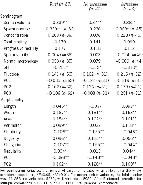
Considering the semen parameters as independent variables, only semen volume (for the whole population [r = 0.339, P < 0.01] and also for each subpopulation [r = 0.374, P < 0.05 and r = 0.362, P < 0.05, respectively]) and sperm count (for the whole population [r = 0.330, P < 0.01] and the population with varicocele [r = 0.369, P < 0.05]) showed a positive correlation with age, while the correlation with pH was negative (for the whole population and with varicocele). Most of the morphometric parameters, including PCs, showed correlation with age, even after Bonferroni correction. In general, the elongation/ellipticity of the cells were reduced with age (Table 5).
Table 5.
Pearson's correlation coefficient (r) of the seminogram and morphometric variables and PCs and the age of the individuals
Sperm head morphometric subpopulations
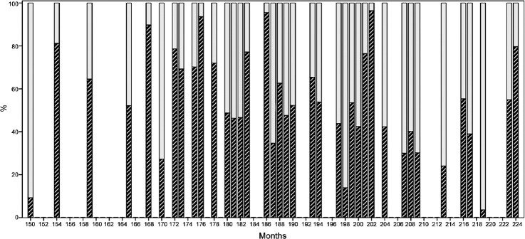
Two sperm subpopulations (SP) or clusters of morphometric characteristics were defined after multivariate cluster analysis for all participants: SP1 and SP2. The characteristics of these subpopulations can be described as follows: SP1 included the largest percentage of spermatozoa (51.2%) and was characterized by a greater head size while SP2 corresponded to 48.8% of the population. In addition to size differences, SP1 contained more elongated cells than SP2 but the other shape parameter values were not different between these subpopulations. Concerning the donors with varicocele, the percentage of SP1 was higher with sperm heads more elongated than in SP2, while SP2 had more elongated sperm head than nonvaricocele donors. MANOVA analysis showed differences between both subpopulations in the total and each diagnostic group (Table 6). No correlation was found between the SP1 and SP2 subpopulations and the age of the donors (Figure 1).
Table 6.
Morphometric SPs values from the total population and divided into no varicocele and varicocele populations, obtained from morphometric data (mean±s.d.)

Figure 1.
Distribution of subpopulations (ordinate, %) for each individual (bars) and age (abscissa). Solid bar corresponds to SP1, cross-hatched to SP2. SPs: subpopulations.
DISCUSSION
The oigarche has a similar physiological meaning to that of the menarche, or first menstruation, in women. However, in latter case, the information about how and when it takes place is considerable, while in the case of oigarche, the age of its onset has barely been studied. The timing of the first ejaculation is mainly related to the development of the sex accessory glands, the prostate and the seminal vesicles, in response to the testosterone produced mainly by the testis following stimulation of the Leydig cells by the LH which is released by the hypophysis during sexual maturation.8
In this study, the questionnaire used in our adolescent population showed a range of 10–15 years of age for the onset of the oigarche with a mean value of 12.8 years. This value is slightly lower than that reported for adolescents from Israel (13.5 years1) and Belgium (13.1 years9) and significantly lower than that reported for adolescents from Mexico (14.0 years10) and China (14.2 years in the cities and 14.8 in rural areas11). Certainly, these studies were carried out many years ago and the current values might have changed. Spermarche (the first appearance of spermatozoa in urine) has been recorded in a few cases (2%) at the age of 11 years,12 being more frequent between 13.4 and 13.8 years of age13,14 or even later, after 14 years of age.15,16 In contrast to our results, adolescents of 12 years old had a relatively high concentration of spermatozoa in the ejaculate, implying that spermarche in our study population probably occurs before this age. This disparity in the age of oigarche could be explained by the ethnic diversity found in populations from different continents, subjacent genetics, socioeconomic status, overall health and welfare status, certain type of physical exercise, and environmental factors. There are no scientific data showing that the age of oigarche occurred earlier in the past decades, as in the case of menarche.17,18
The fact that participants with no varicocele were better identified as such by their sperm morphometric characteristics after discriminant analysis than those presenting with varicocele, indicates that the varicocele is asymptomatic. Even when comparing the whole population of adolescents, with and without varicocele, there are differences in their semen characteristics.3,19 Most of the existing scientific literature on semen quality during adolescence is related to the presence of various pathologies, and not to the evolution of seminal characteristics in healthy men.3,19,20 We have found only one study performed in healthy adolescents, but it was related to body maturation and classified the individuals according to the Tanner index. Tanner Stage 5 adolescents present higher testicular volume and sperm count than Tanner Stage 4.2 Despite differences in the method of analysis, these data are consistent with our findings of a positive correlation between these parameters.
PCA of seminal variables in the whole population showed sperm motility as the most important parameter defining the population. Morphometry PCs were the same in the whole population and the diagnostic groups, indicating that these did not depend on varicocele, but PCA from seminal variables in the whole population indicated that sperm motility was the most relevant semen parameter. Although participants with no varicocele had the same pattern as the whole population, the values of PC2 and PC3 were inverted. The most relevant and novel finding emerging from this study is that morphometric parameters (sperm head size and shape) were highly correlated with sperm quality while standard subjective semen morphology parameter values were not. This means that to evaluate the quality of a semen sample, it is more important to perform a CASA-Morph analysis than to perform conventional semen analysis.
Many clinicians believe that sperm morphology is not relevant in the evaluation of male fertility, especially the subjective assessment of sperm morphology. The subjective assessment of sperm morphology following WHO7 criteria was present only in PC5 and accounted for only 9.2% of the total variance. However, sperm morphometric analysis by CASA-Morph can explain more than 40% of the variance. This suggests that the subjective assessment of sperm morphology is not an efficient method for the evaluation of semen quality, in sharp contrast to CASA-Morph morphometric analysis.21,22
Regarding the changes during adolescence, the increase in semen volume and total sperm count indicate a likely improvement in testicular and accessory glands function during this period. The only variable that decreased as a function of time was pH, reflecting an increase in seminal fluid acidity. No seminal PC showed correlation with age, indicating that the correlations observed for volume, sperm count, and pH were masked when these variables were considered as part of the general mathematical model.
It has been noticed that the secretion of seminal plasma increases with age after oigarche,23 but this does not explain the increase in total sperm count which implies a gradual increase in testicular sperm production.24 The average sperm count in our study was greater than that reported for Italian25 and German26 adolescents between 14 and 17 years of age. Using Bonferroni correction, significant morphometric correlations were found for individual parameters and for PCs. In general, spermatozoa showed an increase in their head size and a reduction in elongation during adolescence. The reduction of the seminal parameter values by PCA showed that three PCs were needed to explain 69.4% of the variance for the whole population, which showed slight variations between controls and varicocele donors. The eight morphometric parameters were reduced to only two PCs, one referring to shape and the other to size, explaining 86.5% of the variance. No differences were found in PCs between control and varicocele groups.
Our study shows that using CASA-Morph technology with multivariate cluster analyses, two sperm subpopulations of spermatozoa with different morphometric characteristics can be distinguished in ejaculates from adolescents. These results support the hypothesis that human ejaculates, like those of other species, comprise heterogeneous populations of spermatozoa, and so not all spermatozoa should be considered as a homogeneous population. To our knowledge, this is the first description of subpopulations of spermatozoa reported in adolescents on the basis of morphometric analysis.
Even though subpopulation studies have become a topic of utmost interest, human sperm subpopulation studies are scarcer than those reported in other mammalian species, and more focused on the kinematics properties of spermatozoa.27,28,29 Despite several studies suggesting that variations in these populations can be related to fertility patterns in other species,30,31,32 to date, there are no reports describing sperm morphometric subpopulations in humans. We did not find any relationship between sperm head subpopulation structure and age. The variations observed herein could be related to the fact that each individual presents a well-differentiated subpopulation structure, as it has been observed in other species. Furthermore, more controlled studies on age and degree of individual maturity are necessary to shed some light into maturation during adolescence starting, if possible with younger donors, looking for the onset of sperm production and follow-up for longer periods to evaluate the individual evolution.
The effects of varicocele on male fertility must be analyzed by taking into account as many parameters as possible. More recently, sperm DNA fragmentation analysis has been introduced as a new diagnostic tool for the evaluation of male fertility.33 However, a more holistic approach, using as many variables as possible, will be required for a better assessment of male fertility potential.
CONCLUSIONS
The results of this study show that CASA-Morph technology combined with PC and multivariate cluster analyses is a more useful, simple, and objective approach for the evaluation of male fertility. The thorough analysis of semen from adolescents requires substantial research and the absence of ethical barriers. This would allow developing new studies to provide additional data as in the case of adult males. The information derived from our study could have practical applications for future studies related to subpopulation analysis of human ejaculates in populations of males of different ages with and without varicocele.
AUTHOR CONTRIBUTIONS
FV, CS and EBO conceived and designed the experiments; PC and AG-M performed the experiments; AV and CS analyzed the data; CS wrote the paper.
COMPETING INTERESTS
CS is Professor at Valencia University and acts as Scientific Director of Proiser R+D S.L Research and Development Laboratory. Neither he nor the other authors have interests that influenced the results presented in this paper.
ACKNOWLEDGMENTS
The authors would like to thank the biologist Álvaro Manotas, the psychologist Ana Elvira Navarro and the microbiologist Marbel López, for their valuable contribution to the realization of this project; to Dr. Eduardo Bustos-Obregón, for his participation in the design of the experiments; to Dr. José María Pomerol and Dr. Oswaldo Rajmil; to the adolescents who participated in this study; to Dr. Jennifer Vergara for the original management of the semen analysis databases; and to Dr. Sogol Fereidounfar and Dr. Juan Álvarez for their critical review of this manuscript. This research was funded by the Universidad del Norte and the Centro Médico del Hombre, Barranquilla, Colombia. AV was granted by CONICIT and MICITT, Costa Rica.
REFERENCES
- 1.Laron Z, Arad J, Gurewitz R, Grunebaum M, Dickerman Z. Age at first conscious ejaculation: a milestone in male puberty. Helv Paediatr Acta. 1980;35:13–20. [PubMed] [Google Scholar]
- 2.Mori MM, Cedenho AP, Koifman S, Srougi M. Sperm characteristics in a sample of healthy adolescents in São Paulo, Brazil. Cad Saude Pública (Rio de Janeiro) 2002;18:525–30. doi: 10.1590/s0102-311x2002000200018. [DOI] [PubMed] [Google Scholar]
- 3.Zampieri N, Zuin V, Corroppolo M, Cervellione RM, Camoglio FS. Varicocele and adolescents: semen quality after 2 different laparoscopic procedures. J Androl. 2007;28:727–33. doi: 10.2164/jandrol.107.002600. [DOI] [PubMed] [Google Scholar]
- 4.Keene DJ, Saijad Y, Making G, Cervellione RM. Sperm banking in the United Kingdom is feasible in patients 13 years old or older with cancer. J Urol. 2010;188:594–7. doi: 10.1016/j.juro.2012.04.023. [DOI] [PubMed] [Google Scholar]
- 5.Leitzmann MF, Platz EA, Stampfer MJ, Willet WC, Giovannucci E. Ejaculation frequency and subsequent risk of prostate cancer. JAMA. 2004;291:1578–86. doi: 10.1001/jama.291.13.1578. [DOI] [PubMed] [Google Scholar]
- 6.Kurtz MP, Zurakowski D, Rosoklija I, Bauer SB, Borer JG, et al. Semen parameters in adolescents with varicocele: association with testis volume differential and total testis volume. J Urol. 2015;193:1843–7. doi: 10.1016/j.juro.2014.10.111. [DOI] [PubMed] [Google Scholar]
- 7.World Health Organization. Laboratory Manual for the Examination of Human Semen and Semen-Cervical Mucus Interaction. 3rd ed. Cambridge: Cambridge University Press; 1992. [Google Scholar]
- 8.Coffey D. Knobil E, Neil J. The Physiology of Reproduction. Ch 24. New York: Raven Press Ltd; 1988. Androgen action and the accessory tissues. [Google Scholar]
- 9.Carlier JG, Steeno OP. Oigarche: the age at first ejaculation. Andrologia. 1985;17:104–6. doi: 10.1111/j.1439-0272.1985.tb00969.x. [DOI] [PubMed] [Google Scholar]
- 10.García-Baltazar J, Figueroa-Perea J, Reyes-Zapata H, Brindis C, Pérez-Palacios G. The reproductive characteristics of adolescents and young adults in Mexico City. Salud Pública Mex. 1993;35:682–91. [PubMed] [Google Scholar]
- 11.Ji C, Ohsawa S. Onset of the release (spermarche) in Chinese male youth. Am J Human Biol. 2000;12:577–87. doi: 10.1002/1520-6300(200009/10)12:5<577::AID-AJHB1>3.0.CO;2-1. [DOI] [PubMed] [Google Scholar]
- 12.Richardson DW, Short RV. Time of the onset of sperm production in boys. J Biosoc Sci. 1978;10(Suppl 5):15–25. doi: 10.1017/s0021932000024044. [DOI] [PubMed] [Google Scholar]
- 13.Nielsen C, Skakkebäek N, Richardson D, Darlin J, Hunter W, et al. Onset of the release of spermatozoa (spermarche) in boys in relation to age, testicular growth, pubic hair and heigh. J Clin Endocrinol Metab. 1986;62:532–5. doi: 10.1210/jcem-62-3-532. [DOI] [PubMed] [Google Scholar]
- 14.Hirsch M, Shemesh J, Modan M, Lunenfeld B. Emission of spermatozoa: age of onset. Int J Androl. 1979;2:289–98. [Google Scholar]
- 15.Kulin H, Frotera M, Demmers L, Bartholomew M, Lloyd T. The onset of sperm production in puberal boys. Relationship to gonadotropin excretion. Am J Dis Child. 1989;143:190–3. [PubMed] [Google Scholar]
- 16.Schaefer F, Marr J, Seidel C, Tilgen W, Schärer K. Assessment of gonadal maturation by evaluation of spermaturia. Arch Dis Child. 1990;65:1205–7. doi: 10.1136/adc.65.11.1205. [DOI] [PMC free article] [PubMed] [Google Scholar]
- 17.Bagga A, Kulkarni S. Age at menarche and secular trend in Maharashtrian (Indian) girls. Acta Biol Szegediensis. 2000;44:53–7. [Google Scholar]
- 18.Clavel-Chapelon F EN-EPIC group. European Prospective Investigation into Cancer. Evolution of age at menarche and at onset of regular cycling in a large cohort of French women. Hum Reprod. 2002;17:228–32. doi: 10.1093/humrep/17.1.228. [DOI] [PMC free article] [PubMed] [Google Scholar]
- 19.Bertolla RP, Cedenho AP, Hassun PA, Lima SB, Ortiz V, et al. Sperm nuclear DNA fragmentation in adolescents with varicocele. Fertil Steril. 2006;85:625–8. doi: 10.1016/j.fertnstert.2005.08.032. [DOI] [PubMed] [Google Scholar]
- 20.Lacerda JI, Del Guidice PT, da Silva BF, Nichi M, Fariello RM, et al. Adolescent varicocele: improved sperm function after varicocelectomy. Fertil Steril. 2011;95:994–9. doi: 10.1016/j.fertnstert.2010.10.031. [DOI] [PubMed] [Google Scholar]
- 21.Soler C, Gaßner P, Nieschlag E, de Monserrat JJ, Gutiérrez R, et al. Use of the Integrated Semen Analysis Sistem (ISAS®) for morphometric analysis and its role in assisted reproduction technologies. Rev Int Androl. 2005;3:112–9. [Google Scholar]
- 22.Bellastella G, Cooper TG, Battaglia M, Stróse A, Torres I, et al. Dimensions of human ejaculated spermatozoa in Papanicolau-stained seminal and swim-up smears obtained from the Integrated Semen Analysis Sistem (ISAS®) Asian J Androl. 2010;12:871–9. doi: 10.1038/aja.2010.90. [DOI] [PMC free article] [PubMed] [Google Scholar]
- 23.Janczewski Z, Bablok L. Semen characteristics in pubertal boys. I semen quality after first ejaculation. Arch Androl. 1985;15:199–205. doi: 10.3109/01485018508986912. [DOI] [PubMed] [Google Scholar]
- 24.Bablock L, Janczewski Z. Development of biological value of sperm in delayed puberty. Pol Tyg Lek. 1992;47:537–9. [PubMed] [Google Scholar]
- 25.Paris E, Menchetti A, De Lazaro L, Marrocco M, Nuzzo C, et al. Lo spermiogramma nell'adolescenza. Minerva Prdiatr. 1998;56:303. [PubMed] [Google Scholar]
- 26.Kliesch S, Behre H, Jürgens H, Nieschlag E. Cryopreservation of semen from adolescent patients with malignancies. Med Pediatr Oncol. 1996;26:20–7. doi: 10.1002/(SICI)1096-911X(199601)26:1<20::AID-MPO3>3.0.CO;2-X. [DOI] [PubMed] [Google Scholar]
- 27.Buffone MG, Doncel GF, Marín Briggiler CI, Vazquez-Levin MH, Calamera JC. Human sperm subpopulations: relationship between functional quality and protein tyrosine phosphorylation. Hum Reprod. 2004;19:139–46. doi: 10.1093/humrep/deh040. [DOI] [PubMed] [Google Scholar]
- 28.Chantler E, Abraham-Peskir J, Roberts C. Consistent presence of two normally distributed sperm subpopulations within normozoospermic semen: a kinematic study. Int J Androl. 2004;27:350–9. doi: 10.1111/j.1365-2605.2004.00498.x. [DOI] [PubMed] [Google Scholar]
- 29.Sousa AP, Amaral A, Baptista M, Tavares R, Caballero Campo P, et al. Not all sperm are equal: functional mitochondria characterize a subpopulation of human sperm with better fertilization potential. PLoS One. 2011;6:e18112. doi: 10.1371/journal.pone.0018112. [DOI] [PMC free article] [PubMed] [Google Scholar]
- 30.Peña FJ, Saravia F, García-Herreros M, Núñez-Martínez I, Tapia JA, et al. Identification of sperm morphometric subpopulations in two different portions of the boar ejaculate and its relation to post-thaw quality. J Androl. 2005;26:716–23. doi: 10.2164/jandrol.05030. [DOI] [PubMed] [Google Scholar]
- 31.de Paz P, Mata-Campuzano M, Tizado EJ, Alvarez M, Alvarez-Rodríguez M, et al. The relationship between ram sperm head morphometry and fertility depends on the procedures of acquisition and analysis used. Theriogenology. 2011;76:1313–25. doi: 10.1016/j.theriogenology.2011.05.038. [DOI] [PubMed] [Google Scholar]
- 32.Ramón M, Soler AJ, Ortiz JA, Gaía-Alvarez O, Maroto-Morales A, et al. Sperm population structure and male fertility: an intraspecific study of sperm design and velocity in red deer. Biol Reprod. 2013;89:1–7. doi: 10.1095/biolreprod.113.112110. [DOI] [PubMed] [Google Scholar]
- 33.Majzoub A, Esteves S, Gosálvez J, Agarwal A. Specialized sperm function tests in varicocele and the future of andrology laboratory. Asian J Androl. 2016;18:205–12. doi: 10.4103/1008-682X.172642. [DOI] [PMC free article] [PubMed] [Google Scholar]


