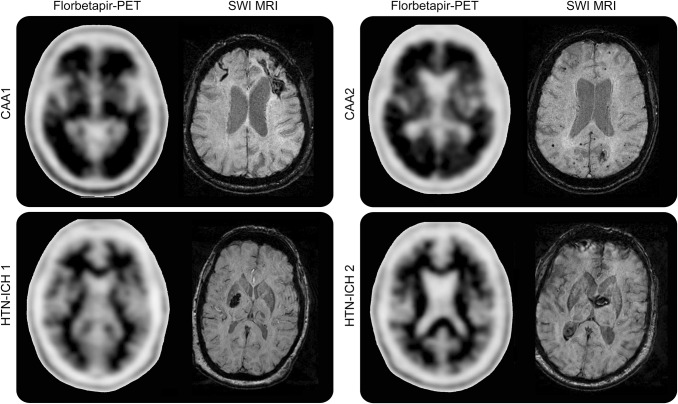
Figure 4. Examples of florbetapir-positive and -negative scans from patients with cerebral amyloid angiopathy (CAA) and hypertensive intracerebral hemorrhage (HTN-ICH).
The upper panel shows florbetapir-positive PET scans from 2 patients with CAA. On susceptibility-weighted imaging (SWI) MRI of the first patient (CAA 1), a left frontal lobar ICH and right frontal superficial siderosis are seen, whereas the SWI MRI of the second patient (CAA 2) shows a left occipital ICH and multiple lobar microbleeds. The florbetapir scans for these patients with CAA show 2 or more brain areas (each larger than a single cortical gyrus) in which there is reduced or absent gray-white contrast, corresponding to intense gray matter radioactivity. The lower panel shows SWI MRIs displaying deep hypertensive ICHs (HTN-ICH 1 and 2). The corresponding negative florbetapir scans show more radioactivity in white matter than in gray matter, creating clear gray-white contrast.

