ABSTRACT
The genome of infectious hematopoietic necrosis virus (IHNV), a salmonid novirhabdovirus, has been engineered to modify the gene order and to evaluate the impact on a possible attenuation of the virus in vitro and in vivo. By reverse genetics, eight recombinant IHNVs (rIHNVs), termed NxGy according to the respective positions of the nucleoprotein (N) and glycoprotein (G) genes along the genome, have been recovered. All rIHNVs have been fully characterized in vitro for their cytopathic effects, kinetics of replication, and profiles of viral gene transcription. These rIHNVs are stable through up to 10 passages in cell culture. Following bath immersion administration of the various rIHNVs to juvenile trout, some of the rIHNVs were clearly attenuated (N2G3, N2G4, N3G4, and N4G1). The position of the N gene seems to be one of the most critical features correlated to the level of viral attenuation. The induced immune response potential in fish was evaluated by enzyme-linked immunosorbent spot assay (ELISPOT) and seroneutralization assays. The recombinant virus N2G3 induced a strong antibody response in immunized fish and conferred 86% of protection against wild-type IHNV challenge in trout, thus representing a promising starting point for the development of a live attenuated vaccine candidate.
IMPORTANCE In Europe, no vaccines are available against infectious hematopoietic necrosis virus (IHNV), one of the major economic threats in fish aquaculture. Live attenuated vaccines are conditioned by a sensible balance between attenuation and pathogenicity. Moreover, nonsegmented negative-strain RNA viruses (NNSV) are subject to a transcription gradient dictated by the order of the genes in their genomes. With the perspective of developing a vaccine against IHNV, we engineered various recombinant IHNVs with reordered genomes in order to artificially attenuate the virus. Our results validate the gene rearrangement approach as a potent and stable attenuation strategy for fish novirhabdovirus and open a new perspective for design of vaccines against other NNSV.
INTRODUCTION
Infectious hematopoietic necrosis virus (IHNV) is a major pathogen for salmonid aquaculture belonging to the Novirhabdovirus genus. Although inactivated and live attenuated viruses have been proven to be effective to protect fish against IHNV (1), to date, only a unique DNA vaccine administrable by injection has been licensed in Canada (2). A live attenuated vaccine usable by bath immersion for mass delivery is needed and would greatly lower the economic loss for fish farmers. All the Mononegavirales genomes consist of a nonsegmented negative-strand RNA molecule with a highly conserved gene order. For novirhabdovirus, the genome encodes six proteins in the following order from the 3′ to the 5′ end: the nucleoprotein (N), the polymerase-associated phosphoprotein (P), the matrix protein (M), the unique glycoprotein (G), the nonstructural protein (NV), and the RNA-dependent RNA polymerase (L) (Fig. 1A) (3). This gene order is crucial for virus replication due to a decreasing gradient of transcription from the 3′ to the 5′ end. The viral polymerase binds to the 3′ end of the genome and starts transcription in a sequential start-stop mechanism, resulting in one mRNA species for each viral gene (4, 5). Between each gene, the polymerase can dissociates from the genome, resulting in a gradient of expression in which the 3′-proximal genes are more transcribed than those located at the 5′ end (5, 6). The modification of the gene order has an important impact on virus replication and pathogenicity, as demonstrated by Wertz and colleagues on vesicular stomatitis virus (VSV) (7, 8). Moving the N gene downstream on the VSV genome decreased the amount of N protein and delayed the kinetics of replication, leading to an attenuated phenotype (9). These recombinant viruses were less pathogenic but maintained their immunogenicity in vivo, providing a new approach for vaccine development (7–10). Interestingly, although there was no direct accordance between in vitro replication efficiency and virulence in mice when the P, M, and G genes were shuffled in the VSV genome (11), the N gene position seems to be one of the most critical factors for the virus pathogenicity.
FIG 1.
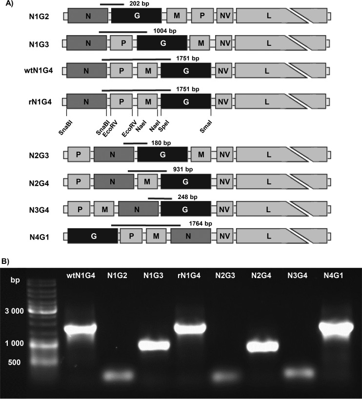
IHNV genome rearrangement. (A) Schematic representation of the engineered rIHNV genomes with rearranged gene order and expected sizes of RT-PCR products. Unique restriction enzyme sites inserted by site-directed mutagenesis at the beginning and the end of the N, P, M, and G ORF are indicated on the rN1G4 genome. (B) Confirmation of the gene order of each rIHNV after two passages on EPC cells. Agarose gel electrophoresis of RT-PCR products amplified from the rIHNV genome was performed using specific N and G primers.
Based on this observation and using the reverse genetics system for IHNV (12, 13), we have generated a panel of recombinant IHNVs (rIHNVs) with modified gene order. Based on the previous results obtained for VSV (14, 15), we have mainly focused on the rearrangement of the G and N genes, since the G protein is the main target for neutralizing antibodies (16) and the N protein has been demonstrated to be a key factor in regulating RNA synthesis and replication in rhabdoviruses (7, 17). We have characterized the impact of gene order rearrangement on rIHNV replication kinetics and viral gene expression by quantitative reverse transcription-PCR (qRT-PCR). Moreover, we have also evaluated the capacity of these rIHNVs to induce a type I interferon (IFN) response in vitro. The degree of attenuation of each rIHNV has been evaluated in rainbow trout through bath immersion infection. Some of the rIHNVs were shown to be attenuated, and their potential as live vaccines has been tested. The capacity of these viruses to confer high protection rates was correlated to a high antibody response. These data demonstrate that moving the N and G genes along the novirhabdovirus genome is a promising approach for vaccine development in fish.
MATERIALS AND METHODS
Cells and viruses.
EPC (epithelioma papulosum cyprini) cells were maintained in Glasgow minimum essential medium (GMEM)/HEPES (25 mM) supplemented with 2 mM l-glutamine (complete medium; PAA Laboratories, France) and with 10% fetal bovine serum (FBS; Eurobio). Wild-type IHNV 32/87 and rIHNV were grown in monolayer cultures of EPC cells at 15°C in complete medium supplemented with 2% FBS. Virus titers were determined by plaque assay on EPC cells maintained under a 0.35% agarose overlay. At 5 to 6 days postinfection, cells were fixed with 10% Formol and stained with crystal violet. For replication kinetics of the rIHNVs, EPC cells were infected at a multiplicity of infection (MOI) of 0.01. At 1, 2, 4, and 7 days postinfection, supernatants were harvested and virus titers were determined in duplicate. Studies on rIHNV induction of type I IFN response were performed in RTG2 fibroblasts from rainbow trout (Oncorhynchus mykiss). RTG2 cells were maintained in L-15 medium (Invitrogen) supplemented with 10% FBS and incubated at 20°C.
Plasmid constructs and recovery of rIHNV.
A cDNA clone containing the N, P, and M genes (pIHNV1) and a cDNA clone containing the G gene (pIHNV2) (12) were used as templates to introduce unique restriction enzyme sites at the beginning and the end of the N, P, M, and G open reading frames (ORF) (Fig. 1A). That was achieved by site-directed mutagenesis (QuikChange site-directed mutagenesis kit; Stratagene) with specific primers (Table 1). These modified cDNA fragments with rearranged gene order were reintroduced in the T7-driven expression plasmid encoding a full-length cDNA copy of the IHNV genome. The recombinant viruses were recovered as previously described (12, 13).
TABLE 1.
Primers used in this study
| Primer name | Sequence (5′–3′) |
|---|---|
| IHNVCDNA | GTATAAAAAAAGTAACTTGACTAAGCTCAG |
| M-Nae1 For | CCAGAGTCAGTTCAAACGCCGGCATGTCTATTTTCAAGAGAGC |
| M-Nae1 Rev | GCGGGGGAAGGAAAAATAGGAGCCGGCCATGCCGTCTCTC |
| N-Snab1 For | GAGACAGAAACGGATTACGTACGATGACAAGCGCACTC |
| N-Snab1 Rev | CCGATTCATTCCGCTGAATACGTACAGCCCCCCCCTCC |
| P-EcoR1 For | GCTTTTCAACTCAAACGATATCAATGTCAGATGGAGAAGG |
| P-EcoR1 Rev | GAAGCAAGGTCAATAGATATCCTCCTCCAGGATCCC |
| G-Spe1 For | CTCGCAGAGACCCACCACTAGTATGGACACCACGATCACC |
| G-Sma1 Rev | GGCAAACCGGTCCTAACCCGGGTCAATCCTCACTTCCTCC |
| N-end For | CAGTGAGCCAGGAGACTCCGATTCATTCC |
| N-start Rev | CTGAGTCCAGTGAACGTCTCTCTGAGTGC |
| G-start Rev | AATGAGAATGAGCGGAGTGGTGATCGTGG |
| G-end For | TCCCCATGTATCACCTGGCAAACCGGTCC |
| N for | TTAACTTCAACGCCAACAGG |
| N rev | TCGGACAGGTTGATGAGAATG |
| P for | AATTGCTATTCGGTCCAACC |
| P rev | ATACCCAATTCCGAATCCAAG |
| M for | ACTCTATGAATCTGGCGGCT |
| M rev | GTTGACCTTTTCTTGGGTGC |
| G for | AACCGATGAGCATCAAATCAG |
| G rev | ATTGAAGGTCGAATGAGTCC |
| NV for | GCGATACAAGAACAAGGTGG |
| NV rev | AGATCAGTGCGTTGACGACA |
| L for | CTTCCCATTGTCATCCTCCT |
| L rev | AGCATTTCCACATGGTCACAGG |
| 18S for | AAACGGCTACCACATCCAAG |
| 18S rev | TTACAGGGCCTCGAAAGAGA |
| IFN1 for | AAAACTGTTTGATGGGAATATGAAA |
| IFN1 rev | CGTTTCAGTCTCCTCTCAGGTT |
| Mx for | AGCGTCTGGCTGATCAGATT |
| Mx rev | ACCCCACTGAAACACACCTG |
| MDA5 for | AGAGCCCGTCCAAAGTGAAGT |
| MDA5 rev | GTTCAGCATAGTCAAAGGCAGGTA |
| LGP2b for | GTGGCAGGCAATGGGGAATG |
| LGP2b rev | CCTCCAGTGTAATAGCGTATCAATCC |
Nucleotide sequences of the rearranged recombinant IHNV.
To determine the nucleotide sequence of each rearranged IHNV genome, genomic RNAs were extracted from infected-cell supernatant using a QIAamp viral RNA purification kit (Qiagen, Courtaboeuf, France) according to the manufacturer's instructions. Viral RNA was then reverse transcribed using reverse transcriptase II (Thermo Fisher Scientific) with IHNVCDNA primer (Table 1) and then amplified by PCR using specific primers covering sequences from the 3′ end to the beginning of NV gene (primer sequences are available upon request). The viral genomes were amplified as 4 overlapping PCR products. PCR products of roughly 1,500 to 2,300 nucleotides (nt) were purified with a PCR purification kit (Qiagen) by following the manufacturer's instructions. Purified PCR products were then subjected to sequencing with specific primers. Sequences were assembled using Vector NTI Advance 11 (Invitrogen).
RNA isolation, RT-PCR, and qPCR analysis.
EPC cells in 24-well plates were infected at an MOI of 0.1 with rIHNV. At 8 h postinfection, total RNA was extracted with an RNEasy kit (Qiagen). Five micrograms of RNA was used to obtain cDNA using Superscript reverse transcriptase II (Invitrogen Life Technologies) and oligo(dT)12-18 primers (150 ng/reaction). Quantitative real-time PCR (qPCR) was performed using SYBR green one-step kit reagent (Bio-Rad) and a Master Cycler Realplex thermal cycler (Eppendorf) according to the manufacturer's instructions with specific primers (Table 1) and the minimum information for publication of quantitative real-time PCR experiments (MIQE) guidelines (18). Conditions for qPCR amplifications were 20 min at 50°C and then 10 min at 95°C, followed by 40 amplification cycles (15 s at 95°C, 20 s at 60°C, and 30 s at 72°C). The specificity of each primer pair was confirmed via melting-curve analysis. The reference gene, carp 18S RNA, was used as a normalizing gene. The expression levels were calculated using the threshold cycle (2−ΔΔCT) method, where ΔCT is determined by subtracting the 18S value from the CT of the targeted gene and ΔΔCT is determined by subtracting the ΔCT of the NV gene from the ΔCT of the target gene in order to normalize the level of expression of each gene by genome. The levels of expression in the samples were then expressed as the percentage of expression relative to that of the first gene on the genome (N or P gene). The data presented are averages from a minimum of four independent experiments.
Analysis of the type I IFN response.
RTG2 cells in 24-well plates were infected at an MOI of 0.1 with rIHNV. At 24 or 48 h of incubation at 14°C, RNA was extracted from the cells using Tri reagent (Ambion) by following the manufacturer′s instructions. One microgram of RNA was used to obtain cDNA in each sample using Bioscript reverse transcriptase (Bioline Reagents Ltd.) and oligo(dT)12-18 (0.5 μg/ml) by following the manufacturer's instructions. To evaluate the levels of transcription of the different genes, real-time PCR was performed in a LightCycler 96 System instrument (Roche) using FastStart essential DNA green master (Roche) and specific primers (Table 1). Each sample was measured under the following conditions: 10 min at 95°C, followed by 45 amplification cycles (15 s at 95°C and 1 min at 60°C). The expression of individual genes was normalized to relative expression of trout EF-1α, and the expression levels were calculated using the 2−ΔCT method, where ΔCT was determined by subtracting the EF-1α value from the CT of the targeted gene. Negative controls with no template were included in all the experiments. A melting curve for each PCR was determined by reading fluorescence at every degree between 60°C and 95°C to ensure that only a single product had been amplified.
Experimental fish infection and virus isolation from tissue samples.
All animal studies were carried out in strict accordance with the European guidelines and recommendations on animal experimentation and welfare. All animal experiment procedures were approved by the local ethics committee on animal experimentation (COMETHEA INRA no. 45) and registered under permit no.12/111. For experimental fish infections, 50 virus-free juvenile rainbow trout (Oncorhynccus mykiss) were infected by bath immersion with rIHNV at a final titer of 5 × 10⁴ PFU/ml for 2 h at 10°C in 3 liters of freshwater. Aquaria were then filled up to 30 liters with freshwater. Fish immunizations were done in three distinct experiments, twice on 50 fish by groups (average weight, 0.7 g) with mortality monitored for 35 days (wild-type N1G4 [wtN1G4], N1G3, N2G4, N2G3, and rN1G4) or 50 days (wtN1G4, N1G3, N2G4, N1G2, N4G1, and N3G4) and once on 50 fish by groups (average weight, 1 g) with mortality monitored for 33 days (wtN1G4, N1G2, and N2G3). Control fish were mock infected with cell culture medium under the same conditions. Challenges were performed under similar conditions. For virus isolation (N2G3 and N2G4), the heads or bodies of random dead fish were homogenized in a mortar with a pestle and sterile sea sand in complete medium with amphothericin B (Sigma). The volume added was proportional to fish head or body weight (1 ml/g to get viral titer in PFU/g). Crushed tissues were then centrifuged at 4,000 rpm for 15 min at 4°C to remove tissue debris and sea sand, and virus titers in supernatants were determined by inoculation to EPC cells as described above.
Quantification of antibody-secreting cells (ASCs) in the head kidney in response to N2G3 by ELISPOT assay.
Enzyme-linked immunospot (ELISPOT) assay was used to quantify the number of IgM-secreting B cells in the head kidney after immunization with N2G3. For this, rainbow trout were infected with N2G3 as described above or mock infected. After 30 days, four control and six infected fish were sacrificed and the head kidney was removed. Head kidney leukocytes were isolated as described before (19). ELISPOT plates containing Inmobilon-P membranes (Millipore) were activated with 70% ethanol for 30 s, coated with anti-trout IgM monoclonal antibody (MAb; clone 4C10) at 2 μg/ml diluted in phosphate-buffered saline (PBS), and incubated overnight at 4°C. To block nonspecific binding to the membrane, plates were then incubated with 2% bovine serum albumin (BSA)–PBS for 2 h at room temperature. Head kidney leukocytes from individual fish were added to the wells in triplicate at a concentration of 1 × 10⁵ per well. After 72 h of incubation at 20°C, cells were washed 5 times with PBS and plates were blocked again with 2% BSA–PBS for 1 h at room temperature. After blocking, biotinylated anti-trout IgM MAb (clone 4C10) was added to the plates and incubated at 1 μg/ml for 1 h at room temperature. Following additional washing steps (5 times in PBS), the plates were developed using streptavidin-horseradish peroxidase (HRP) (Thermo Scientific) for 1 h at room temperature, washed again with PBS, and incubated with 3-amino 9-ethylcarbazole (Sigma-Aldrich) for 30 min at room temperature in the dark. Substrate reaction was stopped by washing the plates with tap water. Once the membranes dried, they were digitally scanned and spot counts determined by the ImmunoSpot Series 45 Micro ELISPOT analyzer.
IHNV seroneutralization assays.
Seroneutralization in the presence of trout complement (from ANSES-Brest) was performed by a simplified endpoint technique. Clotted trout blood samples harvested from rIHNV N2G3 fish 35 days postinfection were centrifuged for 10 min at 2,000 rpm at 4°C and the supernatants were collected, heated at 45°C for 30 min, and cooled to 4°C. Three sera from mock-infected fish and 23 sera from rIHNV N2G3-infected fish were collected and pooled in 1 group and 5 groups, respectively. A recombinant IHNV expressing green fluorescent protein (rIHNV-GFP; 500 PFU) was incubated with serial 2-fold dilutions of sera (from 48- to 6,144-fold dilutions) in GMEM-HEPES medium containing trout complement (40-fold final dilution) at 4°C overnight. As a control, rIHNV-GFP was incubated with medium containing trout complement under the same conditions and rIHNV-GFP was also incubated with a known IHNV-positive serum (from ANSES-Brest). The mixtures were then used to inoculate EPC cells in 96-well plates (3 × 10⁵ cells per well) and incubated for 6 days at 15°C. Finally, the medium was removed and the monolayers were fixed and stained with crystal violet. The neutralizing antibody titer was expressed as the reciprocal value of the highest serum dilution that reduced the cytopathic effect by 50%. The neutralizing antibody titer after staining was correlated to analysis of rIHNV-GFP infection under a UV light microscope (data not shown). Each microplate encompassed 10 assays plus one positive- and one negative-control serum.
Accession number(s).
Sequences covering the rearranged part of the genomes (from the 3′ end to the beginning of the NV gene) have been deposited in GenBank with accession numbers KX860117, KX860118, KX860119, KX860120, KX860121, and KX860122.
RESULTS
Recovery of rIHNV with rearranged gene order.
Unique restriction enzyme sites were introduced by site-directed mutagenesis immediately upstream and downstream of the start and the stop codons of each ORF in the IHNV genome. Restriction enzyme sites were SnaBI for the N gene, EcoRV for the P gene, NaeI for the M gene, and SpeI and SmaI at the start and end of the G gene, respectively (Fig. 1A). The recombinant viruses were readily recovered as previously described (12, 13). Recombinant viruses with rearranged gene order were named according to their respective N and G gene positions: N1G2 (3′-N-G-M-P-NV-L5′) (for this virus the M and P gene order has also been modified), N1G3 (3′-N-P-G-M-NV-L-5′), N1G4 (recombinant or wild type) (3′-N-P-M-G-NV-L-5′), N2G3 (3′-P-N-G-M-NV-L-5′), N2G4 (3′-P-N-M-G-NV-L-5′), N3G4 (3′-P-M-N-G-NV-L5′), and N4G1 (3′-G-P-M-N-NV-L-5′) (Fig. 1A). Compared to wild-type N1G4 IHNV (wtN1G4), the recombinant N1G4 IHNV (rN1G4) has a similar gene order but contains additional restriction enzyme sites introduced to each ORF. The gene order of each recovered rIHNV was ascertained by reverse transcription-PCR (RT-PCR) on RNA genomes extracted from viral stocks from infected cells after two passages. PCR was carried out using primers targeting the N and G genes (Table 1). As shown in Fig. 1B, the sizes of the amplified DNA fragments confirmed the expected genome structure of each rIHNV. Moreover, the structure of the rearranged genomes was shown to be conserved after 10 passages in cell culture (Fig. 2). The sequences of the rearranged genomes were partially determined from the 3′ end to the beginning of the NV gene (see Materials and Methods), thus covering the genes and gene junctions which were shuffled. The nucleotide sequencing confirmed that except some silent mutations, no other incidental mutations were introduced in the rIHNV genomes.
FIG 2.
Genome stability of selected rIHNVs after 2 and 10 passages (P2 and P10) in cell culture. rIHNVs were passaged up to 10 times on EPC cells. At the second and tenth passages, viral RNA was extracted from infected cell supernatants to confirm the gene order of each rIHNV. Agarose gel electrophoresis of RT-PCR products amplified from the rIHNV genome was performed using specific N and G primers.
Comparison of replication kinetics of rIHNV.
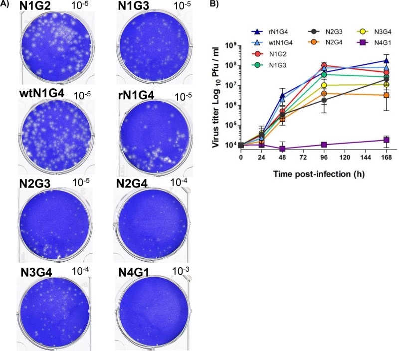
To characterize the in vitro phenotype of each rIHNV, EPC cells were infected and the morphology of virus-induced plaques was observed (Fig. 3A). Differences in the size of the plaques were visualized for the different viruses. The viruses in which the N gene has been moved (N2G3, N2G4, N3G4, and N4G1) produced smaller plaque sizes than those with the N gene at its original position (N1G2, N1G3, wtN1G4, and rN1G4). This probably reflects an alteration in viral replication. To better measure the replication kinetics of each rIHNV, multiple-step growth curves were performed on EPC cells infected at an MOI of 0.01 (Fig. 3B). Comparison of the growth curves for rN1G4 and wtN1G4 demonstrated that the addition of restriction enzyme sites in the IHNV genome did not affect virus replication. At 4 days postinfection, both N1G2 and N1G3 showed replication kinetics similar to that of the wild-type virus, from 3.5 × 10⁷ to 108 PFU/ml. N2G3, N2G4, and N3G4 exhibited slower growth kinetics and reduced viral titers ranging from 1.8 × 10⁶ to 107 PFU/ml. The N4G1 virus showed the most impaired replication capacity in fish cells (1.1 × 10⁴ PFU/ml) at 4 days postinfection. These data emphasize that the N gene position is crucial for virus replication efficiency.
FIG 3.
(A) Plaque size differences among rIHNVs. EPC cells were infected at an MOI of 0.01 for 7 days, fixed, and stained with crystal violet. Corresponding well dilutions are given at the top corner of each well. (B) rIHNV kinetics of replication. Growth curves were determined for EPC cells in triplicate with an MOI of 0.01. Averages and standard deviations are shown.
Impact of gene order on viral gene expression.
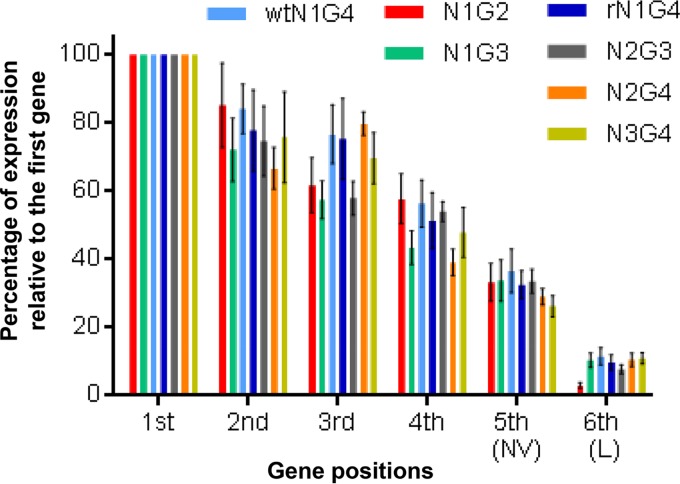
To evaluate the impact of viral gene shuffling on their levels of expression, viral mRNAs were quantified by qRT-PCR (Fig. 4). For this, EPC cells were infected with each recombinant virus at an MOI of 0.1. N4G1 was excluded due to its strongly altered replication cycle. At 8 h postinfection, total RNA from infected cells was used for RT and qPCR with oligo-dT12-18 and specific primers, respectively (Table 1). A gradient of transcription was observed for all rIHNVs, with an average of an 18% decrease between upstream and downstream genes as shown in Fig. 4. No statistical differences between viruses were observed in the levels of expression of the genes based on their positions within the genome. The level of transcription of each viral gene was dependent on the gene position in the genome, while the gene by itself does not seem to significantly impact its transcription rate.
FIG 4.
Relative level of expression of each viral mRNA expressed at 8 h postinfection in EPC cells. The level of expression of each viral mRNA was compared to that of the first gene (N or P). No significant difference could be measured between each virus compared to wtN1G4 based on a two-way analysis of variance (ANOVA) using Tukey's multiple-comparison test (30).
Impact of gene order on viral induction of type I IFN expression.
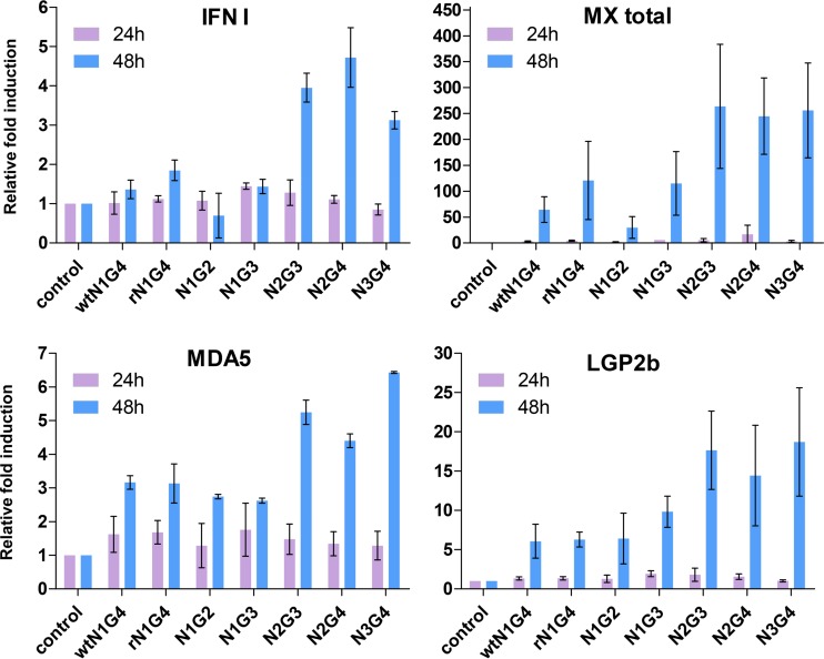
As a first step toward an evaluation of the immunogenicity of rIHNV, the capacity of the various recombinant viruses to induce a type I IFN response in vitro was studied in RTG2 cells (Fig. 5). N4G1 was excluded from this study because it did not reach a sufficient titer for this assay. After 48 h of infection, both wtN1G4 and rN1G4 induced the transcription of IFN1 (a type I IFN gene), Mx (a type I induced gene), and the RIG-I-like receptors MDA5 and LGP2b in comparison to that in mock-infected control cells. This induction was similarly observed in rIHNVs that maintained the N gene in position 1 (N1G3 and N1G2) but was strongly increased for all four genes with rIHNVs in which the N gene had been moved to position 2 or 3 (N2G4, N2G3, and N3G4) in the viral genome. No differences were observed among viruses based on the position of the G gene; therefore, the capacity of these rIHNV to induce a type I IFN response seems to be more influenced by their replication kinetics than by the amount of G protein produced.
FIG 5.
Level of transcription of genes related to the IFN response in RTG2 cells infected with the different rIHNVs. RTG2 cells were infected with 1 × 10⁵ PFU/ml of the different viruses and incubated for 24 h or 48 h at 14°C. Mock-infected controls were also included. The levels of transcription of IFN1, total Mx, MDA5, and LGP2b were evaluated through real-time PCR. Transcript levels were calculated relative to that of an endogenous control (EF-1α). Modulated expression was then expressed as fold change, calculated as the mean expression levels in infected cells normalized to that of controls. Means ± SDs of two replicates are shown.
Virus pathogenicity in juvenile trout.
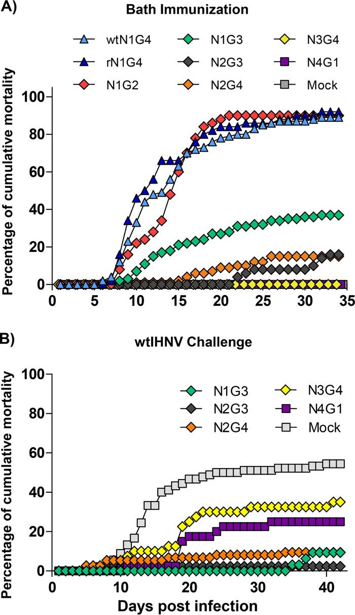
The pathogenicity of rIHNV was evaluated by infecting juvenile rainbow trout (mean weight of 0.7 g) with the selected viruses and recording the virus-induced mortality rate for up to 35 days. As shown in Fig. 6A, no significant differences in fish mortality were observed between wtN1G4 and rN1G4. For both viruses, the mortality started at day 7 postinfection and reached roughly 90% of cumulative mortality at day 35. These data indicated that the addition of 8 restriction enzyme sites (20 nucleotides changes in total) in the IHNV genome has no deleterious effect on virus pathogenicity. A similar mortality rate was observed with N1G2. Interestingly, N1G3 was attenuated in vivo, inducing only 37% of the cumulative mortality at day 35. N2G4 and N2G3 were almost completely attenuated. The mortality started at day 15 and day 22 postinfection for N2G4 and N2G3, respectively. The cumulative mortality rate at day 35 was around 15% for both viruses. All dead fish in the N2G3 and N2G4 groups were positive for IHNV infection, as confirmed by virus isolation and immunofluorescence assays on cell culture (data not shown). N3G4 and N4G1 were completely attenuated in juvenile trout. No mortality was recorded for both viruses during the entire experimental period as for the mock-infected fish.
FIG 6.
Fish mortality curves following infection by bath immersion with rIHNV. (A) Fifty virus-free juvenile rainbow trout (mean weight of 0.7 g) were infected with each of the rIHNVs as described in Materials and Methods. Fish mortality was recorded every day for 35 days. The averages from two independent experiments are represented. (B) Challenge of rIHNV-immunized fish with wtIHNV. Thirty-five or 50 days after immunization with rIHNV, fish were all challenged at the same time with wtIHNV (see Materials and Methods). Fish mortality was recorded every day for 42 days.
Evaluation of the potential of rIHNV as live attenuated vaccine.
At 35 or 50 days postimmunization, as animal experiments were done in two batches, the potential of rIHNV as live vaccine was tested by challenging the surviving fish with wtIHNV. As shown in Fig. 6B, the mortality rate in the mock-vaccinated group reached 55.6%, whereas it reached 35% for the N3G4 group, 25% for the N4G1 group, around 9% for both the N2G4 and N1G3 groups, and 2% for the N2G3 group. N1G3 was the worst group in terms of relative percent survival (RPS) (17%), due to the high induced mortality rate during the immunization step (Table 2). In terms of overall protection, three groups showed RPSs ranging from 37% to 56%. Two groups, N3G4 and N4G1, induced no mortality during the immunization but were poorly protected against the wtIHNV challenge. In contrast, mortality was observed during the immunization for the N2G4 group but surviving fish were rather well protected against the wtIHNV. The highest calculated RPS was 68% for N2G3. Although mortality was not negligible, surviving fish were highly protected in this group, with only 2% mortality after wtIHNV challenge. Despite a residual virulence in fish fry, these results highlight an interesting potential of the attenuated N2G3 as live vaccine.
TABLE 2.
Summary of rIHNV-infected fish from the experiment whose results are presented in Fig. 6a
| Virus | % cumulative mortality |
||
|---|---|---|---|
| Immunization | Challenge | RPSb | |
| N1G2 | 90 | ||
| N1G3 | 37 | 9.4 | 17 |
| wtN1G4 | 88 | ||
| rN1G4 | 92 | ||
| N2G3 | 16 | 2 | 68 |
| N2G4 | 15 | 9.5 | 56 |
| N3G4 | 0 | 35 | 37 |
| N4G1 | 0 | 25 | 55 |
| Mock | 0 | 55.6 | |
wtIHNV challenge was performed at day 35 and day 50 postimmunization and ended 42 days later.
Relative percent survival (RPS) = 1 − (percent mortality in group/percent mortality in control) × 100 (31).
Virus infectivity and vaccine potential of N2G3 virus in trout.
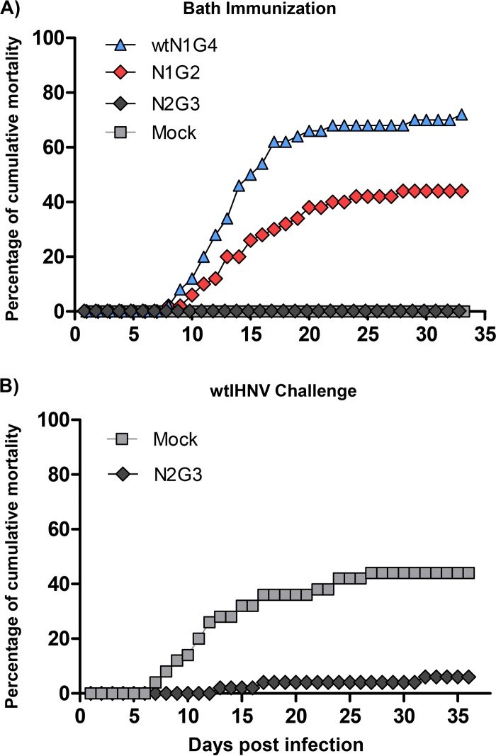
Age- and weight-dependent susceptibility of rainbow trout (Oncorhynchus mykiss) to IHNV has been well documented (20). In the current study, N2G3 presented the best balance between attenuation and protection. Thus, this immunostimulatory potential was evaluated by infecting older fish (mean weight of 1 g) by bath immersion. As shown in Fig. 7A, a 72% cumulative mortality was observed for wtN1G4-infected fish, whereas only 44% of the fish died from N1G2 infection, although this virus was as pathogenic as wild-type virus in younger fish (Fig. 6A). Interestingly, N2G3 did not lead to any mortality at 35 days postinfection. These N2G3-immunized fish were then challenged with wtIHNV (Fig. 7B). While 44% of the fish died in the nonimmunized group, only 6% of cumulative mortality was recorded in N2G3-vaccinated fish group. The overall protection of 86% induced upon vaccination with N2G3 by bath immersion makes this virus a promising vaccine candidate.
FIG 7.
(A) Fish mortality curves following infection by bath immersion with wtN1G4, N1G2, or N2G3. Fifty virus-free juvenile rainbow trout (average weight of 1 g) were infected and the mortality was recorded for 33 days. (B) Protection of fish immunized with N2G3. N2G3-infected fish were challenged with wtIHNV.
Antibody response elicited by the N2G3 virus.
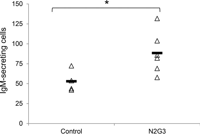
To evaluate the impact of the antibody response induced by immunization with N2G3 virus, juvenile rainbow trout were infected with N2G3 virus or mock infected. After 30 days, fish were sacrificed, the head kidney was removed, and blood samples taken. Following the isolation of head kidney leukocytes, an ELISPOT assay was performed to quantify the induced amount of IgM-secreting B cells. As shown in Fig. 8, a significant induction of IgM-secreting B cells in the kidney was apparent following N2G3 immunization. This suggested that the immune response triggered by N2G3 recombinant virus is humoral. That was further confirmed by serum neutralization assays showing that neutralizing antibodies correlated to the protection induced by the N2G3 virus since neutralizing antibodies titers ranged from 96 to 768 depending on the fish pools (see Materials and Methods).
FIG 8.
ELISPOT analysis of IgM-secreting cells in head kidney leukocytes following N2G3 or mock immunization. Thirty days postimmunization, fish were sacrificed and the head kidney was removed. ELISPOT assay was performed on isolated kidney leukocytes. Values are expressed as IgM-secreting cells in 105 leukocytes from individual fish tested. Bars indicate mean values in each group, and the asterisk denotes significant differences between groups (P < 0.05).
DISCUSSION
By reverse genetics, six recombinant viruses have been produced with gene rearrangement focusing on the N, P, M, and G genes. In Mononegavirales, gene rearrangement had initially been studied for VSV by Wertz's group (9). Despite the fact that in vitro attenuation was observed after shuffling of the G, P, and M genes, no major attenuation could be seen in vivo in mice (11). However, the impact of moving the N gene was more drastic both in vitro and in vivo, with a strong attenuation in mice in correlation with a more distal position of this gene within the genome (9). In the current study, we confirmed these observations for a fish novirhabdovirus. Moving the G gene upstream (N1G2 and N1G3) had only a low impact on virus replication kinetics in vitro, whereas moving the N gene downstream (N2G4 and N3G4) led to a reduced capacity of replication and a drastic effect when the N and G genes were inverted (N4G1). This delay in replication might be explained by the necessity for the virus to have a minimal amount of N proteins to initiate the genome replication (21). Moreover, in the current study, it has been demonstrated by qRT-PCR that during the viral replication, the amount of viral mRNA synthesized is not dependent on the gene itself but is strictly reliant on its position within the genome. These data are in accordance with what was first observed by Finke and colleagues through Northern blot assays (17).
Live attenuated viruses are known to be more efficient than inactivated vaccines to stimulate durably the immune response (22, 23). In addition, live attenuated vaccines are less expensive than inactivated virus to produce since there is no requirement for an inactivation step and for an additional adjuvant to boost the immune response. For aquaculture, this kind of vaccine is the best option since bath immersion administration would allow mass delivery to fish. Therefore, rIHNV with rearranged gene order could be a promising vaccine candidate. Gene arrangement leads to a modification in the transcription gradient of the gene, which could offer interesting levels of attenuation of virus replication and thus virus pathogenicity. Moreover, with the exception of a single report on respiratory syncytial virus (24), genetic recombination for Mononegavirales appears to be a very rare event. Thus, from a safety point of view, strategies aiming to modify the gene order in the viral genome are probably the most attractive approaches, avoiding any risk of reversion to a wild-type phenotype. In this study, we investigated whether through several passages in cell culture, the rearranged genome of rIHNV still maintained the expected gene order. As shown in Fig. 2, no unexpected gene order changes were observed for the five viruses that were tested (wtN1G4, N1G3, rN1G4, N2G3, and N2G4) after 10 successive passages in fish cells. This was further confirmed by the nucleotide sequencing of all rIHNV genomes. All the rIHNVs were used to infect trout to determine their level of attenuation. Analysis of the data emphasizes that when the N gene remains in the first position in the viral genome, the corresponding rIHNVs (N1G2, N1G3, and N1G4) are highly virulent. When the N gene is moved in second position, the respective rIHNVs (N2G3 and N2G4) are almost totally attenuated. In contrast, at the third and fourth positions, the resulting viruses (N3G4 and N4G1) are totally attenuated. However, the overattenuated N3G4 and N4G1 were not protective at all against the wtIHNV, probably due to a decreased immunogenicity as a consequence of its low replication capacity. Interestingly, N2G3 and N2G4 were able to induce a protective immune response in immunized fish. Even more, N2G3 was fully attenuated and protective in older fish. This protection can be correlated with the induction of IgM-secreting B cells seen in the kidney following immunization and also the induction of neutralization antibodies.
The type I IFN system constitutes the first line of antiviral defense until adaptive immune responses are mounted, being particularly important to fish in which an adaptive immune system is limited and is dependent on environmental parameters such as temperature. In fact, DNA vaccines against novirhabdoviruses confer protection through IFN induction in the first weeks postvaccination until a specific immune response is achieved (25). On the other hand, it is well known that in mammals IFN stimulates different B lymphocyte functions (26). Therefore, we also studied the potential of each of these viruses to induce the transcription of a selection of genes implicated in the type I IFN response in rainbow trout cells. We observed that the capacity of these rIHNVs to induce the transcription of genes related to this IFN response was not affected by the position of the G gene in the genome, as would had been expected based on the fact that the G gene is a strong inducer of type I IFN (25, 27). Instead, N2G3, N2G4, and N3G4, in which the N transcription levels were reduced, were clearly much stronger inducers of type I IFN. It could be possible that a less efficient replication potential of these attenuated viruses gives the cells a higher chance to mount a strong IFN response. On the other hand, this increase might imply that the N protein interferes with the cellular IFN system, as occurs in other Mononegavirales, such as Borna disease virus (28) and rabies virus (29). Consequently, we could also hypothesize that the fact that these viruses that transcribe smaller amounts of N are not able to block the IFN system in the cell may be the reason they are attenuated in vitro. To confirm any of these possibilities, further studies have to be undertaken.
To conclude, gene rearrangement in Mononegavirales genomes is a powerful strategy to rapidly generate recombinant viruses with an interesting balance between attenuation and immunogenicity. In the case of IHNV, the N2G3 virus is a good example of a compromise between attenuation and induced protection, although the residual virulence has to be abrogated by additional genome modifications.
ACKNOWLEDGMENTS
We thank Thierry Morin (ANSES-Brest) for the gift of trout complement and IHNV-positive serum and members of the fish facilities (Infectiologie Expérimentale des Rongeurs et Poissons, INRA) for taking care of experimental fish. The technical assistance of Laury Baillon (INRA), Julie Bernard (INRA), Louison Beugnies (INRA), and Rosario Castro (CISA-INIA) is greatly appreciated.
REFERENCES
- 1.Biacchesi S, Bremont M. 2014. Vaccination against viral hemorrhagic septicemia and infectious hematopoietic necrosis, p 289–302. In Gudding R, Lillehaug A, Evensen Ø (ed), Fish vaccination. John Wiley & Sons, Ltd, Chichester, UK. [Google Scholar]
- 2.Alonso M, Leong JA. 2013. Licensed DNA vaccines against infectious hematopoietic necrosis virus (IHNV). Recent Pat DNA Gene Seq 7:62–65. doi: 10.2174/1872215611307010009. [DOI] [PubMed] [Google Scholar]
- 3.Kurath G, Ahern KG, Pearson GD, Leong JC. 1985. Molecular cloning of the six mRNA species of infectious hematopoietic necrosis virus, a fish rhabdovirus, and gene order determination by R-loop mapping. J Virol 53:469–476. [DOI] [PMC free article] [PubMed] [Google Scholar]
- 4.Abraham G, Banerjee AK. 1976. Sequential transcription of the genes of vesicular stomatitis virus. Proc Natl Acad Sci U S A 73:1504–1508. doi: 10.1073/pnas.73.5.1504. [DOI] [PMC free article] [PubMed] [Google Scholar]
- 5.Whelan SP, Barr JN, Wertz GW. 2004. Transcription and replication of nonsegmented negative-strand RNA viruses. Curr Top Microbiol Immunol 283:61–119. [DOI] [PubMed] [Google Scholar]
- 6.Iverson LE, Rose JK. 1981. Localized attenuation and discontinuous synthesis during vesicular stomatitis virus transcription. Cell 23:477–484. doi: 10.1016/0092-8674(81)90143-4. [DOI] [PubMed] [Google Scholar]
- 7.Flanagan EB, Ball LA, Wertz GW. 2000. Moving the glycoprotein gene of vesicular stomatitis virus to promoter-proximal positions accelerates and enhances the protective immune response. J Virol 74:7895–7902. doi: 10.1128/JVI.74.17.7895-7902.2000. [DOI] [PMC free article] [PubMed] [Google Scholar]
- 8.Flanagan EB, Zamparo JM, Ball LA, Rodriguez LL, Wertz GW. 2001. Rearrangement of the genes of vesicular stomatitis virus eliminates clinical disease in the natural host: new strategy for vaccine development. J Virol 75:6107–6114. doi: 10.1128/JVI.75.13.6107-6114.2001. [DOI] [PMC free article] [PubMed] [Google Scholar]
- 9.Wertz GW, Perepelitsa VP, Ball LA. 1998. Gene rearrangement attenuates expression and lethality of a nonsegmented negative strand RNA virus. Proc Natl Acad Sci U S A 95:3501–3506. doi: 10.1073/pnas.95.7.3501. [DOI] [PMC free article] [PubMed] [Google Scholar]
- 10.Flanagan EB, Schoeb TR, Wertz GW. 2003. Vesicular stomatitis viruses with rearranged genomes have altered invasiveness and neuropathogenesis in mice. J Virol 77:5740–5748. doi: 10.1128/JVI.77.10.5740-5748.2003. [DOI] [PMC free article] [PubMed] [Google Scholar]
- 11.Ball LA, Pringle CR, Flanagan B, Perepelitsa VP, Wertz GW. 1999. Phenotypic consequences of rearranging the P, M, and G genes of vesicular stomatitis virus. J Virol 73:4705–4712. [DOI] [PMC free article] [PubMed] [Google Scholar]
- 12.Biacchesi S, Thoulouze MI, Bearzotti M, Yu YX, Bremont M. 2000. Recovery of NV knockout infectious hematopoietic necrosis virus expressing foreign genes. J Virol 74:11247–11253. doi: 10.1128/JVI.74.23.11247-11253.2000. [DOI] [PMC free article] [PubMed] [Google Scholar]
- 13.Biacchesi S, Bearzotti M, Bouguyon E, Bremont M. 2002. Heterologous exchanges of the glycoprotein and the matrix protein in a novirhabdovirus. J Virol 76:2881–2889. doi: 10.1128/JVI.76.6.2881-2889.2002. [DOI] [PMC free article] [PubMed] [Google Scholar]
- 14.Arnheiter H, Davis NL, Wertz G, Schubert M, Lazzarini RA. 1985. Role of the nucleocapsid protein in regulating vesicular stomatitis virus RNA synthesis. Cell 41:259–267. doi: 10.1016/0092-8674(85)90079-0. [DOI] [PubMed] [Google Scholar]
- 15.Patton JT, Davis NL, Wertz GW. 1984. N protein alone satisfies the requirement for protein synthesis during RNA replication of vesicular stomatitis virus. J Virol 49:303–309. [DOI] [PMC free article] [PubMed] [Google Scholar]
- 16.Lorenzen N, Olesen NJ, Jorgensen PE. 1990. Neutralization of Egtved virus pathogenicity to cell cultures and fish by monoclonal antibodies to the viral G protein. J Gen Virol 71(Part 3):561–567. [DOI] [PubMed] [Google Scholar]
- 17.Finke S, Cox JH, Conzelmann KK. 2000. Differential transcription attenuation of rabies virus genes by intergenic regions: generation of recombinant viruses overexpressing the polymerase gene. J Virol 74:7261–7269. doi: 10.1128/JVI.74.16.7261-7269.2000. [DOI] [PMC free article] [PubMed] [Google Scholar]
- 18.Bustin SA, Benes V, Garson JA, Hellemans J, Huggett J, Kubista M, Mueller R, Nolan T, Pfaffl MW, Shipley GL, Vandesompele J, Wittwer CT. 2009. The MIQE guidelines: minimum information for publication of quantitative real-time PCR experiments. Clin Chem 55:611–622. doi: 10.1373/clinchem.2008.112797. [DOI] [PubMed] [Google Scholar]
- 19.Abós B, Castro R, Pignatelli J, Luque A, Gonzalez L, Tafalla C. 2013. Transcriptional heterogeneity of IgM+ cells in rainbow trout (Oncorhynchus mykiss) tissues. PLoS One 8:e82737. doi: 10.1371/journal.pone.0082737. [DOI] [PMC free article] [PubMed] [Google Scholar]
- 20.Bergmann SM, Fichtner D, Skall HF, Schlotfeldt HJ, Olesen NJ. 2003. Age- and weight-dependent susceptibility of rainbow trout Oncorhynchus mykiss to isolates of infectious haematopoietic necrosis virus (IHNV) of varying virulence. Dis Aquat Organ 55:205–210. doi: 10.3354/dao055205. [DOI] [PubMed] [Google Scholar]
- 21.Gupta AK, Shaji D, Banerjee AK. 2003. Identification of a novel tripartite complex involved in replication of vesicular stomatitis virus genome RNA. J Virol 77:732–738. doi: 10.1128/JVI.77.1.732-738.2003. [DOI] [PMC free article] [PubMed] [Google Scholar]
- 22.Graham BS, Crowe JE Jr. 2007. Immunization against viral diseases, p 487–539. In Knipe DM, Howley PM, Griffin DE, Lamb RA, Martin MA, Roizman B, Straus SE (ed), Fields virology, 5th ed, vol I Lippincott Williams & Wilkins, Philadelphia, PA. [Google Scholar]
- 23.Minor PD. 2015. Live attenuated vaccines: historical successes and current challenges. Virology 479-480:379–392. [DOI] [PubMed] [Google Scholar]
- 24.Spann KM, Collins PL, Teng MN. 2003. Genetic recombination during coinfection of two mutants of human respiratory syncytial virus. J Virol 77:11201–11211. doi: 10.1128/JVI.77.20.11201-11211.2003. [DOI] [PMC free article] [PubMed] [Google Scholar]
- 25.McLauchlan PE, Collet B, Ingerslev E, Secombes CJ, Lorenzen N, Ellis AE. 2003. DNA vaccination against viral haemorrhagic septicaemia (VHS) in rainbow trout: size, dose, route of injection and duration of protection-early protection correlates with Mx expression. Fish Shellfish Immunol 15:39–50. doi: 10.1016/S1050-4648(02)00137-7. [DOI] [PubMed] [Google Scholar]
- 26.Braun D, Caramalho I, Demengeot J. 2002. IFN-alpha/beta enhances BCR-dependent B cell responses. Int Immunol 14:411–419. doi: 10.1093/intimm/14.4.411. [DOI] [PubMed] [Google Scholar]
- 27.Chico V, Martinez-Lopez A, Ortega-Villaizan M, Falco A, Perez L, Coll JM, Estepa A. 2010. Pepscan mapping of viral hemorrhagic septicemia virus glycoprotein G major lineal determinants implicated in triggering host cell antiviral responses mediated by type I interferon. J Virol 84:7140–7150. doi: 10.1128/JVI.00023-10. [DOI] [PMC free article] [PubMed] [Google Scholar]
- 28.Song W, Kao W, Zhai A, Qian J, Li Y, Zhang Q, Zhao H, Hu Y, Li H, Zhang F. 2013. Borna disease virus nucleoprotein inhibits type I interferon induction through the interferon regulatory factor 7 pathway. Biochem Biophys Res Commun 438:619–623. doi: 10.1016/j.bbrc.2013.08.006. [DOI] [PubMed] [Google Scholar]
- 29.Masatani T, Ito N, Shimizu K, Ito Y, Nakagawa K, Sawaki Y, Koyama H, Sugiyama M. 2010. Rabies virus nucleoprotein functions to evade activation of the RIG-I-mediated antiviral response. J Virol 84:4002–4012. doi: 10.1128/JVI.02220-09. [DOI] [PMC free article] [PubMed] [Google Scholar]
- 30.Livak KJ, Schmittgen TD. 2001. Analysis of relative gene expression data using real-time quantitative PCR and the 2(−Delta Delta C(T)) method. Methods 25:402–408. doi: 10.1006/meth.2001.1262. [DOI] [PubMed] [Google Scholar]
- 31.Amend DF. 1981. Potency testing of fish vaccines. Dev Biol Stand 49:447–454. [Google Scholar]


