

Significance
Since the discovery of bat influenza A-like genomic sequences (provisionally designated HL17NL10 and HL18NL11), it was uncertain whether these sequences encode infectious viruses and, if so, which cells might support propagation of these viruses. Using chimeric vesicular stomatitis virus (VSV) encoding HL18 or HL17, a particular cell line of canine origin was found to be highly susceptible to infection. Taking advantage of this cell line, we succeeded to generate recombinant HL17NL10 and HL18NL11. These viruses revealed marked differences to conventional influenza A viruses because they do not use the same receptor, in addition to initiating infection from the basolateral site of polarized epithelial cells. The established reverse genetic system will undoubtedly help characterizing these hitherto uncultivable viruses.
Keywords: influenza, orthomyxoviridae, emerging viruses, bat, chiroptera
Abstract
Two novel influenza A-like viral genome sequences have recently been identified in Central and South American fruit bats and provisionally designated “HL17NL10” and “HL18NL11.” All efforts to isolate infectious virus from bats or to generate these viruses by reverse genetics have failed to date. Recombinant vesicular stomatitis virus (VSV) encoding the hemagglutinin-like envelope glycoproteins HL17 or HL18 in place of the VSV glycoprotein were generated to identify cell lines that are susceptible to bat influenza A-like virus entry. More than 30 cell lines derived from various species were screened but only a few cell lines were found to be susceptible, including Madin–Darby canine kidney type II (MDCK II) cells. The identification of cell lines susceptible to VSV chimeras allowed us to recover recombinant HL17NL10 and HL18NL11 viruses from synthetic DNA. Both influenza A-like viruses established a productive infection in MDCK II cells; however, HL18NL11 replicated more efficiently than HL17NL10 in this cell line. Unlike conventional influenza A viruses, bat influenza A-like viruses started the infection preferentially at the basolateral membrane of polarized MDCK II cells; however, similar to conventional influenza A viruses, bat influenza A-like viruses were released primarily from the apical site. The ability of HL18NL11 or HL17NL10 viruses to infect canine and human cells might reflect a zoonotic potential of these recently identified bat viruses.
The second largest order of mammals, the Chiroptera, comprises more than 1,240 different species worldwide, constituting approximately 20% of all mammalian species known (1). In the last few years, it has become increasingly evident that bats harbor a plethora of viruses from various families (2–4). Some bat-borne viruses including Marburg virus, Nipah virus, Hendra virus, SARS-CoV, and MERS-CoV have been able to cross the species barrier and cause severe disease in humans (5).
In 2012, a novel influenza virus genome sequence was identified in rectal swab samples and other tissue specimens from the fruit bat Sturnira lilium in Guatemala (6). Because the sequences encoding the corresponding hemagglutinin-like (HL) and neuraminidase-like (NL) proteins were found to be phylogenetically related to, but distinct from, the hemagglutinin (HA) and neuraminidase (NA) of conventional influenza A viruses, the new virus was initially classified as subtype H17N10 (6) and suggested to be renamed to HL17NL10, based on the lack of hemagglutination and neuraminidase activities of the viral glycoproteins (7). Shortly after this discovery, another influenza A-like virus genome was identified in feces from the flat-faced fruit bat Artibeus planirostris in Peru, this virus being designated H18N11 (8) or HL18NL11 (7). Serological analyses demonstrated that up to 50% of serum samples collected from different bat species in Peru contained antibodies directed against HL18, whereas HL17-specific antibodies were detected in 38% of serum samples collected from eight different bat species in Southern Guatemala (8).
Structural and biochemical analysis revealed that HL17, HL18, NL10, and NL11 proteins have overall similar structures compared with conventional HA and NA glycoproteins, respectively (8–10). However, most of the amino acids required for sialidase activity are substituted in NL10 and NL11 and, therefore, a sialidase activity is not associated with these proteins. The putative receptor-binding pockets of HL17 and HL18 contain several acidic amino acid residues, rendering the binding to negatively charged molecules such as sialic acid unlikely (8, 11). Accordingly, infection of bat cells with HL-pseudotyped vesicular stomatitis virus (VSV) was not affected if the cells were pretreated with sialidase (12, 13). In an attempt to identify the putative receptor for HL17, a chip covering more than 600 different glycans was screened with recombinant soluble HL17 protein, but no binding to carbohydrates was observed (14). Exposure of recombinant HL17 and HL18 to low pH did not render the proteins sensitive to degradation by trypsin, in contrast to conventional HA subtypes (11). However, infection of bat cells with HL-pseudotyped VSV occurred in a pH-dependent manner (12, 13). Using the same approach, it was also shown that proteolytic activation of the viral glycoproteins is essential to obtain infectious pseudotyped viruses (12, 13). Moreover, HL17- and HL18-pseudotyped viruses revealed a restricted cell tropism, because only certain bat cell lines were found to be susceptible to infection. In one study, Madin-Darby canine kidney (MDCK) cells were successfully infected with HL-pseudotyped VSV (12); however, this finding was not confirmed by others (13). Susceptible human cells could not be identified yet.
Experiments with polymerase reconstitution assays or recombinant chimeric viruses revealed that the internal proteins and the M2 protein of influenza A-like viruses encoded by six of the eight viral RNA segments were functional in mammalian and avian cells (15, 16). Nevertheless, infectious HL17NL10 or HL18NL11 influenza A-like viruses could be neither isolated nor cultivated, most likely because the cellular receptor and appropriate host cells have not been identified yet.
To identify cell lines that support replication of bat influenza A-like viruses, more than 30 cell lines from various species were screened by inoculation with chimeric VSV-expressing HL17 or HL18 in place of the VSV-G glycoprotein. This approach allowed the generation by reverse genetics and propagation of infectious recombinant HL18NL11 and HL17NL10 bat influenza A-like viruses. The preliminary characterization of these hitherto uncultivable viruses revealed similarities but also dissimilarities to conventional influenza A viruses. Our findings may help to further assess the zoonotic potential of these newly identified viruses.
Results
Recombinant VSV-Expressing HL Proteins of Bat Influenza A-Like Viruses.
To identify cells susceptible to bat influenza virus infection, recombinant VSV expressing HL17, HL18, or NL11 were generated (Fig. 1A). Apart from these envelope proteins, the recombinant viruses also expressed the green fluorescent protein (GFP) or the secreted Nano luciferase (sNLuc) from an extra transcription unit downstream of the glycoprotein genes (Fig. 1A). In VSVΔG-HL18-NL11, this extra transcription unit was used to express NL11 along with HL18. The hemagglutinin-like glycoproteins HL17 and HL18 contain monobasic proteolytic cleavage sites upstream of the highly conserved hydrophobic fusion peptide (Fig. 1B) and require exogenous trypsin for activation (12). To allow virus entry without supplementing the culture medium with trypsin, we also generated recombinant VSV, designated VSV*ΔG-HL17pb and VSV*ΔG-HL18pb, respectively, expressing HL proteins with a polybasic (pb) proteolytic cleavage site (Fig. 1B). Recombinant VSV were generated and propagated in a trans-complementing helper cell line expressing the VSV-G protein in a conditional manner (17). The envelope of the viral particles produced in this way was equipped with the VSV-G protein, allowing primary infection of a broad range of cells independently of HL17 or HL18 (Table S1). Following infection of Vero cells, expression of the HL proteins at the cell surface was verified by indirect immunofluorescence (Fig. S1A). Immune serum directed against HL17 specifically bound to cells infected with VSV*ΔG-HL17 but not to cells infected with VSV*ΔG-HL18. Vice versa, immune serum raised against HL18 only recognized cells infected with VSV*ΔG-HL18.
Fig. 1.

Chimeric VSV encoding the HL proteins of bat influenza A-like viruses. (A) Genome maps of recombinant VSV vectors. sNLuc, secreted Nano luciferase. (B) Proteolytic cleavage sites of HL17, HL18, and modified versions containing a polybasic (pb) sequence motif. (C) Indicated cell lines were infected with the indicated viruses at an m.o.i. of 0.01 and maintained in serum-free medium either in the absence (-T) or presence (+T) of trypsin. Virus spreading was detected 20 h after infection by monitoring GFP-mediated fluorescence. (D) Subconfluent MDCK II cells were infected with either VSV*ΔG-HL17, VSV*ΔG-HL17pb, VSV*ΔG-HL18, or VSV*ΔG-HL18pb using an m.o.i. of 0.01. Subsequently, input virus was neutralized by incubating the cells with a monoclonal antibody directed to the VSV-G protein. The cells were maintained in serum-free medium in the absence or presence of trypsin as indicated. At the indicated times, aliquots of cell-culture supernatant were collected and titrated on MDCK II cells. Error bars indicate the mean and SD of three independent experiments. (E) Replication kinetics (m.o.i. of 0.01) of VSV*ΔG-NL11 (green circles) and VSVΔG-HL18-NL11 (violet circles) on MDCK II cells in the presence of trypsin. For comparison, replication of VSV*ΔG-HL18 (red circles) from Fig. 1D is also shown. (F) Neutralization of chimeric viruses by immune sera. VSV*ΔG-HL17 and VSV*ΔG-HL18 (100 FFU) were incubated with chicken immune sera (1:100) raised against either HL17 or HL18 or without serum (w/o) and subsequently added to MDCK II cells. Infected cells were detected 20 h after infection by GFP fluorescence.
Table S1.
Susceptibility of mammalian and chicken cells to infection with chimeric VSV or bat influenza A-like viruses
| Cell line | Tissue | VSV* | VSV*ΔG-HL17pb | VSV*ΔG-HL18pb | HL17NL10 | HL18NL11 |
| Dog | ||||||
| MDCK I | Kidney | ++ | − | − | − | − |
| MDCK II | Kidney | ++ | + | +++ | + | +++ |
| D-17 | Osteosarcoma | +++ | − | − | − | − |
| RIE 1495 | Unknown | ++ | + | +++ | ++ | +++ |
| Human | ||||||
| A549 | Lung carcinoma | +++ | + | + | − | − |
| Calu-3 | Lung carcinoma | n.d. | + | + | − | − |
| 16HBE14o | Bronchial epithelium | +++ | + | + | n.d. | n.d. |
| SK-Mel-28 | Malignant melanoma | +++ | + | + | – | + |
| A498 | Kidney carcinoma | +++ | − | − | n.d. | n.d. |
| HEK 293T | Kidney | +++ | − | − | − | − |
| HeLa | Cervix carcinoma | +++ | − | − | − | − |
| Caco-2 | Colorectal adenocarcinoma | +++ | − | − | n.d. | n.d. |
| MCF-7 | Breast carcinoma | +++ | − | − | n.d. | n.d. |
| U-373 MG | Astrocytoma | +++ | − | − | n.d. | n.d. |
| U-87 MG | Malignant glioma | +++ | + | + | – | + |
| U-118 MG | Malignant glioma | +++ | − | − | n.d. | n.d. |
| SH-SY5Y | Neuroblastoma | +++ | − | − | n.d. | n.d. |
| NHDF | Dermal fibroblasts | ++ | − | − | n.d. | n.d. |
| Monkey | ||||||
| Vero | Kidney | +++ | − | − | − | − |
| Pig | ||||||
| PK-15 | Kidney | +++ | − | − | n.d. | n.d. |
| SK-6 | Kidney | +++ | − | − | n.d. | n.d. |
| LLC-PK1 | Kidney | +++ | − | − | n.d. | n.d. |
| PEDSV-15 | Endothelium | +++ | − | − | n.d. | n.d. |
| Bat | ||||||
| EidNi/41 | Kidney | n.d. | − | − | n.d. | n.d. |
| CarLu1 | Lung | n.d. | − | − | n.d. | n.d. |
| EpoNi/22.1 | Kidney | n.d. | − | + | n.d. | – |
| HypNi/1.1 | Kidney | +++ | − | − | n.d. | n.d. |
| RhiLu1 | Lung | + | − | − | n.d. | n.d. |
| TB1Lu | Lung | + | − | − | n.d. | n.d. |
| Carper AEC.B-3 | Lung | + | − | − | n.d. | n.d. |
| Hamster | ||||||
| BHK-21 | Fibroblast | +++ | − | − | n.d. | n.d. |
| CHO | Ovary | +++ | − | − | n.d. | n.d. |
| Chicken | ||||||
| DF-1 | Fibroblast | +++ | − | − | n.d. | n.d. |
VSV*: VSV expressing VSV-G and additionally GFP. Subconfluent cells were inoculated with virus by using an m.o.i. of 0.1. (−) No, (+) <3%, (++) 3–10%, and (+++) >10% of cells infected. n.d., not determined.
Fig. S1.

Analysis of recombinant HL protein expression. (A) Indirect immunofluorescence analysis of Vero cells at 6 h post infection (p.i.) with either VSV*ΔG-HL17 or VSV*ΔG-HL18. Immune sera directed to either HL17 or HL18 were used for immunostaining of the cells. Bound antibodies were detected with Alexa 546-labeled secondary antibodies (red). GFP-mediated fluorescence appears in green. (B) Western blot analysis of recombinant VSV particles. Vero cells were infected with the indicated viruses (m.o.i. of 5) and maintained for 24 h in serum-free medium in the presence or absence of trypsin. The virus particles released into the cell culture supernatant were pelleted by ultracentrifugation, separated by SDS/PAGE under reducing conditions, and transferred to nitrocellulose membranes. The proteins were detected by using anti-HL17 and anti-HL18 immune sera, respectively. The positions of molecular mass markers (in kilodaltons) are shown on the left hand side; the positions of the HL subunits on the right hand side. Asterisks indicate protein bands that are not related to HL proteins.
Subsequently, we tested whether the HL proteins would be actually incorporated into the VSV envelope in the correctly processed form. For this purpose, Vero cells were infected with the trans-complemented viruses [multiplicity of infection (m.o.i.) of 3] and incubated for 24 h in serum-free medium either in the presence or absence of trypsin. Virus particles released into the cell-culture supernatant were concentrated by ultracentrifugation and separated by SDS/PAGE under reducing conditions. Western blot analysis with anti-HL17 immune serum revealed that HL17 had an apparent molecular mass of approximately 75 kDa in the absence of exogenous trypsin (Fig. S1B). In the presence of trypsin, however, most of HL17 appeared as two subunits corresponding to 50 kDa and 25 kDa, respectively, indicating that trypsin was able to recognize the monobasic cleavage site of HL17. As expected, proteolytic processing of HL17pb did not depend on exogenously added trypsin. Similar results were obtained for HL18 and HL18pb (Fig. S1B).
Recombinant VSV-Encoding HL18 Replicates Autonomously in MDCK II Cells.
To study the role of the HL proteins in the replication cycle of the chimeric viruses, Vero and MDCK II cells were infected with either VSV*ΔG-HL17 or VSV*ΔG-HL18 by using an m.o.i. of 0.01. Both viruses were produced on helper cells providing the VSV-G protein in trans. However, because these viruses did not encode the VSV-G protein, we expected that infectious virus would be disseminated in the cell culture only if the HL proteins would exhibit all functions required to mediate virus entry into permissive cells. Vero cells revealed only single GFP-positive cells with no signs of a disseminated infection of VSV*ΔG-HL17 or VSV*ΔG-HL18, even in the presence of trypsin (Fig. 1C, Upper). Likewise, no spreading of VSV*ΔG-HL17pb and VSV*ΔG-HL18pb was observed in Vero cells. Remarkably, however, in MDCK II cells, VSV*ΔG-HL18 spread from cell to cell in the presence of trypsin, leading to the formation of GFP-positive cell foci (Fig. 1C, Lower). In the absence of trypsin, however, formation of infectious foci by VSV*ΔG-HL18 was rarely observed. In contrast, VSV*ΔG-HL18pb showed virus spreading independently of trypsin. Infection with VSV*ΔG-HL17 did not result in the formation of infectious foci in the presence or absence of trypsin. Only when the cells were infected with VSV*ΔG-HL17pb, a few and very small GFP-expressing cell foci were detected (Fig. 1C, Lower). In agreement with this observation, HL17 was unable to induce syncytia in Vero and MDCK II cells even if the cells had been treated with trypsin before a short exposure to low pH of 5.2 (Fig. S2A). However, trypsin-independent formation of small syncytia was observed in MDCK II cells infected with VSV*ΔG-HL17pb. HL18 induced syncytia in MDCK II cells if the protein was activated with trypsin, whereas HL18pb induced syncytia independently of trypsin. Syncytia formation by HL18 and HL18pb was not observed in Vero cells, suggesting that these cells lack appropriate cellular receptors (Fig. S2A). As expected, H5 of the low-pathogenic avian influenza virus A/duck/Hokkaido/Vac-1/04 (H5N1) (lacking a polybasic cleavage site) induced syncytia in both MDCK II and Vero cells if the glycoprotein was activated with trypsin before the pH shift (Fig. S2A). The H5pb of the highly pathogenic avian influenza virus A/chicken/Yamaguchi/7/04 (H5N1) (harboring a polybasic cleavage site) caused syncytia formation in both cell types independently of exogenous trypsin. The pH threshold triggering membrane fusion was determined by shortly exposing infected MDCK II cells to buffers adjusted to pH values ranging from 5.0 to 6.2 (Fig. S2B). Whereas H5pb already induced syncytia formation at pH 6.0, the bat influenza virus hemagglutinin-like protein HL17pb and HL18pb triggered syncytia formation at pH 5.4 and 5.6, respectively. These results indicate that HL17 and HL18 exert a pH-dependent fusion activity similar to the HA of conventional influenza A viruses, but only in specific cell types, such as MDCK II.
Fig. S2.

Analysis of pH-dependent membrane fusion activity of recombinant HL proteins. (A) MDCK II and Vero cells were infected with VSV*ΔG vectors (m.o.i. of 1) expressing the indicated HL and HA proteins containing either a monobasic or polybasic (pb) cleavage site. At 6 h p.i., the cells were treated (+T) or nontreated (-T) with trypsin and shortly exposed to pH 5.2. The cells were subsequently incubated with normal growth medium for 90 min and then fixed with formalin. The nuclei were stained with DAPI (blue). Infected cells express GFP (green). The pH-dependent membrane fusion activity is indicated by the formation of polykaryons. (B) Determination of the pH threshold for induction of membrane fusion by HL17pb, HL18pb, and H5pb. MDCK II cells were infected with recombinant VSV*ΔG vectors expressing the indicated HL and HA proteins and exposed to the indicated pH values. The cells were processed as described above.
To figure out whether chimeric VSV would be released into the cell culture supernatant rather than directly spreading only to adjacent cells, we infected MDCK II cells with helper cell-origin HL17- or HL18-recombinant VSV (m.o.i. of 0.01) and subsequently inactivated remaining extracellular virus with a VSV-G protein-specific neutralizing antibody. Indeed, infectious VSV*ΔG-HL17 was detected in the cell-culture supernatant at low levels [approximately 103 focus-forming units per ml (FFU/mL)] provided that trypsin was present in the medium (Fig. 1D). VSV*ΔG-HL17pb produced even lower titers, indicating that the infectivity of the virus was low although the glycoprotein was efficiently proteolytically processed (Fig. S1B). HL18-chimeric virus revealed a more replicative phenotype. Some infectious VSV*ΔG-HL18 particles (<103 FFU/mL) were already produced in the absence of trypsin. However, the titers reached ∼105 FFU/mL if the medium contained trypsin. VSV*ΔG-HL18pb replicated in the absence of trypsin and reached titers of ∼2 × 10⁶ FFU/mL at 36 h after inoculation (p.i.) (Fig. 1D). In contrast to VSV*ΔG-HL18, no propagation of VSV*ΔG-NL11 expressing the neuraminidase-like protein NL11 was observed. In addition, VSVΔG-HL18-NL11 expressing both HL18 and NL11 replicated to similar titers as VSV*ΔG-HL18 (Fig. 1E). Infection of MDCK II cells with VSV*ΔG-HL18pb was blocked by HL18-specific antibodies, whereas HL17-specific antibodies had not effect (Fig. 1F). Vice versa, VSV*ΔG-HL17pb was completely neutralized by anti-HL17 antibodies but was not affected by anti-HL18 serum. Similarly, using recombinant VSV encoding HL17 or HL18 and in addition the secreted Nano luciferase (Fig. 1A), the concentration-dependent and subtype-specific neutralizing activity of immune sera was demonstrated (Fig. S3).
Fig. S3.

Neutralization of chimeric VSV using specific immune sera. The chimeric virus VSVΔG-HL17pb-sNLuc (A) and VSVΔG-HL18pb-sNLuc (B) were incubated with serially diluted immune sera directed to HL17, HL18, or H5 and then added to MDCK II cells. Luciferase activity was determined in cell culture supernatants at 20 h p.i. Percentage luciferase activity is shown. Activity of Nano luciferase produced by cells following infection with virus that had been treated with preimmune serum served as reference and was set to 100%.
Together, these data indicate that chimeric VSV encoding HL18 in place of the VSV-G protein is able to propagate in MDCK II cells and does not rely on the presence of the neuraminidase-like protein NL11. Chimeric VSV encoding HL17 also produces infectious particles, however, to a much lower extent.
Both VSV*∆G-HL17pb and VSV*∆G-HL18pb Reveal a Restricted Cell Tropism.
To further explore the cell tropism, several established cell lines of human, monkey, bat, canine, porcine, and avian origin were inoculated with VSV*ΔG-HL17pb or VSV*ΔG-HL18pb. Both chimeric viruses had been passaged on MDCK II cells in the presence of a VSV-G protein neutralizing antibody. Most cell lines, including MDCK I cells, which are related to MDCK II cells (18), did not show any GFP expression, indicating that they are completely resistant to infection (Table S1). Apart from MDCK II and RIE 1495 cells, only a few other cell lines could be infected including the human cell lines U-87 MG (malignant glioma cells), SK-Mel-28 (melanoma cells), and A549 (lung carcinoma cells) (Fig. 2A and Table S1). Interestingly, VSVΔG-HL18pb-sNLuc propagated productively in MDCK II and RIE 1495 cells but poorly in U-87 MG and SK-Mel-28 cells (Fig. 2B). Among the seven bat cell lines tested, only EpoNi/22.1 cells derived from the African bat species Epomops buettikoferi were susceptible to infection with VSV*ΔG-HL18pb (Table S1). In general, VSV*ΔG-HL17pb exhibited a similar cell tropism as VSV*ΔG-HL18pb. However, infection of MDCK II and RIE 1495 cells with VSVΔG-HL17pb-sNLuc resulted in significantly lower levels of luciferase activity (two orders of magnitude; Fig. 2C), indicating that this virus replicated less efficiently than VSVΔG-HL18pb-sNLuc.
Fig. 2.

Susceptibility of mammalian cell lines to infection with chimeric VSV. (A) The indicated cell lines were inoculated with VSV*ΔG-HL17pb or VSV*ΔG-HL18pb by using an m.o.i. of 1. At 20 h p.i., GFP expression was monitored. (B and C) The indicated cell lines were infected with either VSVΔG-HL18pb-sNLuc (B) or VSVΔG-HL17pb-sNLuc (C) by using an m.o.i. of 0.01. At the indicated times, the activity of the Nano luciferase reporter secreted into the cell-culture supernatant was determined. Emission of luminescence is expressed as relative light units (RLU). Error bars indicate the mean and SD of three independent experiments.
Generation of Infectious Recombinant Bat Influenza A-Like Viruses.
Encouraged by the observation that VSV*ΔG-HL18pb efficiently replicated in MDCK II cells, we generated recombinant HL18NL11 (containing HL18 with the authentic proteolytic cleavage site) by first transfecting human 293T cells with plasmids driving transcription of viral RNAs and expression of viral proteins (15). Virus released into the cell culture supernatant was concentrated by ultracentrifugation and incubated with MDCK II cells that had been treated with bacterial sialidase (13). After 2 d in the presence of trypsin, several HL18-positive cells were observed, which were also positive for the NP antigen (Fig. 3A). In the absence of trypsin, infected cells were not detected. Likewise, recombinant HL17NL10 (with the authentic proteolytic cleavage site) could be successfully propagated on canine RIE 1495 cells (Fig. 3B). Sequencing of the HA, NA, and M coding regions of both bat influenza virus subtypes revealed wild-type sequences. Sialidase treatment robustly enhanced the number of focus-forming units of HL18NL11 in MDCK II cells (Fig. 3C) and HL17NL10 in RIE 1495 cells (Fig. 3D). Of note, the two recombinant bat influenza A-like viruses HL17NL10 and HL18NL11 showed a similar cell tropism as the chimeric VSV viruses, although some cell lines were susceptible to chimeric VSV but appeared to be resistant to infection with bat influenza A-like viruses (Table S1). Infection of MDCK II cells with HL18NL11 was prevented by HL18-specific antibodies and, to a lesser extent, by HL17-specific antibodies, whereas H5-specific antibodies had minimal effect (Fig. 3E). Vice versa, infection of RIE 1495 cells with HL17NL10 was partially prevented by HL17-specific antibodies and, to a lesser extent, by HL18-specific antibodies (Fig. 3F). Again H5-specific antibodies had no effect. MDCK II cells infected with HL18NL11 (m.o.i. of 0.001) released increasing amounts of infectious viral particles in a time-dependent manner in the presence of trypsin (Fig. S4). In summary, both subtypes of the newly discovered bat influenza A-like viruses could be successfully recovered from susceptible cell lines that had been identified before using chimeric VSV.
Fig. 3.

Generation of infectious bat influenza A-like viruses. Canine MDCK II cells (A) or RIE 1495 cells (B) were infected with either HL18NL11 or HL17NL10 both harboring an authentic proteolytic cleavage site by using an m.o.i. of 0.01. After 2 d in the presence of trypsin, the cells were fixed and stained with antibodies directed to NP and either HL18 or HL17. A merged image including phase contrast and diamidino-2-phenylindole (DAPI; Sigma; 0.1 μg/mL in ethanol) staining is shown (Right). MDCK II cells (C) or RIE 1495 cells (D) were treated or left untreated with bacterial sialidase (200 mU/mL) for 1 h at 37 °C before infection with HL18NL11 or HL17NL10 (m.o.i. of 0.01), respectively. Two days p.i., the number of infectious particles in the cell culture supernatant was determined by a focus-forming unit assay using antibodies directed against HL18 or HL17 for immunostaining of infected cell foci. HL18NL11 (2,000 FFU) (E) or HL17NL10 (2,000 FFU) (F) were incubated for 1 h with immune sera raised against HL18, HL17, or H5 (serum dilution 1:800) and then added to RIE 1495 cells. Two days after inoculation, cells were stained with antibodies directed to either HL18 (E) or HL17 (F). Error bars indicate the mean and SD of three independent experiments.
Fig. S4.
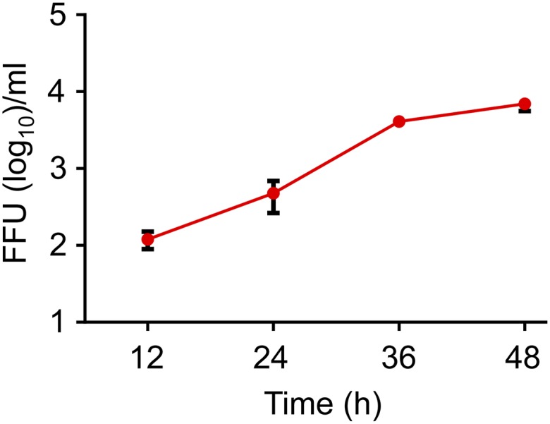
Release of HL18NL11 from infected MDCK II cells. MDCK II cells were infected with HL18NL11 (m.o.i. of 0.001). At the indicated times, infectious virus released into the cell culture supernatant was titrated on RIE 1495 cells.
HL18 Mediates Infection of Polarized MDCK II Cells from the Basolateral Site.
Because infection of MDCK II cells with HL18NL11 occurred predominantly at the edges of the cell cultures and inefficiently in confluent cell layers (Fig. 3A), we hypothesized that the basolateral site might be preferentially used for viral entry. Indeed, following intentional disruption of confluent MDCK II cell monolayers, infection with VSV*ΔG-HL18pb predominantly occurred at the site of scratches (Fig. S5A). To provide further evidence for this phenomenon, MDCK II cells were cultured for 3 d on porous Transwell filters, which allowed the formation of a polarized cell monolayer. Filter-grown cells were subsequently infected from either the apical or basolateral site, and infection was monitored by the expression of GFP (Fig. 4A). In line with previous findings (19), recombinant VSV* expressing the original VSV-G envelope protein (Fig. 1A) predominantly entered the cells via the basolateral membrane (Fig. 4A), indicating that the cells were actually fully polarized. When the cells were infected with VSV*ΔG-H5pb-N1, encoding HA and NA of a highly pathogenic avian H5N1 influenza virus (20), virus entry occurred from both sites. In contrast, VSV*ΔG-HL18bp entered polarized MDCK II cells predominantly from the basolateral membrane (Fig. 4A). A similar directional infection pattern was observed with recombinant HL18NL11 (Fig. 4B). Next, we infected MDCK II cells with HL18NL11 (m.o.i. of 0.1) for 50 h and processed the cells for electron-microscopic analysis. Using this approach, release of HL18NL11 particles was found to take place at the apical plasma membrane (Fig. 4C). Spherical but also filamentous HL18NL11 particles were detected. Consistently, HL18 was predominantly found at the apical plasma membrane of polarized MDCK II cells after infection with HL18NL11 (Fig. 4D and Fig. S5B). Similar apical HA localization was observed in polarized MDCK II cells after infection with the mouse-adapted influenza A virus of the H7N7 subtype (A/SC35M) (Fig. S5B). Apical surface expression of HL18 was also evident when polarized MDCK II cells were infected with VSV*ΔG-HL18, whereas the VSV-G protein was specifically transported to the basolateral site of the cell (Fig. S5C), in accordance with a previous report (21). This observation suggests that the apical transport of HL18 is an inherent property of this glycoprotein, which does not depend on other viral factors. Together, these findings indicate that entry and egress of bat influenza A-like virus HL18NL11 occur at opposite sites in polarized MDCK II cells.
Fig. S5.

Directional virus entry and egress. (A) MDCK II cells were grown on cell culture dishes until a confluent monolayer was formed. The cells were scratched with a pipette tip (indicated by the gray lightning symbol) and infected with the indicated chimeric viruses. GFP expression by infected cells was recorded at 7 h p.i. (B) Serial sections of polarized filter-grown MDCK II cells grown on Transwell filter inserts (pore size 0.4 μm) and infected with either HL18NL11 (m.o.i. of 0.3) at the basolateral site for 48 h (Upper) or the conventional influenza A virus A/SC35M (H7N7) at an m.o.i. of 5 at the apical site for 8 h (Lower). HL18 and H7 were detected at the cell surface by HL18- and H7-specific antibodies, respectively. The indicated z scans were taken by using a confocal laser scanning microscope. Numbers indicate the distance from the basal cell border. (C) Localization of newly synthesized HL18 at the apical plasma membrane of polarized MDCK II cells after infection via the basolateral membrane with VSV* or VSV*ΔG-HL18 using an m.o.i. of 10. At 8 h p.i., HL18 was detected by indirect immunofluorescence (red fluorescence). Infected cells are indicated by GFP expression (green fluorescence). XZ scans were taken by using a confocal laser scanning microscope.
Fig. 4.

Entry and egress of chimeric VSV and HL18NL11 in polarized epithelial cells. (A) MDCK II cells grown on porous filter supports (pore size 0.4 μm) were infected with the indicated viruses from either the apical or basolateral site by using an m.o.i. of 10. GFP fluorescence was detected 20 h p.i. (B) Polarized filter-grown MDCK II cells were infected with HL18NL11 (m.o.i. of 0.3) at the basolateral or apical site. The number of infectious foci in the cell layer was determined 2 d p.i. Error bars indicate the mean and SD of two independent experiments. (C) Electron-microscopic analysis of MDCK II cells infected for 50 h with HL18NL11 (m.o.i. of 0.1). Ultrathin section showing pleomorphic virus particles released from the apical cell surface. (D) Localization of HL18 at the apical plasma membrane of polarized filter-grown MDCK II cells 48 h after infection with HL18NL11 (m.o.i. of 0.01). Dashed lines indicate locations of the x and y sections. (E) Cartoon depicting the directional entry and egress of bat influenza A-like viruses in polarized epithelial cells.
Discussion
In this work, we succeeded in propagating infectious HL18NL11 and HL17NL10 bat influenza A-like viruses, demonstrating that the recently identified genomic sequences (6, 8) encode fully infectious viruses. This achievement was possible after screening for susceptible cell lines by using autonomously replicating VSV chimeras lacking the VSV entry protein G and encoding instead either HL17 or HL18.
Although the HL17 and HL18 envelope glycoproteins lack sialic acid-binding activity, they share a similar structure with the HA of conventional influenza A viruses (8, 11). Based on VSV chimeras encoding HL18 or HL17, our data suggest that HL proteins and classical influenza A virus HA also share functional characteristics. Like HA, HL proteins are bifunctional proteins exhibiting receptor-binding and membrane-fusion activities. Likewise, fusion activity is triggered by a low pH, suggesting that virus particles enter the cells by receptor-mediated endocytosis. However, the role of the NL proteins is still an enigma. VSV chimeras expressing either NL10 or NL11 instead of the VSV-G protein could be propagated on VSV-G protein-expressing helper cells but were unable to propagate on MDCK II cells. Moreover, expression of HL18 along with NL11 had no effect on viral titers, suggesting that VSV*∆G-HL18 does not rely on NL11 for release from infected cells.
Classical influenza A viruses normally enter polarized epithelial cells via the apical plasma membrane (22), in line with the airborne transmission of these viruses. It was therefore surprising to note that VSV*∆G-HL18pb and recombinant HL18NL11 viruses preferentially entered polarized MDCK II cells from the basolateral site. This characteristic probably reflects the basolateral localization of the putative cellular receptor of HL17 and HL18 in MDCK II cells. Several other viruses are also known to use basolateral receptors for entry into epithelial cells, including VSV (19), hepatitis B virus (23, 24), hepatitis C virus (25), adenovirus type 2 and 5 (26), vaccinia virus (27), and measles virus (28). Interestingly, many of these viruses exploit junctional and adhesion proteins of the host cell for attachment and entry (29). However, to invade the host, the epithelial barrier must be overcome. Some viruses therefore make use of the parenteral route (30, 31) or first replicate in tissue macrophages or other nonepithelial cells (32). Experimental infection of bats with recombinant influenza A-like viruses will show what the most likely route of transmission is and in which organ these viruses preferentially replicate.
Despite basolateral entry, newly synthesized HL18 was found to be transported to the apical plasma membrane of polarized MDCK II cells where budding and release of the virus took place. In this regard, HL18NL11 (and probably HL17NL10) resembles conventional influenza A viruses, which also bud from the apical domain, specifically from cholesterol-enriched plasma membrane domains where the envelope glycoproteins HA and NA accumulate (33). The opposite sites of virus entry and release may explain, at least partially, why infection of the MDCK II cell monolayer with HL18NL11 was less robust than infection with conventional influenza A viruses. Following release from the apical site, viral particles may have only limited access to the basolateral site to initiate another round of infection (Fig. 4E).
Many of the cell lines tested turned out to be resistant to infection with the VSV chimeras or the recombinant bat influenza A-like viruses HL17NL10 and HL18NL11 (Table S1), indicating that these viruses have a restricted cell tropism, at least in vitro. HL17 and HL18 mediated a similar cell tropism, suggesting that they bind to the same receptor. However, compared with VSV-expressing HL17, VSV-expressing HL18 always performed better in terms of virus spreading, low pH-induced syncytia formation, and production of infectious virus titers. This difference might be attributed to a slight conformational defect of HL17 as the primary sequence may still contain cloning errors. The lack of both VSV*ΔG-HL17 spreading in MDCK II cells and low pH-induced syncytia formation in the presence of trypsin may also be attributed to an improper conformation of HL17.
Our observation that cell lines derived from kidney epithelium, bronchial epithelium, malignant glioma, and malignant melanoma are susceptible to bat influenza A-like viruses or VSV*ΔG-HL17pb/VSV*ΔG-HL18pb also suggests that expression of the putative receptor might not be restricted to a certain tissue or cell type only. Our finding that at least two human cell lines were susceptible to infection with HL18NL11 and HL17NL10 suggests that a potential infection of humans with influenza A-like viruses cannot be ruled out.
The nature of the cellular receptor or receptors that mediate infection of bat influenza A-like viruses is still unknown. However, it is evident from our data and data of others (8, 11–13) that sialic acid does not serve as the receptor determinant for these viruses. Rather than destroying any cellular receptors, sialidase treatment significantly enhanced the infection of MDCK II cells by VSV*∆G-HL18pb and recombinant bat influenza A-like viruses. The reason for this phenomenon is not yet known. However, many cell surface molecules contain complex oligosaccharides with sialic acids. Because the putative receptor-binding domain of the HL proteins contains negatively charged amino acids (8, 11), the removal of the negatively charged sialic acids may facilitate binding of the HL proteins to the putative cellular receptor. Interestingly, the related MDCK I cell line turned out to be resistant to infection with HL18NL11 or HL17NL10. MDCK I cells have been isolated from low passage parental MDCK cells and form tight monolayers displaying a high transepithelial electrical resistance (TER) of more than 4,000 Ω⋅cm2. MDCK II cells originate from higher passage MDCK cells and are characterized by TER values of less than 300 Ω⋅cm2 (34, 35). The two MDCK sublines also differ in other aspects such as glycosphingolipid composition (36, 37), protein composition (38, 39), and susceptibility to influenza C virus infection (40). The two MDCK sublines might be instrumental for the identification of the cellular receptor by comparative transcriptomics and ectopic expression of candidate genes.
In summary, we identified several mammalian cell lines, which allowed recovery and propagation of recombinant HL17NL10 and HL18NL11. These influenza A-like viruses revealed similarities but also striking dissimilarities to conventional influenza A viruses. The established reverse genetic system will undoubtedly help to further characterize these hitherto uncultivable viruses. Experimental infection of bats will certainly provide deeper insight into the tropism of the viruses in the natural host and their mode of transmission. The identification and characterization of cellular receptors mediating virus attachment and entry is important to understand the tissue tropism and zoonotic potential of these viruses. Finally, the mysterious role of the NL protein in the viral replication cycle could now be investigated by taking advantage of reverse genetics.
Materials and Methods
Generation of Chimeric VSV with a Foreign Envelope.
MDCK II cells were inoculated for 2 h with VSV*ΔG-HL or VSVΔG-HL-sNLuc (m.o.i. of 5) that had been propagated on BHK-G43 helper cells (expressing VSV-G protein). The cells were washed once with minimal essential medium (MEM) and then incubated for 30 min with a VSV-G protein-specific monoclonal antibody (1:100) with wild-type VSV neutralizing activity (clone I1, ATCC) and subsequently washed and maintained for 24 h in serum-free medium optionally supplemented with 1 µg/mL of acetylated trypsin (Sigma). The cell culture supernatant was collected and clarified by low-speed centrifugation, and viral titers were determined on subconfluent MDCK II cells. All work with recombinant VSV expressing authentic and modified HL proteins were performed according to biosafety level 2.
Generation of Recombinant Bat Influenza A-Like Viruses.
Recombinant bat influenza A-like viruses were generated on HEK 293T cells by using the eight plasmids reverse-genetics system as described (15, 16) and concentrated by ultracentrifugation. Recombinant influenza A-like viruses HL17NL10 and HL18NL11 containing the authentic HL proteolytic cleavage site were handled in laboratories complying to biosafety level 2.
Ethics Statement.
Animal trials were performed in compliance with the Swiss animal protection law and approved by the animal welfare committee of the Canton of Bern (authorization no. BE76/10 and BE119/13).
SI Materials and Methods
Cells.
BHK-21, A549, and A498 cells were obtained from the German Cell Culture Collection (DSZM); HEK 293T, PK-15, Vero cells, HeLa, Caco-2, SH-SY5Y, SK-Mel-28, D-17, CHO, Calu-3, and DF-1 cells were from the American Type Culture Collection (ATCC); and MCF-7 cells were purchased from the European cell culture collection (ECACC). MDCK I, MDCK II, RIE 1495, LLC-PK1, and 16HBE14o cells were obtained from Georg Herrler, University of Veterinary Medicine Hannover, Hannover, Germany. MDCK I cells are characterized by expression of gp40/podoplanin, whereas MDCK II cells lack this protein (38). MDCK II cells produce the Forssman glycosphingolipid, which is absent from MDCK I cells (36). RIE 1495 cells [stored in the “Collection of Cell Lines in Veterinary Medicine” (CCLV) at the Friedrich-Loeffler Institute in Greifswald-Insel Riems, Germany with the code CCLV-RIE 1495] are canine epithelial cells. Morphologically, they resemble MDCK II cells and express the Forssman antigen; however, they do not establish polarized monolayers when grown on Transwell filter systems. U-373 MG, U-87 MG, and U-118 MG cells were originally from ATCC and provided by Dorothee Holm-von Laer, University of Innsbruck, Innsbruck, Austria. Normal human dermal fibroblasts (NHDF) were purchased from Lonza, Basel, Switzerland. The bat cell lines EidNi/41, EpoNi/22.1, CarLu1, CarperAEC.B-3, HypNi/1.1, RhiLu1, and TB1 Lu were obtained from Marcel Müller, University of Bonn, Bonn, Germany. SK-6 cells were provided by M. Pensaert, Ghent University, Merelbeke, Belgium; and PEDSV cells by J. D. Seebach, University of Zürich, Zurich. BHK-G43, a transgenic BHK-21 cell clone expressing the VSV G protein in a regulated manner, has been described (17). The cell lines were maintained as proposed by the ATCC, ECACC, and DSZM.
cDNA.
CDNA (cDNA) encoding the eight RNA segments of A/little yellow-shouldered bat/Guatemala/164/2009 (HL17NL10) and A/flat-faced bat/Peru/033/2010 (HL18NL11) were kindly provided by Ruben Donis, Centers for Disease Control and Prevention, Atlanta (GenBank accession nos. CY103880–CY103888 and CY125943–CY125949, respectively).
Generation of Recombinant VSV Vectors.
The ORFs of either the HL17 or the HL18 protein were amplified by PCR and inserted into the pVSV* plasmid using single MluI and BstEII restriction sites upstream and downstream of the fourth transcription unit, thereby replacing the VSV G gene (41). The resulting plasmids are referred to as pVSV*ΔG-HL17 and pVSV*ΔG-HL18. Polybasic proteolytic cleavage sites were introduced into the HL genes by overlapping PCR method (42), resulting in the plasmids pVSV*ΔG-HL17pb and pVSV*ΔG-HL18pb. Additional genomic cDNAs were generated by replacing the GFP gene in these plasmids by the secreted Nano luciferase gene (sNLuc, Promega). The identity of the recombinant VSV genomes was confirmed by DNA sequencing. Infectious VSV was recovered from transfected cells and propagated in BHK-G43 helper cells (43). Infectious virus titers were determined on BHK-21 cells and expressed as focus-forming units per milliliter (FFU/mL).
Antibodies and Generation of Immune Sera.
Immune sera directed against HL17 and HL18 were produced by immunizing specific pathogen-free white Leghorn chickens with 108 FFU of VSV*ΔG-HL17 or pVSV*ΔG-HL18 via the intramuscular route. Chicken immune sera directed against H5 or H7 has been described recently (44). Polyclonal NP-specific antibodies were purchased from Thermo Fischer (catalog no. PA5-32242).
Immunofluorescence Analysis.
For staining of HL proteins, infected cells were fixed with 4% (wt/vol) paraformaldehyde for 10 min, subsequently washed with PBS, permeabilized with 0.5% Triton X-100 for 5 min, and incubated for 1 h at room temperature with either anti-HL17 or anti-HL18 immune serum (1:250). For staining of the influenza virus nucleoprotein (NP), permeabilized cells were incubated for 1 h at room temperature with polyclonal anti-NP antibody (1:2,000). H7 antigen was detected by anti H7 immune serum (1:250). For cell surface staining of HL or HA, treatment with Triton X-100 was omitted. For detection of the primary antibodies, Alexa Fluor 488 or Alexa Fluor 546-labeled anti-chicken IgY (1:500) or anti-rabbit IgG (1:200) were used. Nuclei were stained for 5 min with diamidino-2-phenylindole (DAPI; Sigma; 0.1 μg/mL in ethanol) (1:10,000). Finally, the cells were washed with distilled water and embedded in Mowiol 4–88 (Sigma) mounting medium.
Electron Microscopy.
MDCK II cells infected with HL18NL11 were fixed with aldehydes, postfixed with 1% osmium tetraoxide, dehydrated in a graded ethanol series, and embedded in a mixture of Epon and Araldite. Ultrathin sections were contrast-stained with uranyl acetate and lead citrate, and electron micrographs were acquired by using a JEM1400 or Zeiss 109 transmission electron microscope as described (45).
Western Blot Analysis.
Vero cells grown in T-75 cell culture flasks were infected with recombinant HL-expressing VSV (m.o.i. of 5) and incubated in serum-free GMEM in the presence of 1 µg/mL of acetylated trypsin (Sigma). At 24 h p.i., the cell-culture supernatant was harvested and clarified by low-speed centrifugation. Virus particles were pelleted through a 20% (wt/wt) sucrose cushion by ultracentrifugation (105,000 × g, 60 min, 4 °C) and dissolved in 100 µL of SDS sample buffer containing 100 mM DTT. The viral proteins were separated by SDS 12% (wt/vol) polyacrylamide gel electrophoresis (PAGE) and transferred to nitrocellulose membranes by semidry blotting. The nitrocellulose membranes were blocked overnight at 4 °C with Odyssey Blocking Reagent (LI-COR Biosciences) and incubated with chicken immune sera directed to HL17 or HL18 (1:1,000) and subsequently with IRDye 800CW-labeled donkey anti-chicken IgY serum (1:5,000). The membranes were thoroughly washed with PBS containing 0.1% Tween-20 and then imaged with the Odyssey Infrared Imaging System (LICOR).
Virus Neutralization.
All immune sera were heated for 30 min to 56 °C to inactivate complement factors. Twofold serial dilutions of the immune sera were prepared in MEM containing 1% FBS. The diluted immune sera (50 µl) were incubated for 30 min at 37 °C with 50 µl of chimeric virus containing 100 FFU and subsequently inoculated for 60 min at 37 °C with subconfluent MDCK II cells that were grown in 96-well microtiter plates. The cells were washed twice with MEM and maintained for 20 h at 37 °C with 100 µl of MEM containing 5% (vol/vol) FBS. For detection of secreted Nano luciferase activity, 25 µL of cell culture supernatant was mixed with 25 µL of Nano-Glo luciferase substrate (Promega) and transferred to black 96-well microplates. Luminescence was recorded with a Centro LB 960 microplate luminometer (Berthold Technologies). HL17NL10 and HL18NL11 (2,000 FFU) were incubated with diluted antibodies for 1 h at 37 °C and subsequently inoculated with confluent sialidase-treated RIE 1495 cells in 96-well plates. Virus neutralization was measured 2 d p.i. by reduction of FFU.
Sialidase Treatment.
Confluent cells were treated with 200 mU/mL of bacterial sialidase (Sigma, catalog no. N3001) diluted in PBS for 1 h at 37 °C.
Sequencing of Bat Influenza A-Like Segments.
Cell culture supernatant of RIE 1495 cells pretreated with sialidase and infected with either HL18NL11 or HL17NL10 (m.o.i of 0.001 FFU) was harvested 48 h after infection, and RNA was extracted by using Direct-zol RNA MiniPrep Kit (Zymo Research). HA, NA, and M segments were amplified by using OneStep RT-PCR Kit (QIAGEN) and the following forward (F) and reverse (R) primers sequences and melting temperature (Tm): HL18 (F: 5′-AGCAGAAGCAGGGTGATTATT-3′, R: 5′-TTTTTGCAATAATAGATTGACAT-3′, Tm: 61 °C); HL17 (F: 5′-AAGGAGCTGATTGTCCTACTAATCCTTCTC-3′, R: 5′-CTATATACATATTTTACATTGGATTGAGCC-3′, Tm: 61 °C); NL11 (F: 5′-AGCAGAAGCAGGAGTTTTTCA-3′, R: 5′-AGTAGAAACAAGGAGTTTT-3′, Tm: 58 °C); NL10 (F: 5′-ATGTCTATCAACGGAACGACATGTCTACTC-3′, R: 5′-TCATGAATCCCAATTTATATTCTGAACAGG-3′, Tm: 58 °C); M for HL18NL11 (F: 5′-AGCAGAAGCAGGCATTATTCA-3′, R: 5′-CGATGATAGTCATAATTGGATCTGG-3′, Tm: 61 °C); and M for HL17NL10 (F: 5′-AGCAGAAGCAGGCATTATCCA-3′, R: 5′-CCATGATTACTATGATAGGGTCCGG-3′, Tm: 61 °C). PCR amplification products were purified from agarose gels by using a DNA extraction kit (Zymo Research) and directly sequenced (GATC Biotech).
Polykaryon Formation Assay.
Vero and MDCK II cells were infected with chimeric VSV by using an m.o.i. of 1 (1 FFU per cell). At 5 h p.i., cells were treated for 1 h at 37 °C with acetylated trypsin (10 µg/mL) or were left untreated. The cells were exposed for 10 min to low pH buffer (50 mM Mes, 150 mM NaCl, pH 5.2) and subsequently incubated for 90 min at 37 °C with MEM containing 5% (vol/vol) FBS. The cells were fixed with 3% (wt/vol) paraformaldehyde and nuclei stained for 5 min at 37 °C with DAPI. The cells were washed with distilled water, embedded in Mowiol 4–88 mounting medium, and investigated with an inverted fluorescence microscope (Zeiss, Cell Observer).
Acknowledgments
We thank Claudia Kastenholz, Sebastian Giese, Ran Wei, and Vincent Idstein (University of Freiburg) for technical assistance; Wenjun Ma (Kansas State University) for kindly providing the HL18NL11 rescue plasmids; Quinnlan David (University of Freiburg) for critical reading of the manuscript; and Marcel Müller (University of Bonn) for providing the bat cells. This work was supported by Deutsche Forschungsgemeinschaft SCHW 632/17-1 (to M.S.) and the Excellence Initiative of the German Research Foundation (GSC-4, Spemann Graduate School) (E.A.M. and H.B.). This work was also partially supported by the Center for Research on Influenza Pathogenesis and National Institute of Allergy and Infectious Diseases-funded Center of Excellence on Influenza Research and Pathogenesis via Contract HHSN272201400008C (to A.G.S.) and by the Swiss Federal Food Safety and Veterinary Office project 1.13.09 (to G.Z.).
Footnotes
The authors declare no conflict of interest.
This article is a PNAS Direct Submission.
This article contains supporting information online at www.pnas.org/lookup/suppl/doi:10.1073/pnas.1608821113/-/DCSupplemental.
References
- 1.Jones KE, Purvis A, MacLarnon A, Bininda-Emonds OR, Simmons NB. A phylogenetic supertree of the bats (Mammalia: Chiroptera) Biol Rev Camb Philos Soc. 2002;77(2):223–259. doi: 10.1017/s1464793101005899. [DOI] [PubMed] [Google Scholar]
- 2.Li W, et al. Bats are natural reservoirs of SARS-like coronaviruses. Science. 2005;310(5748):676–679. doi: 10.1126/science.1118391. [DOI] [PubMed] [Google Scholar]
- 3.Turmelle AS, Olival KJ. Correlates of viral richness in bats (order Chiroptera) EcoHealth. 2009;6(4):522–539. doi: 10.1007/s10393-009-0263-8. [DOI] [PMC free article] [PubMed] [Google Scholar]
- 4.Smith I, Wang LF. Bats and their virome: An important source of emerging viruses capable of infecting humans. Curr Opin Virol. 2013;3(1):84–91. doi: 10.1016/j.coviro.2012.11.006. [DOI] [PMC free article] [PubMed] [Google Scholar]
- 5.Plowright RK, et al. Ecological dynamics of emerging bat virus spillover. Proc Biol Sci. 2015;282(1798):20142124. doi: 10.1098/rspb.2014.2124. [DOI] [PMC free article] [PubMed] [Google Scholar]
- 6.Tong S, et al. A distinct lineage of influenza A virus from bats. Proc Natl Acad Sci USA. 2012;109(11):4269–4274. doi: 10.1073/pnas.1116200109. [DOI] [PMC free article] [PubMed] [Google Scholar]
- 7.Ma W, García-Sastre A, Schwemmle M. Expected and unexpected features of the newly discovered bat influenza A-like viruses. PLoS Pathog. 2015;11(6):e1004819. doi: 10.1371/journal.ppat.1004819. [DOI] [PMC free article] [PubMed] [Google Scholar]
- 8.Tong S, et al. New world bats harbor diverse influenza A viruses. PLoS Pathog. 2013;9(10):e1003657. doi: 10.1371/journal.ppat.1003657. [DOI] [PMC free article] [PubMed] [Google Scholar]
- 9.Li Q, et al. Structural and functional characterization of neuraminidase-like molecule N10 derived from bat influenza A virus. Proc Natl Acad Sci USA. 2012;109(46):18897–18902. doi: 10.1073/pnas.1211037109. [DOI] [PMC free article] [PubMed] [Google Scholar]
- 10.Zhu X, et al. Crystal structures of two subtype N10 neuraminidase-like proteins from bat influenza A viruses reveal a diverged putative active site. Proc Natl Acad Sci USA. 2012;109(46):18903–18908. doi: 10.1073/pnas.1212579109. [DOI] [PMC free article] [PubMed] [Google Scholar]
- 11.Zhu X, et al. Hemagglutinin homologue from H17N10 bat influenza virus exhibits divergent receptor-binding and pH-dependent fusion activities. Proc Natl Acad Sci USA. 2013;110(4):1458–1463. doi: 10.1073/pnas.1218509110. [DOI] [PMC free article] [PubMed] [Google Scholar]
- 12.Maruyama J, et al. Characterization of the glycoproteins of bat-derived influenza viruses. Virology. 2016;488:43–50. doi: 10.1016/j.virol.2015.11.002. [DOI] [PMC free article] [PubMed] [Google Scholar]
- 13.Hoffmann M, et al. The hemagglutinin of bat-associated influenza viruses is activated by TMPRSS2 for pH-dependent entry into bat but not human cells. PLoS One. 2016;11(3):e0152134. doi: 10.1371/journal.pone.0152134. [DOI] [PMC free article] [PubMed] [Google Scholar]
- 14.Sun X, et al. Bat-derived influenza hemagglutinin H17 does not bind canonical avian or human receptors and most likely uses a unique entry mechanism. Cell Reports. 2013;3(3):769–778. doi: 10.1016/j.celrep.2013.01.025. [DOI] [PubMed] [Google Scholar]
- 15.Juozapaitis M, et al. An infectious bat-derived chimeric influenza virus harbouring the entry machinery of an influenza A virus. Nat Commun. 2014;5:4448. doi: 10.1038/ncomms5448. [DOI] [PMC free article] [PubMed] [Google Scholar]
- 16.Zhou B, et al. Characterization of uncultivable bat influenza virus using a replicative synthetic virus. PLoS Pathog. 2014;10(10):e1004420. doi: 10.1371/journal.ppat.1004420. [DOI] [PMC free article] [PubMed] [Google Scholar]
- 17.Hanika A, et al. Use of influenza C virus glycoprotein HEF for generation of vesicular stomatitis virus pseudotypes. J Gen Virol. 2005;86(Pt 5):1455–1465. doi: 10.1099/vir.0.80788-0. [DOI] [PubMed] [Google Scholar]
- 18.Dukes JD, Whitley P, Chalmers AD. The MDCK variety pack: Choosing the right strain. BMC Cell Biol. 2011;12:43. doi: 10.1186/1471-2121-12-43. [DOI] [PMC free article] [PubMed] [Google Scholar]
- 19.Fuller S, von Bonsdorff CH, Simons K. Vesicular stomatitis virus infects and matures only through the basolateral surface of the polarized epithelial cell line, MDCK. Cell. 1984;38(1):65–77. doi: 10.1016/0092-8674(84)90527-0. [DOI] [PubMed] [Google Scholar]
- 20.Zimmer G, Locher S, Berger Rentsch M, Halbherr SJ. Pseudotyping of vesicular stomatitis virus with the envelope glycoproteins of highly pathogenic avian influenza viruses. J Gen Virol. 2014;95(Pt 8):1634–1639. doi: 10.1099/vir.0.065201-0. [DOI] [PubMed] [Google Scholar]
- 21.Rindler MJ, Ivanov IE, Plesken H, Rodriguez-Boulan E, Sabatini DD. Viral glycoproteins destined for apical or basolateral plasma membrane domains traverse the same Golgi apparatus during their intracellular transport in doubly infected Madin-Darby canine kidney cells. J Cell Biol. 1984;98(4):1304–1319. doi: 10.1083/jcb.98.4.1304. [DOI] [PMC free article] [PubMed] [Google Scholar]
- 22.Slepushkin VA, Staber PD, Wang G, McCray PB, Jr, Davidson BL. Infection of human airway epithelia with H1N1, H2N2, and H3N2 influenza A virus strains. Mol Ther. 2001;3(3):395–402. doi: 10.1006/mthe.2001.0277. [DOI] [PMC free article] [PubMed] [Google Scholar]
- 23.Schulze A, Mills K, Weiss TS, Urban S. Hepatocyte polarization is essential for the productive entry of the hepatitis B virus. Hepatology. 2012;55(2):373–383. doi: 10.1002/hep.24707. [DOI] [PubMed] [Google Scholar]
- 24.Okuyama-Dobashi K, et al. Hepatitis B virus efficiently infects non-adherent hepatoma cells via human sodium taurocholate cotransporting polypeptide. Sci Rep. 2015;5:17047. doi: 10.1038/srep17047. [DOI] [PMC free article] [PubMed] [Google Scholar]
- 25.Harris HJ, et al. Claudin association with CD81 defines hepatitis C virus entry. J Biol Chem. 2010;285(27):21092–21102. doi: 10.1074/jbc.M110.104836. [DOI] [PMC free article] [PubMed] [Google Scholar]
- 26.Walters RW, et al. Basolateral localization of fiber receptors limits adenovirus infection from the apical surface of airway epithelia. J Biol Chem. 1999;274(15):10219–10226. doi: 10.1074/jbc.274.15.10219. [DOI] [PubMed] [Google Scholar]
- 27.Vermeer PD, et al. Vaccinia virus entry, exit, and interaction with differentiated human airway epithelia. J Virol. 2007;81(18):9891–9899. doi: 10.1128/JVI.00601-07. [DOI] [PMC free article] [PubMed] [Google Scholar]
- 28.Ludlow M, et al. Wild-type measles virus infection of primary epithelial cells occurs via the basolateral surface without syncytium formation or release of infectious virus. J Gen Virol. 2010;91(Pt 4):971–979. doi: 10.1099/vir.0.016428-0. [DOI] [PubMed] [Google Scholar]
- 29.Torres-Flores JM, Arias CF. Tight junctions go viral! Viruses. 2015;7(9):5145–5154. doi: 10.3390/v7092865. [DOI] [PMC free article] [PubMed] [Google Scholar]
- 30.Hwang SJ. Hepatitis C virus infection: An overview. J Microbiol Immunol Infect. 2001;34(4):227–234. [PubMed] [Google Scholar]
- 31.Candotti D, Allain JP. Transfusion-transmitted hepatitis B virus infection. J Hepatol. 2009;51(4):798–809. doi: 10.1016/j.jhep.2009.05.020. [DOI] [PubMed] [Google Scholar]
- 32.de Vries RD, Mesman AW, Geijtenbeek TB, Duprex WP, de Swart RL. The pathogenesis of measles. Curr Opin Virol. 2012;2(3):248–255. doi: 10.1016/j.coviro.2012.03.005. [DOI] [PubMed] [Google Scholar]
- 33.Rossman JS, Lamb RA. Influenza virus assembly and budding. Virology. 2011;411(2):229–236. doi: 10.1016/j.virol.2010.12.003. [DOI] [PMC free article] [PubMed] [Google Scholar]
- 34.Barker G, Simmons NL. Identification of two strains of cultured canine renal epithelial cells (MDCK cells) which display entirely different physiological properties. Q J Exp Physiol. 1981;66(1):61–72. doi: 10.1113/expphysiol.1981.sp002529. [DOI] [PubMed] [Google Scholar]
- 35.Richardson JC, Scalera V, Simmons NL. Identification of two strains of MDCK cells which resemble separate nephron tubule segments. Biochim Biophys Acta. 1981;673(1):26–36. [PubMed] [Google Scholar]
- 36.Hansson GC, Simons K, van Meer G. Two strains of the Madin-Darby canine kidney (MDCK) cell line have distinct glycosphingolipid compositions. EMBO J. 1986;5(3):483–489. doi: 10.1002/j.1460-2075.1986.tb04237.x. [DOI] [PMC free article] [PubMed] [Google Scholar]
- 37.Nichols GE, Lovejoy JC, Borgman CA, Sanders JM, Young WW., Jr Isolation and characterization of two types of MDCK epithelial cell clones based on glycosphingolipid pattern. Biochim Biophys Acta. 1986;887(1):1–12. doi: 10.1016/0167-4889(86)90115-1. [DOI] [PubMed] [Google Scholar]
- 38.Zimmer G, Lottspeich F, Maisner A, Klenk HD, Herrler G. Molecular characterization of gp40, a mucin-type glycoprotein from the apical plasma membrane of Madin-Darby canine kidney cells (type I) Biochem J. 1997;326(Pt 1):99–108. doi: 10.1042/bj3260099. [DOI] [PMC free article] [PubMed] [Google Scholar]
- 39.Furuse M, Furuse K, Sasaki H, Tsukita S. Conversion of zonulae occludentes from tight to leaky strand type by introducing claudin-2 into Madin-Darby canine kidney I cells. J Cell Biol. 2001;153(2):263–272. doi: 10.1083/jcb.153.2.263. [DOI] [PMC free article] [PubMed] [Google Scholar]
- 40.Szepanski S, Gross HJ, Brossmer R, Klenk HD, Herrler G. A single point mutation of the influenza C virus glycoprotein (HEF) changes the viral receptor-binding activity. Virology. 1992;188(1):85–92. doi: 10.1016/0042-6822(92)90737-A. [DOI] [PMC free article] [PubMed] [Google Scholar]
- 41.Kalhoro NH, Veits J, Rautenschlein S, Zimmer G. A recombinant vesicular stomatitis virus replicon vaccine protects chickens from highly pathogenic avian influenza virus (H7N1) Vaccine. 2009;27(8):1174–1183. doi: 10.1016/j.vaccine.2008.12.019. [DOI] [PubMed] [Google Scholar]
- 42.Ho SN, Hunt HD, Horton RM, Pullen JK, Pease LR. Site-directed mutagenesis by overlap extension using the polymerase chain reaction. Gene. 1989;77(1):51–59. doi: 10.1016/0378-1119(89)90358-2. [DOI] [PubMed] [Google Scholar]
- 43.Berger Rentsch M, Zimmer G. A vesicular stomatitis virus replicon-based bioassay for the rapid and sensitive determination of multi-species type I interferon. PLoS One. 2011;6(10):e25858. doi: 10.1371/journal.pone.0025858. [DOI] [PMC free article] [PubMed] [Google Scholar]
- 44.Halbherr SJ, et al. Biological and protective properties of immune sera directed to the influenza virus neuraminidase. J Virol. 2015;89(3):1550–1563. doi: 10.1128/JVI.02949-14. [DOI] [PMC free article] [PubMed] [Google Scholar]
- 45.Kolesnikova L, et al. Influenza virus budding from the tips of cellular microvilli in differentiated human airway epithelial cells. J Gen Virol. 2013;94(Pt 5):971–976. doi: 10.1099/vir.0.049239-0. [DOI] [PubMed] [Google Scholar]