Abstract
Objective
To quantify the effects of a fully implanted pulse generator to activate or augment actions of hip, knee and ankle muscles after stroke.
Design
The subject was a 64 year old male with left hemiparesis resulting from hemorrhagic stroke 21 months prior to participation. He received an 8-channel implanted pulse generator (IPG) and intramuscular stimulating electrodes targeting unilateral hip, knee, and ankle muscles on the paretic side. After implantation, a stimulation pattern was customized to assist with hip, knee, and ankle movement during gait.
The subject served as his own concurrent and longitudinal control with and without stimulation. Outcome measures included 10m walk and 6 minute timed walk to assess gait speed, maximum walk time and distance to measure endurance, and quantitative motion analysis to evaluate spatial-temporal characteristics. Assessments were repeated under three conditions: 1) volitional walking at baseline, 2) volitional walking after training, and 3) walking with stimulation after training.
Results
Volitional gait speed improved with training from 0.29m/s to 0.35m/s and further increased to 0.72m/s with stimulation. Most spatial-temporal characteristics improved and represented more symmetrical and dynamic gait.
Conclusions
These data suggest a multi-joint approach to implanted neuroprostheses can provide clinically relevant improvements in gait after stroke.
Keywords: Stroke, neuroprosthesis, gait, implanted electrodes, hip control
Introduction
Stroke is a leading cause of disability and approximately 30% of stroke survivors require assistance to walk1. Walking is impaired by compromised volitional joint control2, impaired muscle recruitment3, and abnormal tone4 affecting function at the hip, knee, and ankle. The resulting joint incoordination alters speed, stride length, cadence and all phases of gait5. Gait impairments increase risk of falls6, and decrease independence and ability to participate in the community7. The reduced physical activity after stroke further diminishes general health8. Walking speed is correlated with different levels of community walking7, and improving gait and community interaction can have a significant effect on health, quality of life9 and participation.
Improving gait requires a therapy or intervention that addresses impairments at multiple joints simultaneously. Therapeutic benefits are those maintained after an intervention is removed, while neuroprosthetic benefits are only derived through the use of an assistive device and are not realized when the device is not in use. Neuroprosthetic interventions in stroke employing electrical stimulation have primarily focused on dorsiflexion assistance to prevent foot drop. Although dorsiflexion assist with common peroneal nerve stimulation can positively impact walking, its neuroprosthetic10 and therapeutic11,12 effects are comparable to an ankle foot orthosis. Surface and implantable peroneal nerve stimulators are commercially available to prevent foot drop, but many patients have deficits at the hip, knee, and ankle that require multi-joint stimulation to realize significant benefit13.
The development of multichannel implantable pulse generators (IPG) could provide an opportunity to improve walking function in moderately and severely impaired patients by providing assistance to compensate for multiple gait deficits at once14. For example, activating knee flexors in early swing has the potential to correct stiff-legged gait to improve toe clearance during swing15 and reduce the chance of tripping and falling. Activation of knee and hip extensors during stance improves stability and can prevent the leg from buckling, also reducing the chance of falls. Additionally, activation of hip flexors and extensors and plantarflexors provide the majority of the power to move the body forward to improve walking speed16,17. Thus, the application of multichannel IPG’s could significantly improve post-stroke gait by correcting deficits at the hip, knee and ankle to simultaneously prevent falls, improve stability, and increase speed.
Multi-joint movement by means of stimulation with percutaneous electrodes has therapeutically improved joint kinematics18, and increased walking distance19. Similarly, multi-joint movement with surface stimulation has been shown to have a neuroprosthetic effect by reducing impairments, including increased knee and hip flexion during swing, and knee stability and ankle push-off during stance13.
This is the first report of the neuroprosthetic and therapeutic effects of a fully implanted eight channel pulse generator for multi-joint movement to assist walking after stroke. We hypothesized that there would be both a therapeutic effect from gait training with stimulation over time, as well as a neuroprosthetic effect while interactively using the stimulation during walking. The research protocol was approved by the medical center’s Institutional Review Board and subject’s consent was obtained and documented prior to participation in the study.
Methods
Participant
The participant was a 64 year old male, 1 year and 9 months post hemorrhagic stroke affecting the right lateral basal ganglia and frontal cortex. He exhibited left sided hemiparesis and left hemisensory deficits. Proprioception was absent in the ankle and diminished at the knee. Tactile sensation was impaired but not absent on the affected side and was consistent along the extremities. Although his vision was intact, he had left hemineglect. He was on anti-spasticity medication.
He was a household ambulator with a straight cane or a quad cane with contact guard assistance. However, he was transported in a wheelchair in the community. He did not fall in the year prior to participating in the study. His gait was limited by a combination of decreased strength, limited independent joint movement and moderate hypertonia. He exhibited a lower extremity extensor synergy with coupled hip, knee, and ankle extension. He had weak hip and knee flexors and ankle dorsiflexors, walking with a stiff legged gait that was compensated by hip hiking. He wore a double upright ankle foot orthosis to limit foot drop. Baseline manual muscle test (MMT)20 and modified Ashworth scale21 scores for the affected lower extremity are provided in Table 1. Both limbs displayed weakness, but the left side was significantly more impaired. His left upper extremity was severely impaired with no volitional movement and moderate hypertonia. During gait, the scapula was protracted and elbow extended while the affected arm hung anterior to the body. No pain was reported during walking.
Table 1.
MMT and Modified Ashworth Scores
| Contralesional Manual Muscle Strength |
Modified Ashworth Scale |
Ipsilesional Manual Muscle Strength |
|
|---|---|---|---|
| Hip flexion | 2+ | 1 | 4+ |
| Hip extension | 2 | 1 | 4+ |
| Knee flexion | 0 | 1 | 4 |
| Knee extension | 2− | 1 | 4+ |
| Ankle dorsiflexion | 0 | 0 | 4+ |
| Ankle plantarflexion |
0 | 0 | 4+ |
Neuroprosthesis
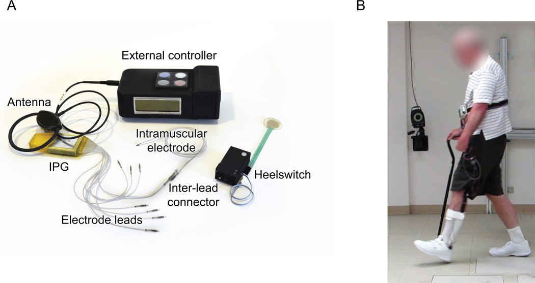
The neuroprosthesis consists of an eight channel IPG22, eight intramuscular electrodes23, and an external control unit (Figure 1a). The IPG produces eight independent channels of current-controlled, charge-balanced biphasic pulses, at a fixed amplitude of 20mA in temporal patterns with variable interpulse interval and pulse duration. The IPG length, width, and depths are 8.9 cm, 3.4 cm, and 1.0 cm respectively. The external control unit transmits power and stimulation commands to the implanted stimulator via a transcutaneous radio-frequency (RF) antenna placed externally over the IPG. Figure 1b shows the participant walking with the system.
Figure 1.
a) Multichannel implantable gait assist system. b) Photograph of the participant walking with the system.
Surgical Planning and Surgical Procedure
Pre-surgical surface and needle stimulation were used to verify that functional lower limb movements could be attained without pain or discomfort. Based on the participant’s motor deficits (Table 1) and the responses to stimulation, the following muscles were targeted for implantation: tensor fasciae latae (hip flexor), sartorius (hip and knee flexor), gluteus maximus (hip extensor), short head of biceps femoris (knee flexor), quadriceps (knee extensor), tibialis anterior/peroneus longus (ankle dorsiflexors), and gastrocnemius (ankle plantar flexor). A single intramuscular electrode was inserted in the quadriceps for knee extension, but the recruitment of specific muscles was not evaluated. One electrode, along with a backup for redundancy, was implanted to activate the common peroneal nerve targeting tibialis anterior and peroneus longus to produce balanced dorsiflexion.
The surgery was performed with the participant under general anesthesia. Motor points for muscles were identified based on anatomical landmarks and neuromuscular anatomy24. A needle probe was used to locate the motor point of each muscle producing a strong isolated contraction. Next a cannula was passed over the probe to a depth just short of the tip; the probe was then removed and the electrode was inserted with a holder that ensured the tip of the electrode ended just beyond the cannula at the motor point. The cannula was removed and the response to the stimulation was confirmed before the lead was tunneled subcutaneously to the lower abdominal area for connection to the IPG. The IPG was inserted into a subfascial pocket on the abdominal wall half way between the anterior superior iliac spine and umbilicus of the subject’s affected side. Prior to closing all incisions, muscle contractions and proper device operation were verified. The subject was discharged after two days post-surgery with unrestricted activity limited only by post-surgical discomfort.
Gait training
After six weeks post-surgery, stimulated responses were measured for each electrode. Joint movements due to stimulation were evaluated while the participant was relaxed and either supine or side-lying. Stimulation produced the expected movements except the electrode targeting the short head of biceps femoris, which produced strong ankle plantarflexion followed by knee flexion, suggesting recruitment of the tibial component of the sciatic nerve. One of the electrodes targeting the common peroneal nerve for dorsiflexion produced a withdrawal response resulting in simultaneous hip and knee flexion. Muscle contraction strength was controlled first by varying pulsewidth to modulate spatial muscle fiber recruitment and second by varying frequency to modulate their temporal summation25. Stimulation frequencies ranged from 20–33Hz with 25Hz used during the early portion of the swing phase and 20Hz during mid and terminal swing. Pulse frequency was 33Hz during the stance phase. Maximum pulsewidths varied from 25–250us, depending on the electrode recruitment properties. Exercise stimulation patterns were created for home use to improve muscle strength and fatigue resistance. Exercise consisted of two patterns that alternated between stimulation to two groups of muscles: 1a) tibialis anterior and peroneus longus, 1b) gastrocnemius and short head of the biceps, and 2a) tensor fasciae latae, sartorius, tibialis anterior and peroneus longus, and 2b) gluteus maximus. Each muscle set was stimulated independently with a burst of pulses at 20HZ for about 200ms followed by a 500ms rest period prior to repeating the cycle. The limb was free to move and no resistance was applied. The participant used each exercise stimulation pattern for half an hour a day while sitting or lying supine.
The participant came to the laboratory once or twice a week for 30 weeks of 2–3 hour sessions of gait training for a total of 46 sessions. The stimulation pattern emulating normal muscle activity during gait was developed and modified heuristically over several sessions according to established protocols26. Multiple changes to the stimulation pattern were made during early sessions of gait training. Stimulation pattern tuning focused on walking coordination and user comfort. During the sessions the therapist worked with the participant to progress from a step-to gait to a two-point gait, learn to shift weight to the affected side, practice starting and stopping walking, and to reduce exaggerated hip and knee flexion and lengthen the step on the unaffected side. Stimulus patterns were adjusted accordingly. During volitional walking the participant used a step-to gait moving his cane, then affected leg, then unaffected leg. He learned to use a two-point gait while walking with stimulation, simultaneously moving the affected leg and the cane.
Step initiation control
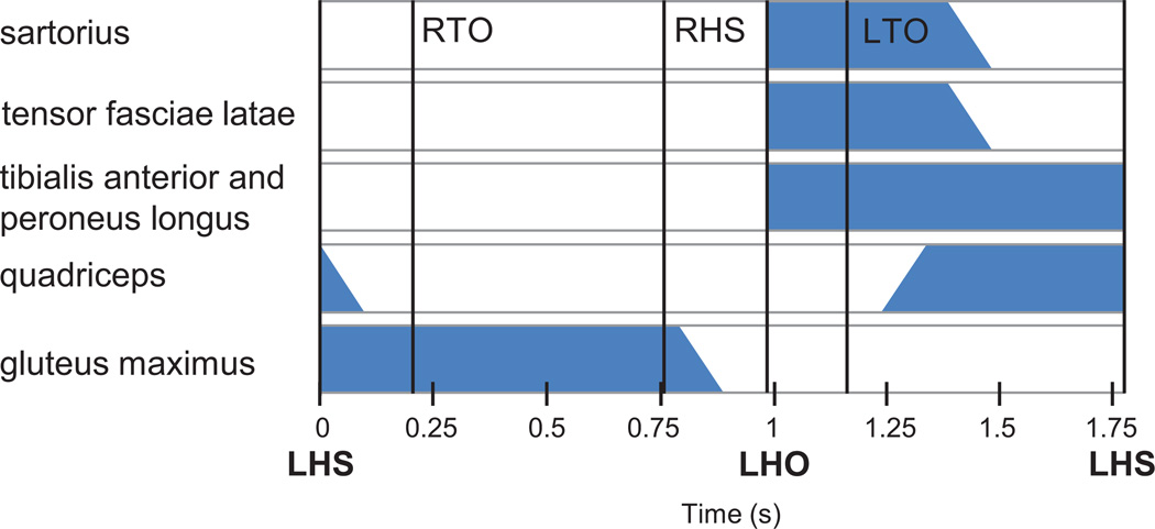
The stimulation pattern is controlled by a heel switch in the sole of the shoe on the affected side. Heel off triggers a pattern associated with the swing phase of gait while initial contact (heel strike) triggers a pattern associated with stance (Figure 2). The swing phase pattern consists of activation of the tensor fasciae latae, sartorius, and tibialis anterior with peroneus longus to provide hip and knee flexion and ankle dorsiflexion. After a delay, tensor fascia latae and sartorius stimulation is ramped down while stimulation to the quadriceps for knee extension is ramped up in preparation for heel strike. Following heel strike, the stance phase pattern consists of turning off tibialis anterior and peroneus longus and ramping down stimulation to quadriceps while initiating gluteus maximus activation for hip extension.
Figure 2.
Stimulation pattern. Left heel off (LHO) and Left heel strike (LHS) trigger the sequential patterns. Left toe off (LTO), right toe off (RTO), and right heel strike (RHS) indicate the average relative timing during the gait cycle.
Outcome assessments
Assessments were performed: 1) at baseline without stimulation prior to gait training, 2) without stimulation after gait training, and 3) with stimulation after gait training. Walking speed was the primary outcome measure. Endurance and spatial-temporal characteristics were secondary measures. Gait speed was measured with a 10m walk test (10MWT) and over a longer distance with a 6 minute walk test (6MWT). The distance walked was recorded at 2 minutes to validate whether walking speed was consistent during the 6MWT or if the participant slowed down. Maximum walking distance until the participant wanted to stop due to fatigue was a measure of endurance. Distance was measured at 2 minute intervals. All measures were taken either in the laboratory (10MWT) or in cleared hospital hallways (6MWT, and maximum distance). Spatial-temporal gait parameters were acquired with a 16 camera Vicon MX40 (Vicon Inc., Oxford, UK) system over an 8m walkway with markers on the heel, toe, and ankle. At least five strides were collected for each trial condition.
Baseline walking only tested volitional effort. Assessments after gait training included walking with and without the IPG turned on. A minimum of five trials were collected for each condition during the 10MWT. Two trials were collected for each condition during the 6MWT. The participant rested 3 minutes between 10MWT and 10 min between 6MWT trials.
Data Analysis
A one way ANOVA with unbalanced replicates was employed to evaluate whether there was a change in the outcome measures between three walking conditions. P values less than 0.05 were considered statistically significant differences. Post-hoc paired t-tests with Bonferroni correction provided comparisons between conditions to determine the contributing effects of the intervention, accounting for the multiple comparisons. Comparison of volitional walking before and after training (conditions 1 and 2) determined the therapeutic effect. Comparing volitional walking and stimulated walking after training (conditions 2 and 3) determined the neuroprosthetic effect. Volitional walking before training and walking with stimulation after training (conditions 1 and 3) were compared to describe the total effect of the multi-joint intervention.
Results
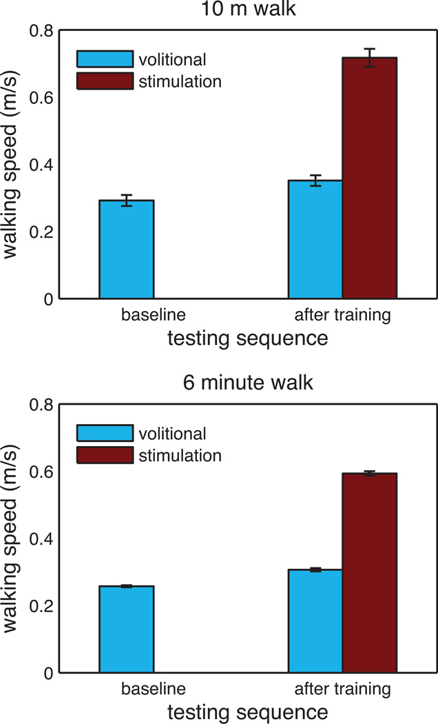
The participant consistently used a two-point gait while walking with stimulation, but it was necessary to complete all volitional assessments using a step-to gait due to safety concerns. Volitional walking with the two-point gait resulted in repeated foot catches and trips which could have produced a fall without providing assistance. There were no toe catches during the assessments, either with or without stimulation using two-point and step-to gait, respectively. There were no falls during the study period. Walking speed was averaged across trials for each walking condition during the 10MWT and 6MWT. Figure 3 shows average walking speed for the three test conditions.
Figure 3.
Gait speed at baseline and after training comparing volitional walking with walking with stimulation. Top graph shows mean (±SD) for 10m walk while the bottom graph shows mean (±SD) for the timed 6 minute walk test.
Comparing the 10MWT baseline walking speed with volitional speed after training showed an increase from 0.29m/s to 0.35m/s (p=0.001). With stimulation, gait speed increased by an additional 0.37m/s to 0.72m/s (p<0.001) during the 10MWT. Thus, there was a total improvement of 0.43m/s (p<0.001) from training and walking with stimulation as compared to baseline volitional walking.
The 6MWT(Figure 3) shows similar effects with a therapeutic increase from 0.26m/s to 0.31m/s (p=0.006), a neuroprosthetic improvement from 0.31m/s to 0.59m/s (p<0.001), and a total improvement from 0.26m/s to 0.59m/s (p<0.001). Walking speed was maintained throughout the 6 minute walk.
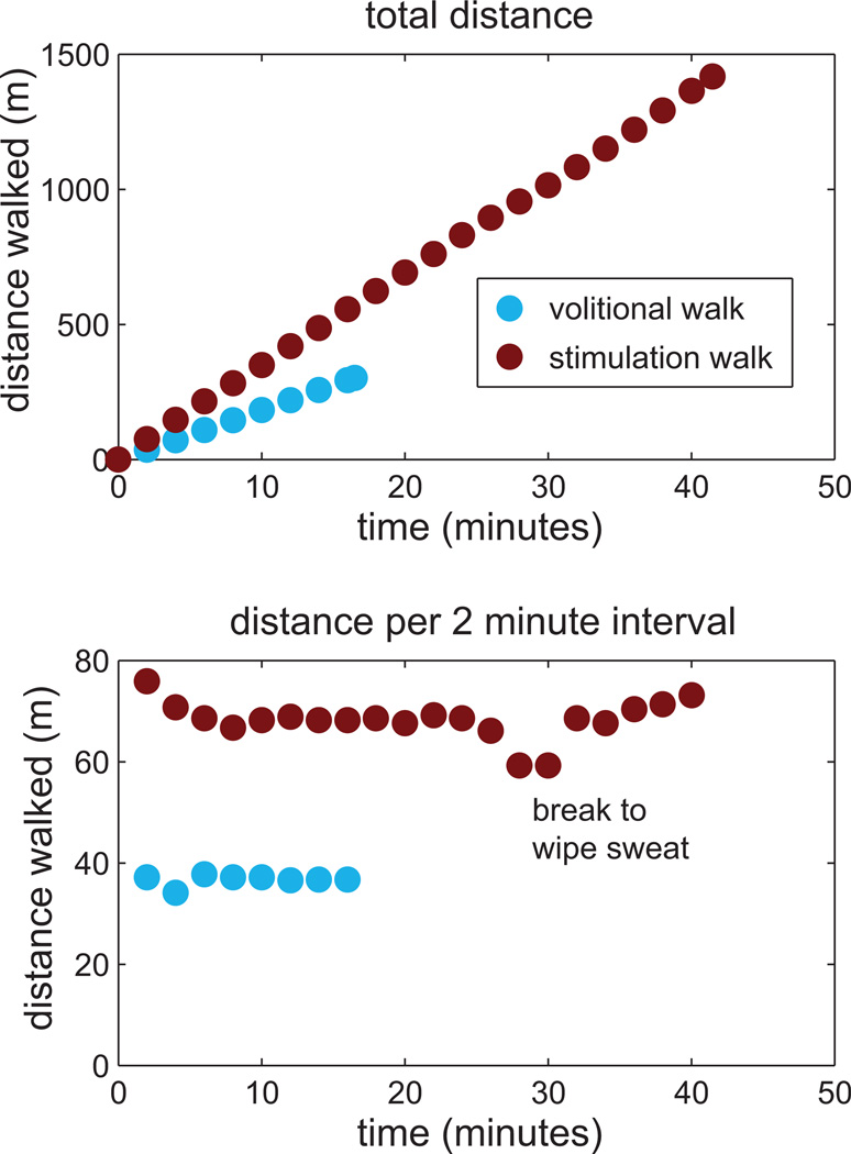
Maximum walking distance during initial training was approximately 76m due to the participant fatiguing. After training, the participant walked 301m in 16:30 minutes (0.30m/s) and 1418m in 41:28 minutes (0.57m/s) without and with stimulation, respectively. Thus, his walking distance increased by 370% with stimulation while walking nearly twice as fast. These data are shown in Figure 4 with the total distance walked over time in the upper panel and the interval distance walked every two minutes in the lower panel. Fluctuations around the 30 minute mark during the stimulation condition were due to the need to stand for a brief rest break.
Figure 4.
Maximum distance walked (top graph) and 2 min incremental distance (bottom graph).
There was a significant difference in most of the spatial-temporal parameters during volitional walking after training as compared to baseline except for affected swing duration, unaffected step length, and affected stance/swing ratio. Similarly, there was a significant change in most of the parameters after training between walking with and without stimulation except for unaffected double support, affected swing duration, and affected step length (Table 2). The largest change was in the affected double support time, which decreased after training and further decreased while walking with stimulation.
Table 2.
Spatial-temporal parameters.
| Spatial-Temporal Parameters | Baseline Mean (±SD) |
Volitional after training Mean (±SD) |
Stimulation after training Mean (±SD) |
|---|---|---|---|
| Unaffected double support (s) | 0.29 (0.02) | 0.23 (0.02)* | 0.20 (0.02)‡ |
| Affected double support (s) | 1.57 (0.11) | 1.31 (0.10)* | 0.40 (0.02)†‡ |
| Unaffected swing (s) | 0.38 (0.03) | 0.46 (0.06)* | 0.56 (0.04)†‡ |
| Affected swing (s) | 0.61 (0.05) | 0.56 (0.03) | 0.61 (0.03) |
| Unaffected stance (s) | 2.47 (0.15) | 2.08 (0.15)* | 1.22 (0.02)†‡ |
| Affected stance (s) | 2.24 (0.13) | 2.00 (0.12)* | 1.16 (0.03)†‡ |
| Unaffected stride length (m) | 0.74 (0.04) | 0.86 (0.03)* | 1.05 (0.05)†‡ |
| Affected stride length (m) | 0.75 (0.02) | 0.84 (0.04)* | 1.02 (0.05)†‡ |
| Unaffected step length (m) | 0.21 (0.03) | 0.20 (0.02) | 0.33 (0.03)†‡ |
| Affected step length (m) | 0.49 (0.03) | 0.59 (0.03)* | 0.63 (0.05)‡ |
| Cadence (steps/min) | 42 | 47* | 67†‡ |
| Unaffected stance/swing ratio (%) | 87:13 | 82:18* | 69:31†‡ |
| Affected stance/swing ratio (%) | 79:21 | 78:22 | 65:35†‡ |
therapeutic effect (p<0.05).
neuroprosthetic effect (p<0.05).
total effect (p<0.05).
Aside from Affected Swing, all other p-values were less 0.01.
Discussion
These results are the first report of the benefits of a fully implanted pulse generator for hip, knee, and ankle control in an individual with stroke. The data demonstrate that stimulation of the hip and knee flexors and extensors and ankle dorsiflexors could improve walking speed, step length, stance and swing times, symmetry, and cadence. While the participant experienced both therapeutic and neuroprosthetic benefits, only the neuroprosthetic effect is representative of a clinically relevant impact on walking speed. A 0.16m/s improvement in gait speed is considered clinically relevant based on improvements correlated with changes in the Modified Rankin Scale, a measure of functional independence27. This threshold is consistent with the generally accepted 0.14m/s change to describe clinically relevant declines in gait speed and function28.
The therapeutic changes to voluntary walking due to gait training with stimulation included reduction in affected double support, increase in unaffected swing duration, and increases in affected and unaffected stride length. These improvements are likely the result of increased strength on the affected side, and improved confidence and stability allowing for longer steps and increased time in single stance. Maximum walking distance at the start of gait training was limited to about 76m due to fatigue. The maximum walk distance of 301m without stimulation after training suggests a clinically relevant improvement in endurance, however, changes in walking speed of 0.05m/s are not considered clinically significant27. Some studies of peroneal nerve stimulation have shown similar therapeutic benefits11,12, while other studies of peroneal nerve stimulation have had a larger therapeutic effect on walking speed29. Although gait training was not the focus of the study, the changes in gait speed are also comparable to those produced by therapist or robot assisted locomotor training on a treadmill (0.06m/s) in a group with similar impairments30. Although therapy is provided over a limited period of time, persistent use of the device during walking may provide ongoing training that maintains both muscle conditioning and cardiovascular health. This use may encourage activity and prevent atrophy and disuse.
The neuroprosthetic benefit produced clinically relevant improvements in gait speed. There was an increase in gait speed of 0.37m/s which exceeds 0.16m/s normally recognized as clinically significant27. An improvement in the speed of walking from 0.35m/s to 0.72m/s represents a potential change in ambulation status from household to limited community ambulator7. One contributor to the increased speed was the transition from a step-to gait to a two-point gait which produced the dramatic decrease in affected double support. The change in gait pattern with stimulation was enabled by the toe clearance produced by the additional knee flexion as evidenced by the foot catches during attempts at volitional two-point gait even while wearing an ankle foot orthosis. In addition to the decrease in affected double support time, there was an increase in affected stride and step length, and an increase in unaffected stride and step length and swing time. The increase in unaffected step length improved step symmetry. The unaffected and affected stance/swing ratios are both approaching more typical ratios (60:40). The improvements in unaffected step swing time and step length indicate better stability resulting from stimulation of hip extensors during stance. Although the participant is not walking with stimulation outside of the laboratory, he reports increased walking in the neighborhood, which is an improvement over previous reports of only household ambulation. These reports suggest that transferring use of the system to the community would further enable community ambulation. The participant reports that he enjoys walking with stimulation and appreciates the exercise as the stimulation enables him to walk significantly faster.
Maximum walk trials showed a consistent increase in walking speed, duration and distance. The therapeutic effect is likely a result of muscle conditioning during stimulated exercise and gait training. However, the study design prevents determining the relative contribution of the stimulation and gait training. The neuroprosthetic effect of walking longer distances at a faster speed suggests that the stimulation did not increase muscle fatigue in a way that limited gait. Home exercise with stimulation presumably contributed to this effect. While walking faster with stimulation uses more energy, the participant effectively maintained a consistent gait speed. Although 43 minute walks may not be typical, the ability to walk at will for long periods of time is useful for community ambulation and participation and demonstrates that walking with stimulation can be maintained for functionally relevant periods of time.
Neuroprosthetic improvements suggest that daily use of an implanted system could have significant clinical relevance to a portion of the stroke population. Prior studies of implanted foot drop stimulators produced neuroprosthetic improvements of 0.08 and 0.03 m/s31,32 while studies of surface peroneal nerve stimulation produced increases of 0.06m/s10 and 0.08m/s29. Only the NESS L300Plus (Bioness Inc., Valencia, CA) incorporates surface stimulation of knee flexors or extensors, which is associated with improvement in gait speed over peroneal nerve stimulation of 0.04m/s33 where 13% of participants used quadriceps stimulation and 87% used hamstrings stimulation. However, there are currently no commercially available systems to provide effective active hip flexion for home use; such an approach would require an IPG. Poststroke impairment covers a broad spectrum of gait deficits and it is important to recognize that presents unique challenges to the design and implementation of assistive devices. Many patients may have sufficient residual function to obviate the potential benefits of a multi-joint neuroprosthesis. However, patients with limited hip and knee control may require multi-joint stimulation to fully realize the benefits of an implanted stimulation system, instead of one that concentrates only at the ankle. In the context of the other studies, the data presented here suggest that there is a spectrum of stroke survivors who would clinically benefit from hip and knee stimulation, in addition to ankle stimulation.
Although this single-subject study provides evidence of efficacy in a single individual with the subject acting as his own concurrent and longitudinal control, the results cannot be generalized without appropriately designed larger scale trial. Future studies would need to recruit additional participants to ensure independent samples and determine what portion of the stroke population would truly benefit from a multi-joint device. Increased sample size would also ensure a priori independence of samples for statistical analysis. While the residuals of the ANOVA were uncorrelated and indicated independence, repeated measures from the same individual in a single-subject study design may not be independent. Although this participant did not have any complications from surgery or device malfunction over the course of the study, these aspects would need to be considered in future assessments within the stroke population.
While statistically significant improvements in voluntary walking were observed post- training, the goal of training was to maximize voluntary function prior to quantifying neuroprosthetic effects of multi-joint stimulation. The current study design does not differentiate between factors contributing to improvements in voluntary function: gait training alone, incorporating stimulation into gait training, and home stimulation exercises. Nevertheless, the positive neuroprosthetic effect of stimulation on the gait of this single subject over voluntary function after gait training with stimulation is clear.
There are many opportunities to refine this intervention and demonstrate its use during ambulation in the home and community, but these data demonstrate that implanted stimulation system for multi-joint control is a promising intervention to provide assistance to stroke survivors during daily walking.
Supplementary Material
Acknowledgments
We gratefully acknowledge Erika Woodrum for creating Figure 1, Dr. Steven Sidik for advising about the statistics, and Drs. Francois Bethoux, John Chae, and Jayme Knutson for their feedback on the manuscript.
This work was supported by the Merit Review Award No. B7692R from the United States (U.S.) Department of Veterans Affairs Rehabilitation Research and Development Service. RJ Triolo was supported by the Senior Research Career Scientist Award No. A9259-L from the U.S. Department of Veterans Affairs Rehabilitation Research and Development Service. NS Makowski was supported in part by the National Institutes of Health (NIH) National Institute of Neurological Disorders and Stroke (NINDS) Award No. U01 NS086872-01. The contents do not represent the views of the U.S. Department of Veterans Affairs or the United States Government.
Footnotes
Author Disclosures: None.
Portions of this work have been accepted for presentation at the American Society for NeuroRehabilitation annual meeting in October 2015.
References
- 1.Go AS, Mozaffarian D, Roger VL, et al. Heart disease and stroke statistics--2014 update: a report from the American Heart Association. Circulation. 2014;129:e28–e292. doi: 10.1161/01.cir.0000441139.02102.80. [DOI] [PMC free article] [PubMed] [Google Scholar]
- 2.Allen JL, Kautz SA, Neptune RR. The influence of merged muscle excitation modules on post-stroke hemiparetic walking performance. Clin Biomech (Bristol, Avon) 2013;28:697–704. doi: 10.1016/j.clinbiomech.2013.06.003. [DOI] [PMC free article] [PubMed] [Google Scholar]
- 3.Arene N, Hidler J. Understanding motor impairment in the paretic lower limb after a stroke: a review of the literature. Top Stroke Rehabil. 2009;16:346–356. doi: 10.1310/tsr1605-346. [DOI] [PubMed] [Google Scholar]
- 4.Watkins CL, Leathley MJ, Gregson JM, Moore AP, Smith TL, Sharma AK. Prevalence of spasticity post stroke. Clin Rehabil. 2002;16:515–522. doi: 10.1191/0269215502cr512oa. [DOI] [PubMed] [Google Scholar]
- 5.De Quervain IA, Simon SR, Leurgans S, Pease WS, McAllister D. Gait pattern in the early recovery period after stroke. J Bone Joint Surg Am. 1996;78:1506–1514. doi: 10.2106/00004623-199610000-00008. [DOI] [PubMed] [Google Scholar]
- 6.Robertson MC, Campbell AJ, Gardner MM, Devlin N. Preventing injuries in older people by preventing falls: a meta-analysis of individual-level data. J Am Geriatr Soc. 2002;50:905–911. doi: 10.1046/j.1532-5415.2002.50218.x. [DOI] [PubMed] [Google Scholar]
- 7.Perry J, Garrett M, Gronley JK, Mulroy SJ. Classification of walking handicap in the stroke population. Stroke. 1995;26:982–989. doi: 10.1161/01.str.26.6.982. [DOI] [PubMed] [Google Scholar]
- 8.Michael KM, Allen JK, Macko RF. Reduced ambulatory activity after stroke: the role of balance, gait, and cardiovascular fitness. Arch Phys Med Rehabil. 2005;86:1552–1556. doi: 10.1016/j.apmr.2004.12.026. [DOI] [PubMed] [Google Scholar]
- 9.Schmid A, Duncan PW, Studenski S, et al. Improvements in speed-based gait classifications are meaningful. Stroke. 2007;38:2096–2100. doi: 10.1161/STROKEAHA.106.475921. [DOI] [PubMed] [Google Scholar]
- 10.Sheffler LR, Bailey SN, Wilson RD, Chae J. Spatiotemporal, kinematic, and kinetic effects of a peroneal nerve stimulator versus an ankle foot orthosis in hemiparetic gait. Neurorehabil Neural Repair. 2013;27:403–410. doi: 10.1177/1545968312465897. [DOI] [PMC free article] [PubMed] [Google Scholar]
- 11.Sheffler LR, Taylor PN, Gunzler DD, Buurke JH, Ijzerman MJ, Chae J. Randomized controlled trial of surface peroneal nerve stimulation for motor relearning in lower limb hemiparesis. Arch Phys Med Rehabil. 2013;94:1007–1014. doi: 10.1016/j.apmr.2013.01.024. [DOI] [PMC free article] [PubMed] [Google Scholar]
- 12.Bethoux F, Rogers HL, Nolan KJ, et al. Long-Term Follow-up to a Randomized Controlled Trial Comparing Peroneal Nerve Functional Electrical Stimulation to an Ankle Foot Orthosis for Patients With Chronic Stroke. Neurorehabil Neural Repair. 2015 doi: 10.1177/1545968315570325. [DOI] [PubMed] [Google Scholar]
- 13.Stanic U, Acimovic-Janezic R, Gros N, Trnkoczy A, Bajd T, Kljajic M. Multichannel electrical stimulation for correction of hemiplegic gait. Methodology and preliminary results. Scand J Rehabil Med. 1978;10:75–92. [PubMed] [Google Scholar]
- 14.Kobetic R. Gait control in stroke. In: Kilgore KL, editor. Implantable Neuroprostheses for Restoring Function. Elsevier; 2015. pp. 281–300. [Google Scholar]
- 15.Little VL, McGuirk TE, Patten C. Impaired Limb Shortening following Stroke: What's in a Name? PLoS One. 2014;9:e110140. doi: 10.1371/journal.pone.0110140. [DOI] [PMC free article] [PubMed] [Google Scholar]
- 16.Turns LJ, Neptune RR, Kautz SA. Relationships between muscle activity and anteroposterior ground reaction forces in hemiparetic walking. Arch Phys Med Rehabil. 2007;88:1127–1135. doi: 10.1016/j.apmr.2007.05.027. [DOI] [PMC free article] [PubMed] [Google Scholar]
- 17.Brincks J, Nielsen JF. Increased power generation in impaired lower extremities correlated with changes in walking speeds in sub-acute stroke patients. Clin Biomech (Bristol, Avon) 2012;27:138–144. doi: 10.1016/j.clinbiomech.2011.08.007. [DOI] [PubMed] [Google Scholar]
- 18.Daly JJ, Barnicle K, Kobetic R, Marsolais EB. Electrically induced gait changes post stroke, using an FNS system with intramuscular electrodes and multiple channels. J Neurol Rehabil. 1993;7:17–25. [Google Scholar]
- 19.Daly JJ, Ruff RL. Feasibility of combining multi-channel functional neuromuscular stimulation with weight-supported treadmill training. J Neurol Sci. 2004;225:105–115. doi: 10.1016/j.jns.2004.07.006. [DOI] [PubMed] [Google Scholar]
- 20.Mendell JR, Florence J. Manual muscle testing. Muscle Nerve. 1990;13(Suppl):S16–S20. doi: 10.1002/mus.880131307. [DOI] [PubMed] [Google Scholar]
- 21.Gregson JM, Leathley M, Moore AP, Sharma AK, Smith TL, Watkins CL. Reliability of the Tone Assessment Scale and the modified Ashworth scale as clinical tools for assessing poststroke spasticity. Arch Phys Med Rehabil. 1999;80:1013–1016. doi: 10.1016/s0003-9993(99)90053-9. [DOI] [PubMed] [Google Scholar]
- 22.Smith B, Peckham PH, Keith MW, Roscoe DD. An externally powered, multichannel, implantable stimulator for versatile control of paralyzed muscle. IEEE Trans Biomed Eng. 1987;34:499–508. doi: 10.1109/tbme.1987.325979. [DOI] [PubMed] [Google Scholar]
- 23.Memberg WD, Peckham H, Keith MW. A surgically-implanted intramuscular electrode for an implantable neuromuscular stimulation system. IEEE Trans Rehabil Eng. 1994;2:80–91. [Google Scholar]
- 24.Marsolais EB, Kobetic R. Implantation techniques and experience with percutaneous intramuscular electrodes in the lower extremities. J Rehabil Res Dev. 1986;23:1–8. [PubMed] [Google Scholar]
- 25.Crago PE, Peckham PH, Thrope GB. Modulation of muscle force by recruitment during intramuscular stimulation. IEEE Trans Biomed Eng. 1980;27:679–684. doi: 10.1109/TBME.1980.326592. [DOI] [PubMed] [Google Scholar]
- 26.Kobetic R, Marsolais EB. Synthesis of paraplegic gait with multichannel functional neuromuscular stimulation. IEEE Trans Rehabil Eng. 1994;2:66–79. [Google Scholar]
- 27.Tilson JK, Sullivan KJ, Cen SY, et al. Meaningful gait speed improvement during the first 60 days poststroke: minimal clinically important difference. Phys Ther. 2010;90:196–208. doi: 10.2522/ptj.20090079. [DOI] [PMC free article] [PubMed] [Google Scholar]
- 28.Perera S, Mody SH, Woodman RC, Studenski SA. Meaningful change and responsiveness in common physical performance measures in older adults. J Am Geriatr Soc. 2006;54:743–749. doi: 10.1111/j.1532-5415.2006.00701.x. [DOI] [PubMed] [Google Scholar]
- 29.Taylor P, Humphreys L, Swain I. The long-term cost-effectiveness of the use of Functional Electrical Stimulation for the correction of dropped foot due to upper motor neuron lesion. J Rehabil Med. 2013;45:154–160. doi: 10.2340/16501977-1090. [DOI] [PubMed] [Google Scholar]
- 30.Hornby TG, Campbell DD, Kahn JH, Demott T, Moore JL, Roth HR. Enhanced gait-related improvements after therapist- versus robotic-assisted locomotor training in subjects with chronic stroke: a randomized controlled study. Stroke. 2008;39:1786–1792. doi: 10.1161/STROKEAHA.107.504779. [DOI] [PubMed] [Google Scholar]
- 31.Burridge JH, Haugland M, Larsen B, et al. Phase II trial to evaluate the ActiGait implanted drop-foot stimulator in established hemiplegia. J Rehabil Med. 2007;39:212–218. doi: 10.2340/16501977-0039. [DOI] [PubMed] [Google Scholar]
- 32.Kottink AI, Tenniglo MJ, de Vries WH, Hermens HJ, Buurke JH. Effects of an implantable two-channel peroneal nerve stimulator versus conventional walking device on spatiotemporal parameters and kinematics of hemiparetic gait. J Rehabil Med. 2012;44:51–57. doi: 10.2340/16501977-0909. [DOI] [PubMed] [Google Scholar]
- 33.Springer S, Laufer Y, Becher M, Vatine JJ. Dual-channel functional electrical stimulation improvements in speed-based gait classifications. Clin Interv Aging. 2013;8:271–277. doi: 10.2147/CIA.S41141. [DOI] [PMC free article] [PubMed] [Google Scholar]
Associated Data
This section collects any data citations, data availability statements, or supplementary materials included in this article.


