Abstract
The aim of this study was to investigate the effects of boric acid in experimental acute sciatic nerve injury. Twenty-eight adult male rats were randomly divided into four equal groups (n = 7): control (C), boric acid (BA), sciatic nerve injury (I), and sciatic nerve injury + boric acid treatment (BAI). Sciatic nerve injury was generated using a Yasargil aneurysm clip in the groups I and BAI. Boric acid was given four times at 100 mg/kg to rats in the groups BA and BAI after injury (by gavage at 0, 24, 48 and 72 hours) but no injury was made in the group BA. In vivo electrophysiological tests were performed at the end of the day 4 and sciatic nerve tissue samples were taken for histopathological examination. The amplitude of compound action potential, the nerve conduction velocity and the number of axons were significantly lower and the myelin structure was found to be broken in group I compared with those in groups C and BA. However, the amplitude of the compound action potential, the nerve conduction velocity and the number of axons were significantly greater in group BAI than in group I. Moreover, myelin injury was significantly milder and the intensity of nuclear factor kappa B immunostaining was significantly weaker in group BAI than in group I. The results of this study show that administration of boric acid at 100 mg/kg after sciatic nerve injury in rats markedly reduces myelin and axonal injury and improves the electrophysiological function of injured sciatic nerve possibly through alleviating oxidative stress reactions.
Keywords: nerve regeneration, peripheral nerve injury, sciatic nerve, boric acid, nerve conduction velocity, axon, myelin, electrophysiology, neural regeneration
Introduction
Compression injuries in the peripheral nerves can cause many different histopathological changes, depending on the severity and duration of the compression pressure (Epstein et al., 1997). Although the causes of these changes are not fully understood, these pathophysiological changes are associated with ischemia, which develops as a result of the direct mechanical damage to peripheral nerves, the cessation of axoplasmic flow or the compression of vascular structures feeding the peripheral nerve due to the compression (Mattson and Camandola, 2001). Many agents have been investigated to eliminate the damaging effects of this pathophysiological process (Roglio et al., 2008; Ma et al., 2015; Guven et al., 2016; Li et al., 2016).
Boric acid, which is a boron compound, is widely used in industry, agriculture and cosmetic applications. Boric acid is quickly absorbed after application and is distributed throughout the body by passive diffusion (Ince et al., 2012). In a study conducted in humans and rats by Murray (1998), boric acid was reportedly to be distributed equally in the blood and tissue after application of boric acid. In addition, boron also exhibited effects on reactive oxygen species, the metabolism of calcium, potassium, xanthine oxidase, cytochrome b5 reductase, and vitamin D (Meacham et al., 1994; Devirian and Volpe, 2003; Türkez et al., 2007; Ince et al., 2010). Boric acid has been shown to be often used in many types of cancer with neutron capture therapy (Barth, 2009). In addition, its effectiveness has been demonstrated in injured liver, lung, cardiac muscle, kidney, brain and bone fracture (Barth, 2009; Ince et al., 2010; Gölge et al., 2015). However, as far as we know, there are no studies on the effectiveness of boric acid in experimental acute peripheral nerve injury.
Therefore, this study aimed to investigate the effectiveness of boric acid in repairing the sciatic nerve following experimental acute sciatic nerve injury electrophysiologically and histologically.
Materials and Methods
Experimental animals
All experimental protocols conducted on the animals were consistent with the National Institutes of Health Guide for the Care and Use of Laboratory Animals (National Institutes of Health Publications No. 80-23) and approved by the Animal Ethics Committee of Adnan Menderes University (approval number: 64583101/2016/52). Twenty-eight adult male Wistar-Albino rats, weighing 250 ± 50 g were used in this study. All of the rats were kept in environmentally controlled conditions at 22–25°C, with appropriate humidity and a 12–hour light/dark cycle. The rats were given free access to food and water. The rats were randomly divided into four groups (n = 7): Control (C), the right sciatic nerves were only exposed; sciatic nerve injury (I): sciatic nerve injury was induced by compressing the right sciatic nerve; no injury + boric acid application (BA): the right sciatic nerves were exposed but not injured, and boric acid was given at 0, 24, 48 and 72 hours after operation; sciatic nerve injury + boric acid treatment (BAI): the right sciatic nerves were injured and boric acid treatment was given at 0, 24, 48 and 72 hours after injury.
Generation of sciatic nerve injury
Rats were anesthetized by an intraperitoneal injection of 10 mg/kg xylazine (Rompun, Bayer, Turkey) and 50 mg/kg ketamine (Ketalar, Parke Davis, Eczacibasi Co., Istanbul, Turkey) and allowed to breathe spontaneously. They were positioned on a heating pad to maintain their body temperature at 37°C and placed in the prone position. All surgical interventions were carried out on the sciatic nerve by the same surgeon using standard microsurgery methods. The rats were kept in an appropriate position on special fixing boards during the surgical intervention, and the operations were carried out. The skin over the surgical site (right gluteal and femur region) was shaved and cleaned with povidone-iodine. An oblique incision was made in the right lower extremity to remove the skin and reveal the biceps femoris in such a way as to allow hip joint movement. Muscle tissue was opened by blunt dissection and then the sciatic nerve was exposed.
In this study, the closing force was defined by a computerized electronic gauged scale according to previous reports (Sarikcioglu and Ozkan, 2003; Sarikcioglu et al., 2007) and an FE-752K aneurysm clip (Aesculap AG & Co., Tutlingen, Germany) was used. The right sciatic nerve was compressed using the Yasargil aneurism clip for 90 seconds, and then the aneurism clip was opened and removed. This process was applied to all rats in groups I and BAI. Also the injured nerve areas were marked with 6-0 Prolene suture. Then, the incision was closed in accordance with the anatomical layers. The right sciatic nerves of rats were reexposed at the end of the day 4 for electrophysiological and histological studies.
Drug treatment
Concentration of the boric acid (H3BO3) was 99.5% and was dissolved in physiological saline because it is highly soluble in water. Then, it was administered intragastrically at 100 mg/kg (Sigma-Aldrich, Chemical Co., St. Louis, MO, USA) using a 16G gavage needle at 0, 24, 48 and 72 hours after injury. This dose was selected as the antioxidant effectiveness of boric acid has been demonstrated (Ince et al., 2010).
In vivo electrophysiological recording
Rats were again anesthetized with a mixture of ketamine/xylazine and the right sciatic nerve was exposed at the end of the day 4 for experimental period to record nerve conduction velocity. Damaged area of the sciatic nerve was proximal to bifurcation (tibial and fibular) section of sciatic nerve (10 mm). The stimulating and recording electrodes were placed on the proximal and distal sides of the damaged area of the sciatic nerve, respectively. The compound action potentials (CAPs) which were generated by applying 10 consecutive electrical impulses (10 V, 0.15 ms) in the proximal side of the sciatic nerve were recorded from the distal side of the sciatic nerve using the PowerLab 26T data acquisition system (ADInstruments Co., Sydney, Australia). All experiments were carried out at room temperature (with a control band of 22 ± 1°C). The amplitudes of CAPs were measured from the baseline to the peak and the latencies were measured from the stimulus artifact to the beginning of the first deflection from the baseline. Nerve conduction velocity (NCV) values were calculated according to the following formula: NCV (m/s) = Distance between stimulating and recording points – 15 mm (m) / latency (second). The average value of NCV and the amplitude of CAP were calculated separately for each rat.
Histopathological evaluation
Sciatic nerve samples were harvested from proximal to bifurcation and they were placed in 10% buffered formalin solution for pathological examination. The samples were taken for routine tissue processing after the tissues were fixed in solution. Then, they were left for 14 hours in the automated tissue processing device. The tissues were fixed in paraffin and 3–4 μm sections were prepared from the paraffin blocks with a microtome. Slides prepared from the sections were stained with hematoxylin-eosin and toluidine blue for routine histopathological examination and the evaluation of axonal degeneration under 40× magnifications. The routine sections were histologically graded for axonal changes and myelin disorganization (Coban et al., 2006). Swelling (pale staining) or shrinkage (dark staining) and vacuolization were observed in the axons due to degeneration. Myelin changes were typically seen, including attenuation, collapse or breakdown. Grading was performed on a scale of 0 to 3 for each section: 0 = normal, 1 = mild, 2 = moderate, 3 = severe (Coban et al., 2006).
One slide from each animal was stained immunohistochemically for nuclear factor kappa B (NF-κB/p50 RB-1648 1/500 dilution; NeoMarkers, Fremont, CA, USA). NF-κB staining was observed in axons and Schwann cells. A semi-quantitative grading system was used to evaluate the staining for NF-κB in both axons and Schwann cells. Grade 0: 2% and below, grade 1: 2–15%, grade 2: 16–25%, grade 3: 26–35%, grade 4: 36% and above (Wang et al., 2006).
The numbers of axons as well as the morphological characteristics of the axons were evaluated in cross-sections of the sciatic nerve. Five images were taken from each slide. All preparations were counted by taking samples from random areas of each sample using the DP-BSW (Microscope digital camera software) program at high magnification (× 100) under light microscope (Olympus BX51, Olympus Co., Tokyo, Japan). This counting was made by the axon counting method which was previously defined (Yang and Bashaw, 2006). All pathological examinations were evaluated by a pathologist blinded to the experimental group information.
Statistical analysis
The data obtained from the animal experiments are expressed as the mean ± SD. The statistical differences between groups were evaluated by one-way analysis of variance and the Tukey's post hoc test using the SPSS 15.0 program (SPSS, Chicago, IL, USA). P values less than 0.05 were considered statistically significant.
Results
Electrophysiological results
The NCV and CAP amplitude values were significantly lower in group I than in groups C and BA (all P values < 0.001). The NCV and CAP amplitude values in group BAI were significantly greater than in group I (all P values < 0.001). However, the NCV and CAP amplitude values in group BAI were significantly lower than in groups C and BA (all P values < 0.001). Moreover, there were no significant differences between NCV or CAP amplitude values between groups C and BA (Figure 1A, B).
Figure 1.
The nerve conduction velocity, amplitude of compound action potential and number of axons in each group.
(A) The values of nerve conduction velocity. (B) The values of amplitude of compound action potential. (C) The number of axons. Data are expressed as a percentage of control. The statistical differences between groups were evaluated by one-way analysis of variance and the Tukey's post hoc test. *P < 0.001, vs. group C; †P < 0.001, vs. group BA; #P < 0.001, vs. group I. C: Control; BA: boric acid; I: sciatic nerve injury; BAI, sciatic nerve injury + boric acid treatment. n = 7 for each group.
Histopathological results
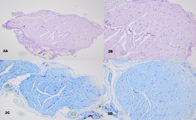
Hematoxylin-eosin and toluidine blue staining showed that the epineurium (a thick fibrous connective tissue) and the perineurium (a thinner connective tissue of nerve fascicles) were seen respectively from outside to inside in the light microscopic examination of the sections obtained by staining the right sciatic nerves with hematoxylin-eosin and toluidine blue in rats of groups C and BA. Schwann cells, which envelop axons, were distinguished by their oval or round nuclei under the endoneurium. Axons were observed to be faded in color in the cytoplasm of Schwann cells. The presence of a myelin sheath, which is made by Schwann cells and wraps around the axon, was seen in myelinated nerve fibers. Unmyelinated nerve fibers, connective tissue cells and blood vessels were distinguished among the myelinated nerve fibers (Figure 2A–D).
Figure 2.
Representative images of the unaffected sciatic nerves of rats in groups C and BA after hematoxylin-eosin and toluidine blue staining.
(A, B) Hematoxylin-eosin staining of the groups C (× 200) and BA (× 400). (C, D) Toluidine blue staining of the groups C (× 200) and BA (× 400). C: Control; BA: boric acid.
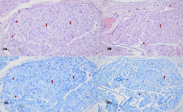
The nerve was seen to be surrounded by epineurium (a thick fibrous connective tissue) in the light microscopic examination of the sections obtained by staining the right sciatic nerves with hematoxylin-eosin and toluidine blue in rats of groups I and BAI. The presence of nerve fascicles, which contain myelinated and unmyelinated nerve fibers, were distinguished under the epineurium. While the axons and myelin sheath were degenerated, the myelin sheath lamellae were separated from each other and the axons were smaller in some nerve fibers, or found to be completely degenerated in other nerve fibers. The presence of degenerated nerve fibers, vacuolization and macrophages, which ingest the myelin sheath, were also distinguished in areas with marked degeneration. In addition, although some myelinated nerve fibers maintained their normal structure, significant decreases in axon diameter and myelin sheath thickness were noted compared to group C. Axonal degeneration, vacuolization and myelin destruction were found to be markedly greater in group I than in group BAI. A larger number of axons and myelin sheaths were maintained in group BAI (Figure 3A–D).
Figure 3.
Representative images of sections obtained from the affected sciatic nerves of the rats in groups I and BAI after hematoxylin-eosin and toluidine blue staining.
(A, B) Hematoxylin-eosin staining of groups I and BAI (×400). (C, D) Toluidine blue staining of groups I and BAI (×400). Axonal degeneration, vacuolization (asterisks) and myelin destruction (arrows) were found to be more severe in group I than in group BAI. I: Sciatic nerve injury; BAI: sciatic nerve injury + boric acid treatment.
NF-κB immunoreactivity
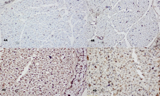
NF-κB immunostaining was observed in axons and Schwann cells. Mild staining was usually observed in the preparations of groups C and BA. NF-κB immunostaining was found to be increased in the preparations of group I, and NF-κB immunoreactivity was observed in the preparations of group BAI, but this staining was less intense in group BAI than in group I (Figure 4A–D, Table 1).
Figure 4.
Representative images of NF-κB immunohistochemical staining (× 400).
In groups C (A) and BA (B), few axons and Schwann cells showed light staining of NF-κB. NF-κB immunoreactivity was weaker in group BAI (D) than in group I (C). C: Control; BA: boric acid; I: sciatic nerve injury; BAI: sciatic nerve injury + boric acid treatment; NF-κB: nuclear factor kappa B.
Table 1.
Axon number and NF-κB immunoreactivity of experimental groups

Number of axons
The number of axons was significantly lower in group I than in groups C and BA, and the number of axons was significantly higher in group BAI than in group I. However, the number of axons was significantly lower in group BAI than in groups C and BA (all P values < 0.001). Moreover, there were no significant differences between groups C and BA regarding the number of axons (Figure 1C).
Discussion
Boron is found as a natural element in nature, and boric acid is a boron compound and contains 17.48% boron. Boron is absorbed by the digestive and respiratory system and is distributed as boric acid to all tissues. In humans, 83–98% of dietary boron is excreted by urine in a few days. Therefore, there is no accumulation of boron in the human body (Tepeden et al., 2016). Boric acid has been reportedly used in many studies (Barth, 2009; Ince et al., 2010). In a study on cyclophosphamide-induced lipid peroxidation and gene toxicity in rats, boric acid was reportedly to reduce cyclophosphamide-induced oxidative stress in a dose-dependent manner. Moreover, the focal gliosis and neuronal degeneration found in the brains of rats administered cyclophosphamide were milder following 20 mg/kg of boric acid treatment (Ince et al., 2014). In another study, 100 mg/kg of boric acid partially reduced the effects of arsenic-induced oxidative stress; focal gliosis was observed in both male and female rats in response to arsenic, but only mild histopathological changes were reported in both groups administered boric acid (Kucukkur et al., 2015). Similarly, while focal gliosis and neuronophagia were found in the brains of rats after malathion-induced oxidative stress, only moderate focal gliosis was found in the groups administered boric acid (5, 10 and 20 mg/kg/d) (Coban et al., 2015). In light of these findings, different doses of boric acid may be able to reduce the destructive effects of oxidative stress in tissues by supporting antioxidant enzymes. A reduction in oxidative stress in nerve tissue may be an important mechanism of injured peripheral nerve healing.
The support Schwann cells in the context of increased expression of neurotrophic factors by the injured nerve may be another healing mechanism of boric acid. Since products such as reactive oxygen species and nitric oxide are increased after peripheral nerve trauma and Schwann cells play an influential role in the release of neurotrophic factors in peripheral nerve healing, apoptosis could be induced via the mitochondrial pathway, depending on the dose and duration of oxidative stress (Bowe et al., 1989; Clarke and Richardson, 1994; Ide, 1996; Zochodne and Levy, 2005; Luo et al., 2010; Ma et al., 2013; Huang et al., 2016). In a study in which hydrogen peroxide-induced oxidative stress in Schwann cells was investigated, the authors reported that ginsenoside Rg1 may have positive effects on peripheral nerve healing by reducing the effect of hydrogen peroxide-induced oxidative stress and by increasing the release of neurotrophic factors such as nerve growth factor and brain derived neurotrophic factor (Luo et al., 2010). In addition, in a study performed in the brains of African ostrich chicks, the use of low concentrations of boric acid (up to 160 mg/dL) was reportedly to increase BDNF expression required for brain development and may also inhibit apoptosis in neuronal cells (Tang et al., 2016).
Another possible effect of boric acid that may mediate peripheral nerve healing is the inhibition of intracellular calcium release. It has been shown in previous studies that intracellular calcium stores can be effective in axonal degeneration. The potential sources of intra-axonal calcium stores in axons are the endoplasmic reticulum and mitochondria (Stirling and Stys, 2010). Cell culture studies reported that boron was a temporary inhibitor of cyclic ADP ribose. Temporary inhibition of cyclic ADP ribose may inhibit the release of calcium ions from the endoplasmic reticulum via ryanodine receptors (Nielsen, 2014).
Boric acid has been shown to have neuroprotective or neurotoxic effects at different doses (Colak et al., 2011). However, there have been no studies investigating the effects of boric acid on the electrophysiological parameters and nerve tissue healing in peripheral nerves. It is well known that the CAP amplitude and NCV decrease in peripheral nerve injury; these electrophysiological parameters indicate axonal and myelin damage, respectively. In our study, electrophysiological findings in group I indicate that myelin damage occurred and the total number of axons was reduced after compression injury. However, both the loss of axons and myelin damage were dramatically reduced when boric acid was applied after injury in group BAI. In addition, the electrophysiological findings in groups BA and C indicated that there were no adverse effects of boric acid on normal and injuried sciatic nerve tissues. In this study, boric acid treatment after injury led to a statistically significant reduction in NF-κB immunoreactivity in group BAI than in group I.
The findings of this study suggest that the dose of boric acid applied in this study did not cause significant axonal or myelin damage. Probably, boric acid treatment appeared to protect nerve morphology by affecting the antioxidant mechanisms at the cellular level and by protecting axons from the destructive effect of oxidative stress. NF-κB is a redox-sensitive transcriptional factor and has been reported in the literature to be activated via hyperglycemia, oxidative stress and proinflammatory cytokines (Epstein et al., 1997; Mattson and Camandola, 2001).
In this study, although the use of boric acid was shown for the first time to have positive effects on peripheral nerve healing in compression type acute peripheral nerve injury, this study has some limitations. First, damage was not generated with variable intensities and durations. Second, different doses of boric acid were not administered at different time points, and the effective dose of boric acid was selected based on studies in the literature. Third, in this initial study, the biochemical and cellular mechanisms were not investigated to reveal the mechanisms of action of boric acid in peripheral nerve healing; this must be investigated in further studies.
In summary, 100 mg/kg of boric acid was shown electrophysiologically and histologically to have positive effects on maintaining the number of axons, axonal structure and myelin structure in peripheral nerves following compression type peripheral nerve injury. Although there are many questions that remain to be answered, the findings of this initial study suggest that boric acid may be a potential therapeutic agent that can be used in peripheral nerve injury.
Footnotes
Conflicts of interest: None declared.
Plagiarism check: This paper was screened twice using CrossCheck to verify originality before publication.
Peer review: This paper was double-blinded and stringently reviewed by international expert reviewers.
Copyedited by Li CH, Song LP, Zhao M
References
- Barth RF. Boron neutron capture therapy at the crossroads: challenges and opportunities. Appl Radiat Isot. 2009;67(7-8 suppl):S3–6. doi: 10.1016/j.apradiso.2009.03.102. [DOI] [PubMed] [Google Scholar]
- Bowe CM, Hildebrand C, Kocsis JD, Waxman SG. Morphological and physiological properties of neurons after long-term axonal regeneration: observations on chronic and delayed sequelae of peripheral nerve injury. J Neurol Sci. 1989;91:259–292. doi: 10.1016/0022-510x(89)90057-9. [DOI] [PubMed] [Google Scholar]
- Clarke D, Richardson P. Peripheral nerve injury. Curr Opin Neurol. 1994;7:415–421. doi: 10.1097/00019052-199410000-00009. [DOI] [PubMed] [Google Scholar]
- Coban FK, Ince S, Kucukkurt I, Demirel HH, Hazman O. Boron attenuates malathion-induced oxidative stress and acetylcholinesterase inhibition in rats. Drug Chem Toxicol. 2015;38:391–399. doi: 10.3109/01480545.2014.974109. [DOI] [PubMed] [Google Scholar]
- Coban YK, Ciralik H, Kurulas EB. Ischemic preconditioning reduces the severity of ischemia-reperfusion injury of peripheral nerve in rats. J Brachial Plex Peripher Nerve Inj. 2006;1:2. doi: 10.1186/1749-7221-1-2. [DOI] [PMC free article] [PubMed] [Google Scholar]
- Colak S, Geyikoglu F, Keles ON, Türkez H, Topal A, Unal B. The neuroprotective role of boric acid on aluminum chlrodie-induced neurotoxicity. Toxicol Ind Health. 2011;27:700–710. doi: 10.1177/0748233710395349. [DOI] [PubMed] [Google Scholar]
- Devirian TA, Volpe SL. The physiological effects of dietary boron. Crit Rev Food Sci Nutr. 2003;43:219–231. doi: 10.1080/10408690390826491. [DOI] [PubMed] [Google Scholar]
- Epstein FH, Barnes PJ, Karin M. Nuclear factor-κB - a pivotal transcription factor in chronic inflammatory diseases. New Engl J Med. 1997;336:1066–1071. doi: 10.1056/NEJM199704103361506. [DOI] [PubMed] [Google Scholar]
- Gölge UH, Kaymaz B, Arpaci R, Kömürcü E, Göksel F, Güven M, Güzel Y, Cevizci S. Effect of boric acid on fracture healing: An experimental study. Biol Trace Elem Res. 2015;167:264–271. doi: 10.1007/s12011-015-0326-3. [DOI] [PubMed] [Google Scholar]
- Guven M, Gölge UH, Aslan E, Sehitoglu MH, Aras AB, Akman T, Cosar M. The effect of aloe vera on ischemia-Reperfusion injury of sciatic nerve in rats. Biomed Pharmacother. 2016;79:201–207. doi: 10.1016/j.biopha.2016.02.023. [DOI] [PubMed] [Google Scholar]
- Huang HC, Chen L, Zhang HX, Li SF, Liu P, Zhao TY, Li CX. Autophagy promotes peripheral nerve regeneration and motor recovery following sciatic nerve crush injury in rats. J Mol Neurosci. 2016;58:416–423. doi: 10.1007/s12031-015-0672-9. [DOI] [PMC free article] [PubMed] [Google Scholar]
- Ide C. Peripheral nerve regeneration. Neurosci Res. 1996;25:101–121. doi: 10.1016/0168-0102(96)01042-5. [DOI] [PubMed] [Google Scholar]
- Ince S, Keles H, Erdogan M, Hazman O, Kucukkurt I. Protective effect of boric acid againist carbon tetrachloride-induced hepatotoxicity in mice. Drug Chem Toxicol. 2012;35:285–292. doi: 10.3109/01480545.2011.607825. [DOI] [PubMed] [Google Scholar]
- Ince S, Kucukkurt I, Cigerci IH, Fidan AF, Eryavuz A. The effect of dietary boric acid and borax supplemantation on lipid peroxidation, antioxidant activity, and DNA damage in rats. J Trace Elem Med Biol. 2010;24:161–164. doi: 10.1016/j.jtemb.2010.01.003. [DOI] [PubMed] [Google Scholar]
- Ince S, Kucukkurt I, Demirel HH, Acaroz DA, Akbel E, Cigerci IH. Protective effects of boron on cyclophosphamide induced lipid peroxidation and genotoxicity in rats. Chemosphere. 2014;108:197–204. doi: 10.1016/j.chemosphere.2014.01.038. [DOI] [PubMed] [Google Scholar]
- Kucukkurt I, Ince S, Demirel HH, Turkmen R, Akbel E, Celik Y. The effect of boron on arsenic-induced lipid peroxidation and antioxidant status in male and famale rats. J Biochem Mol Toxicol. 2015;29:564–571. doi: 10.1002/jbt.21729. [DOI] [PubMed] [Google Scholar]
- Li H, Zhang L, Xu M. Dexamethasone prevents vascular damage in early-stage non-freezing cold injury of the sciatic nerve. Neurol Regen Res. 2016;11:163–167. doi: 10.4103/1673-5374.175064. [DOI] [PMC free article] [PubMed] [Google Scholar]
- Luo X, Chen B, Zheng R, Lin P, Li J, Chen H. Hydogen peroxide induces apoptosis through the mitochondrial pathway in rat schwann cells. Neurosci Lett. 2010;485:60–64. doi: 10.1016/j.neulet.2010.08.063. [DOI] [PubMed] [Google Scholar]
- Ma J, Liu J, Wang Q, Yu H, Chen Y, Xiang L. The benifical effect of ginsenoside R1 on Schwann cells subjected to hyrogen peroxide induced oxidative injury. Int J Biol Sci. 2013;9:624–636. doi: 10.7150/ijbs.5885. [DOI] [PMC free article] [PubMed] [Google Scholar]
- Ma J, Liu J, Yu H, Chen Y, Wang Q, Xiang L. Effect of the metformin on Schwann cells under hypoxia condition. Int J Clin Exp Pathol. 2015;8:6748–6455. [PMC free article] [PubMed] [Google Scholar]
- Mattson MP, Camandola S. NF-kappaB in neuronal plasticity and neurodegenerative disorders. J Clin Invest. 2001;107:247–254. doi: 10.1172/JCI11916. [DOI] [PMC free article] [PubMed] [Google Scholar]
- Meacham S, Taper L, Volpe SL. Effect of boron supplementation on bone mineral density and dietary, blood, and urinary calcium, phosphorus, magnesium, and boron in female athletes. Environ Health Perspect. 102(suppl 7):79–82. doi: 10.1289/ehp.94102s779. [DOI] [PMC free article] [PubMed] [Google Scholar]
- Murray FJ. A comparative review of the pharmacokinetics of boric acid in rodents and human. Biol Trace Elem Res. 1998;66:331–341. doi: 10.1007/BF02783146. [DOI] [PubMed] [Google Scholar]
- Nielsen FH. Update on human health effects of boron. J Trace Elem Med Biol. 2014;28:383–387. doi: 10.1016/j.jtemb.2014.06.023. [DOI] [PubMed] [Google Scholar]
- Roglio I, Bianchi R, Gotti S, Scurati S, Giatti S, Pesaresi M, Caruso D, Panzica GC, Melcangi RC. Neuroprotective effects of dihydroprogesterone and progesterone in an experimental model of nerve crush injury. Neuroscience. 2008;155:673–685. doi: 10.1016/j.neuroscience.2008.06.034. [DOI] [PubMed] [Google Scholar]
- Sarikcioglu L, Ozkan O. Yasargil-Phynox aneurysm clip: a simple and reliable device for making a peripheral nerve injury. Int J Neurosci. 2003;113:455–464. doi: 10.1080/00207450390162218. [DOI] [PubMed] [Google Scholar]
- Sarikcioglu L, Demir Necdet, Demirtop A. A standardized method to create optic nerve crush: Yasargil aneurysm clip. Exp Eye Res. 2007;84:373–377. doi: 10.1016/j.exer.2006.10.013. [DOI] [PubMed] [Google Scholar]
- Stirling DP, Stys PK. Mechanisms of axonal injury: intranodal nanocomplexes and calcium deregulation. Trends Mol Med. 2010;16:160–170. doi: 10.1016/j.molmed.2010.02.002. [DOI] [PMC free article] [PubMed] [Google Scholar]
- Türkez H, Geyiklioğlu F, Tatar A, Keleş S, Özkan A. Effect of some boron compounds on peripheral human blood. Z Naturforsch C. 2007;62:889–896. doi: 10.1515/znc-2007-11-1218. [DOI] [PubMed] [Google Scholar]
- Tang J, Zheng XT, Xiao K, Wang KL, Wang J, Wang YX, Wang K, Wang W, Lu S, Yang KL, Sun PP, Khalig H, Zhong J, Peng KM. effect of boric acid supplementation on the expression of BDNF in african ostrich chick brain. Biol Trace Elem Res. 2016;170:208–215. doi: 10.1007/s12011-015-0428-y. [DOI] [PubMed] [Google Scholar]
- Tepeden BE, Soya E, Korkmaz M. Boric acid reduces the formation of DNA double strand breaks and accelerates wound healing process. Biol Trace Elem Res. 2016 doi: 10.1007/s12011-016-0729-9. DOI: 10.1007/s12011-016-0729-9. [DOI] [PubMed] [Google Scholar]
- Wang Y, Schmeichel AM, Iida H, Schmelzer JD, Low PA. Enhanced inflammatory response via activation of NF-kappaB in acute experimental diabetic neuropathy subjected to ischemia-reperfusion injury. J Neurol Sci. 2006;247:47–52. doi: 10.1016/j.jns.2006.03.011. [DOI] [PubMed] [Google Scholar]
- Yang I, Bashaw GJ. Son of sevenless directly links the robo receptor to rac activation to control axon repulsion at the midline. Neuron. 2006;52:595–607. doi: 10.1016/j.neuron.2006.09.039. [DOI] [PubMed] [Google Scholar]
- Zochodne DW, Levy D. Nitric oxide in damage, disease and repair of the peripheral nervous system. Cell Mol Biol (Noisy-le-grand) 2005;51:255–267. [PubMed] [Google Scholar]


