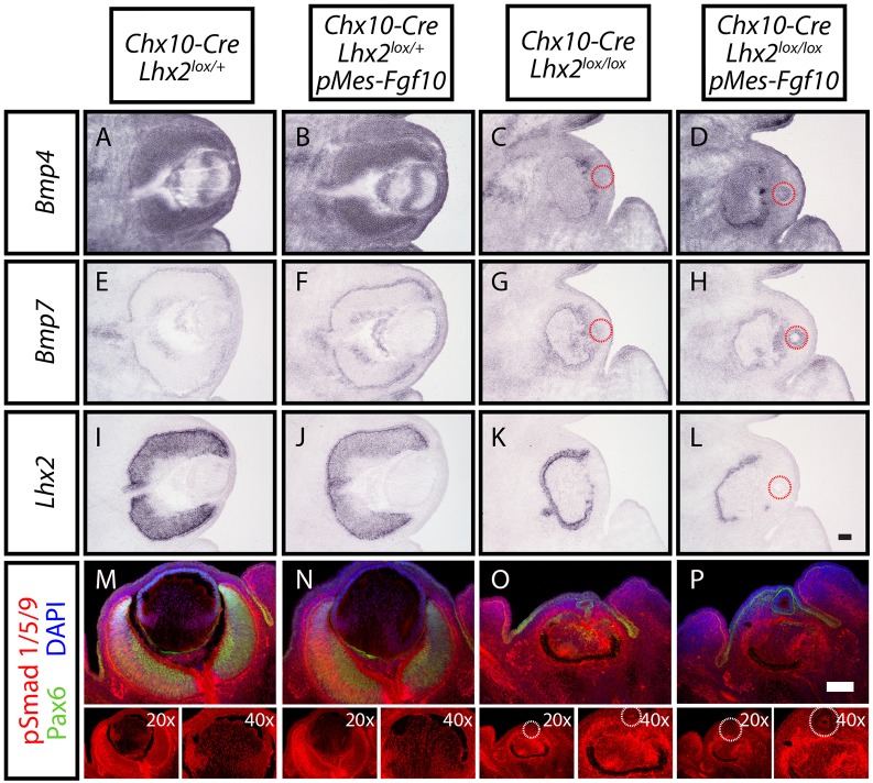
Fig. 7.
Selective deletion of Lhx2 in neuroretina led to downregulation of BMP signaling in the neuroretina and lens. (A-L) In situ hybridization of Bmp4 (A-D), Bmp7 (E-H) and Lhx2 (I-L) mRNA expression levels in Chx10-Cre;Lhx2lox/+ (A,E,I), Chx10-Cre;Lhx2lox/+; pMes-Fgf10 (B,F,J), Chx10-Cre;Lhx2lox/lox (C,G,K) and Chx10-Cre;Lhx2lox/lox;pMes-Fgf10 (D,H,L) eyes at E13.5. Dotted red circles mark the lenses (C,D,G,H,L). (M-P) Immunohistochemical staining for Pax6 (green) and pSmad1/5/9 (red) in lenses of Chx10-Cre;Lhx2lox/+ (M), Chx10-Cre;Lhx2lox/+;pMes-Fgf10 (N), Chx10-Cre;Lhx2lox/lox (O) and Chx10-Cre;Lhx2lox/lox;pMes-Fgf10 animals (P) at E13.5. Nuclei are counterstained with DAPI (blue). The images of pSmad1/5/9 staining in single channel and at higher magnification are included underneath. Dotted white circles mark the lenses (O,P). Scale bars: 100 µm.

