Fig. 6.
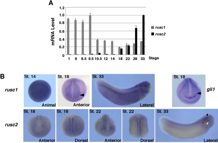
Expression of rusc1 and rusc2 during Xenopus eye development. (A) RT-PCR showing the temporal expression of rusc1 and rusc2 during Xenopus development. The expression level of rusc1 and rusc2 was normalized to that of odc. Data are shown as mean±s.d. (B) Whole-mount in situ hybridization showing the spatial expression pattern of rusc1, rusc2 and gli1. St., stage. Arrowheads point to the eye domains, which express rusc1 but not gli1. Black, red and yellow arrows point to the trigeminal ganglion, middle lateral line placode, and anterodorsal lateral line placode, respectively.

