

Abstract
The nematodes Dispharynx spiralis (Superfamily-Spiruroidea, Family-Acuariidae) parasitising the proventriculus and Heterakis gallinarum (Superfamily-Subuluroidea, Family-Heterakidae) found in the caecum of two backyard poultry birds are described. The usual location of D. spiralis is glandular stomach or proventriculus, where their heads may be deeply buried in the proventricular wall. H. gallinarum occurs in the caecum and commonly called as caecal worm of poultry.
Keywords: Dispharynx spiralis, Heterakis gallinarum, Proventriculus, Caecal worm
Introduction
Backyard poultry rearing under semiscavenging system is found suitable to the villagers as additional source of income and nutrition supplement (Latif 2001). The poultry production is hindered due to infectious diseases caused by parasitic nematodes. This paper describes the natural concurrent infection caused by parasitic nematodes such as Dispharynx spiralis and Heterakis gallinarum in backyard poultry birds (Gallus domesticus) and necessary information regarding the identification of male and female parasites of both species.
Materials and methods
Ten adult chicken from backyard poultry farming were presented for necropsy and with a history of morbidity and mortality at College of Veterianary science, Rajendranagar, Hyderabad. The abdomen of the bird was opened, the entire digestive tract was removed carefully and subdivided into esophagus, crop, proventriculus, gizzard, intestine, caeca and cloaca. At gross necropsy the proventricular wall and mucosa found severely thickened and filled with mucoid material. Numerous short, white, coiled nematode parasites were present within and adhered to the mucosal lining of proventriculus. The various sections of the intestines were separated and the serosal surfaces were examined for gross lesions. After opening up the intestine, faeces was collected. White coloured nematodes were present in the faeces collected from the caecum (Figs. 1, 2, 3).
Fig. 1.

Dispharynx spiralis worms embedded in proventricular mucosa
Fig. 2.
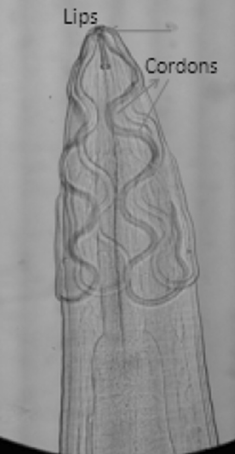
Anterior end of D. spiralis showing 4 convoluted cordons
Fig. 3.
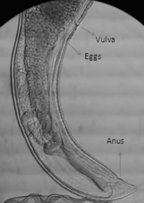
Hind end of female D. spiralis
Parasitological examination
The proventriculus and the faecal sample were collected and transferred to the laboratory. The proventriculus was searched carefully and the nematodes embedded in mucosa were collected with the help of fine forceps. The recovered nematodes were washed thoroughly with normal saline. The male and female worms were differentiated after being subjected for microscopic examination. Few male and female worms (10 each) were first dehydrated in ascending grades of alcohol i.e. 70, 90 %, absolute alcohol, cleared in lactophenol, stained with FAAL (Lucy et al. 2012) and temporary mounting was done. The composition of FAAL stain is 40 % formaldehyde 5 ml, Rectified spirit 20 ml, Lactophenol 75 ml and Azocarmine G 5 mg (Figs. 4, 5, 6).
Fig. 4.

Coiled hind end of male D. spiralis. a Boat shaped right spicule. b Slender left spicule
Fig. 5.
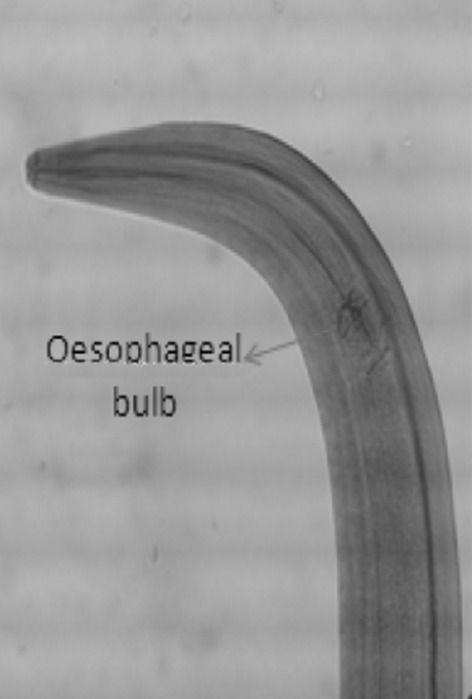
Anterior end of H. gallinarum showing esophageal bulb
Fig. 6.
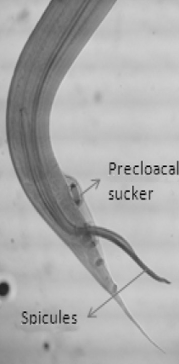
Hind end of male H. gallinarum
Similarly the worms from the faecal sample (collected from caecum) were separated, washed thoroughly with normal saline, dehydrated, cleared, stained with FAAL and finally temporary mounting was done as above.
Results
Diagnosis in this case was based on the characteristic proventricular lesion, eggs, and adult worm identification. The worms collected from the proventriculus were identified as D. spiralis and those which are collected from faecal material collected from caecum were identified as H. gallinarum as per the key provided by (Soulsby 2006a, b).
Dispharynx spiralis
Anterior end—1. Lips are smaller and triangular in shape and pharynx is cylindrical. Anterior part of the body is ornamented with cordons (cuticular ridges or grooves) which are transversely striated. There are four distinct convoluted cordons beginning at dorsal and ventral sides of oral opening and they are recurrent but don’t anastomose. In female the vulva is situated in the posterior part of the body. Eggs have thick shell and contain well developed larvae. In male hind end is more coiled and left spicule is slender, long, right spicule is boat shaped (Fig. 7).
Fig. 7.

Female H. gallinarum
Heterkis gallinarum
Occurs in caeca and anterior end has a strong posterior bulbous oesophagus. In female the vulva opens directly behind the middle of the body. In male the tail is provided with large alae, prominent pre-cloacal sucker and two unequal spicules are present. Right spicule is slender and long, left spicule is brat shaped (Soulsby 2006a, b).
Discussion
Dispharynx nasuta (syn. Dispharynx (Acuaria) spiralis), also known as the ‘proventricular worm’, is a spirurid nematode parasite of the proventriculus of many passerine, columbiform and free ranging gallinaceous birds (Goble and Kutz 1945; Urquhart et al. 1994). The nematodes attach by their anterior end to the mucosal epithelial cells initially causing ulceration at the site of attachment. In most avian species these worms cause only a mild nodular reaction in the mucosa and a small amount of inflammation. However in some species (American wood cock, ruffed grouse, blue grouse) D. nasuta acts as a primary pathogen (Goble and Kutz 1945). When present in large number (10 or more), the infection causes severe hyperplasia of proventricular glands.
Conclusion
Backyard farmers rarely deworm their chickens. Easy access to paratenic and intermediate hosts of H. gallinarum and D. spiralis may be responsible for the infection. Regular deworming at 3 months interval with appropriate anthelmintics is recommended against both the nematode parasites.
Acknowledgments
We all are grateful to Associate Dean, College of Veterinary Science, Rajendra Nagar, Hyderabad for his cooperation regarding this work.
Footnotes
G. S. Sreenivasa Murthy—Assistant Professor (Senior Grade) & Head and Rasmita Panda—MVSc Scholar in Parasitology.
References
- Goble FC, Kutz HL. The genus Dispharynx (Nematoda: Acuariidae) in galliform and passeriform birds. J Parasitol. 1945;31:323–331. doi: 10.2307/3273088. [DOI] [PubMed] [Google Scholar]
- Latif MA. Microcredits and savings of rural household of Bangladesh. Bangladesh Dev Stud. 2001;27:51–71. [Google Scholar]
- Lucy S, Devada K, Sreekrishnam R. A single step processing including staining of nematodes in FAAL solution. J Vet Parasitol. 2012;26(1):91–92. [Google Scholar]
- Soulsby EJL. Helminth arthropods and protozoa of domesticated animals. London: Bailliere Tindall; 2006. pp. 298–300. [Google Scholar]
- Soulsby EJL. Helminth arthropods and protozoa of domesticated animals. London: Bailliere Tindall; 2006. pp. 162–163. [Google Scholar]
- Urquhart GM, Armour J, Duncan JL, Dunn AM, Jennings FW. Veterinary parasitology. London: Churchill Livingstone Inc; 1994. pp. 82–83. [Google Scholar]