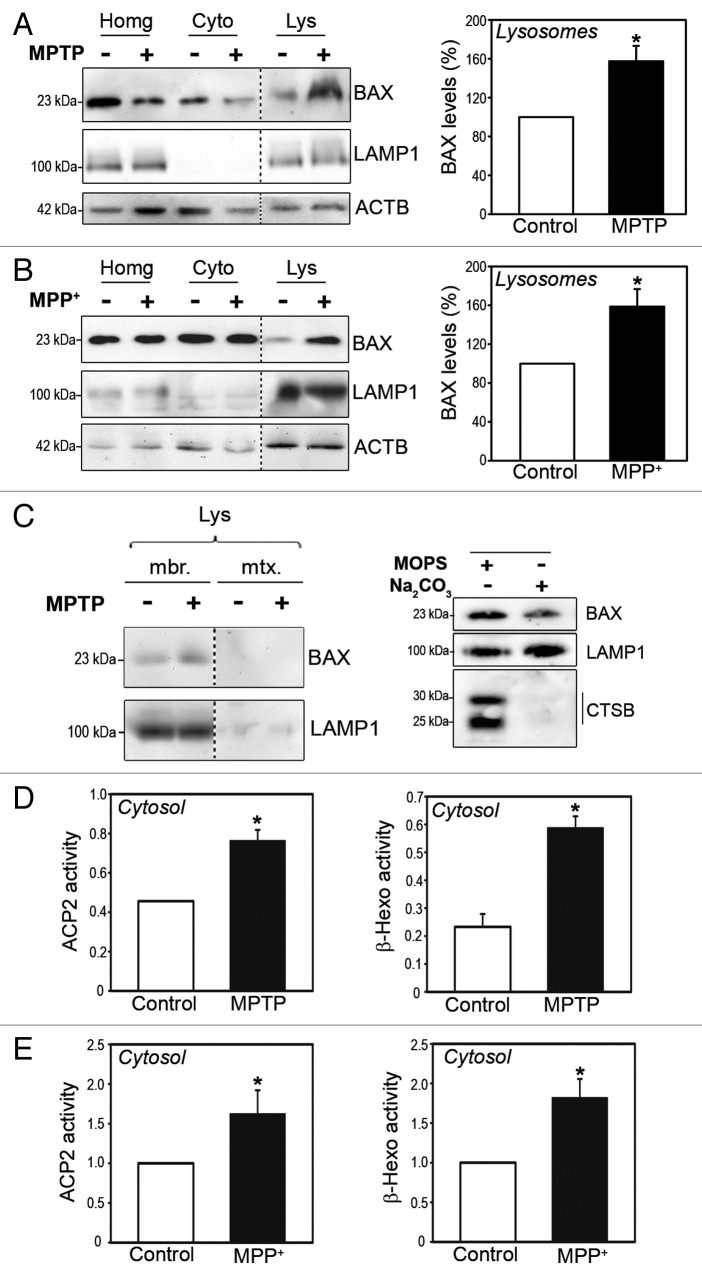
Figure 1. BAX lysosomal translocation early following MPTP treatment in vitro and in vivo. (A and B) BAX immunoblot levels in purified lysosomal (Lys), cytosolic (Cyto) and total protein homogenate (Homg) fractions from ventral midbrain of saline- and MPTP-treated mice (A) and control and MPP+-treated BE(2)-M17 cells (B). Mice received daily injections of 30 mg/kg/day of MPTP for 5 consecutive d and were euthanized 2 h after the last MPTP injection. Cells were incubated with 250 μM MPP+ for 3 h. Quantifications correspond to BAX levels in lysosomal fractions. Lysosomal transmembrane protein LAMP1 is shown as a lysosomal marker. (C, left panel) BAX immunoblot levels in lysosomal membrane and lumen fractions obtained by hypotonic shock from ventral midbrain of saline- and MPTP-treated mice. (C, right panel) BAX immunoblot levels in lysosomal pellet fractions from MPP+-treated (250 μM for 3 h) BE(2)-M17 cells following alkaline extraction with Na2CO3 (0.1 M, pH 11.5, for 30 min). Alkali-resistant transmembrane protein LAMP1 and alkali-sensitive CTSB are shown as experimental controls. (D and E) Enzymatic activities of lysosomal enzymes ACP2 and β-hexosaminidase (β-hexo) in cytosolic, lysosomal-free fractions from the ventral midbrain of MPTP-treated mice, 2 h post-MPTP (D) and BE(2)-M17 cells treated with 250 μM MPP+ for 3 h (E). In all panels, data represent mean ± SEM. In (B and E), at least 3 independent experiments were performed. In (A,C, and D) each experiment corresponds to pooled ventral midbrains from 9 animals (either saline- or MPTP-treated). *P < 0.05, compared with respective control groups.

An official website of the United States government
Here's how you know
Official websites use .gov
A
.gov website belongs to an official
government organization in the United States.
Secure .gov websites use HTTPS
A lock (
) or https:// means you've safely
connected to the .gov website. Share sensitive
information only on official, secure websites.
