Abstract
Aim
To assess the role of breast US in diagnosing and classifying gynecomastia as the primary imaging modality and to compare US findings and classification system with the mammographic ones.
Patients and methods
48 patients suspected of having gynecomastia underwent mammography and US. Two radiologists in consensus retrospectively evaluated mammograms and sonograms. Both US and mammographic images were evaluated categorizing gynecomastia into non-mass, nodular and flame shaped patterns. The two category assignations were compared in order to find any difference. The reference standard for both the classification systems was represented by the cytological examination in 18 out of 44 cases (41%) and the six-month US follow-up in the remaining cases.
Results
The US examination revealed pseudo-gynecomastia in 4/48 (8%) and true gynecomastia in the remaining 44 (92%). Gynecomastia was bilateral in 25/44 cases (57%) and unilateral in the remaining 19 (43%). The cases of true gynecomastia included non mass shape in 26/44 cases (59%), nodular shape in 12 (27%) and flame shape in 6 (14%). The mammographic examination revealed the same results as compared with US findings. 18/44 (41%) patients affected by nodular or dendritic gynecomastia underwent cytological examination confirming the presence of glandular tissue and the benign nature of the clinical condition.
Conclusions
US could be proposed as the primary imaging tool for diagnosing and classifying gynecomastia, avoiding unnecessary X-ray examinations or invasive procedures in case of diffuse gynecomastia. In case of nodular or dendritic patterns, biopsy remains mandatory for a definitive diagnosis.
Keywords: Ultrasound, Gynecomastia, Mammography, Patterns
Introduction
Gynecomastia is the most common disease of the male breast, defined as benign proliferation of the male breast glandular tissue. It is most prevalent in the newborn, adolescent and the elderly (1–3).
Palpation usually demonstrates a soft, mobile and elastic mass in the subareolar region, centered directly under the nipple. Pain may be present in gynecomastia of less than 6 months duration. Over time, gynecomastia can become fibrotic and patients often present with a painless firm, even hard mass that is difficult to differentiate from breast carcinoma. Furthermore gynecomastia could be coexistent with breast cancer and obscuring it (4–6).
Although breast cancer is rare in men (estimated as 1% of all breast cancer), the differential diagnosis between gynecomastia and male breast cancer or the exclusion of concurrent cancer with gynecomastia represents the primary aim for the clinician (7–10).
Breast imaging provides many useful and accurate techniques for studying the breast tissue and characterizing breast diseases both in female and male patients (11–15).
Diagnostic evaluation of patients with gynecomastia can be costly and can involve numerous radiographic tests, including mammography, sonography and sometimes biopsy.
Mammography is usually the primary modality of diagnosis and classification of this clinical condition when imaging is indicated (2, 3).
With regard to sonography, a few medical literature has been reported especially about the sonographic patterns of gynecomastia (16, 17). In particular, it is still unclear the real benefit of sonography for diagnosing and classifying gynecomastia and also if it could be used alone or in combination with mammography in all cases (2, 18). Besides, the different series are limited, largely descriptive, with limited number of cases (8). It is also reported that sonography adds positively to clinical management when mammography reveals other findings in addition to gynecomastia (3) and the medical literature recommends the use of sonography as the initial examination for patients younger than 30 years or for men who refuse mammography (2, 19).
Biopsy is recommended in case of suspicious lesions or if a coexistent lesion is clinically or radiologically suspected; therefore, it should not be performed routinely (6, 18).
The aim of this study is to assess the role of breast ultrasound in diagnosing and classifying gynecomastia as the primary imaging modality and to compare US findings and classification system with the mammographic ones.
Patients and methods
In the period from between May 2011 and June 2014, a total of 56 male patients with breast symptoms presenting for imaging were identified, and a retrospective analysis was performed searching for the diagnosis of gynecomastia. The breast consultation electronic archives were searched for male patients and the computerized files were searched by the key word “gynecomastia”, “breast nodule”. Eight out of 56 patients were excluded from the study because of the biopsy results during data analysis (2 hematoma, 4 lipoma and 2 breast cancer). Therefore, only 48 patients were included and both mammography and sonography were performed in all cases.
Sonographic examinations were performed using a linear 13 MHz probe (MicroMaxx Ultrasound System, Sonosite, Bothell, WA, US) in a standard supine position with arms above the head. Two investigators in consensus reviewed each imaging study without knowledge of the clinical history or any accompanying imaging study. The mammograms were reviewed separately from the sonograms without knowledge of the clinical or correlative sonographic finding.
Basing on the aim of our study, sonograms were assessed searching for the presence of gland tissue, first of all distinguishing true gynecomastia from pseudogynecomastia.
Pseudogynecomastia is a breast enlargement characterized by increased subareolar fat tissue without enlargement of the breast glandular component. The medical literature describes the mammographic and ultrasound criteria used to diagnose gynecomastia, pseudogynecomastia and normal male breast as follow: the absence of dense retroareolar tissue in an enlarged breast with predominance of fat tissue allows to differentiate pseudogynecomastia from true gynecomastia (5).
In case of true gynecomastia, the sonographic findings were categorized into three groups as follows:
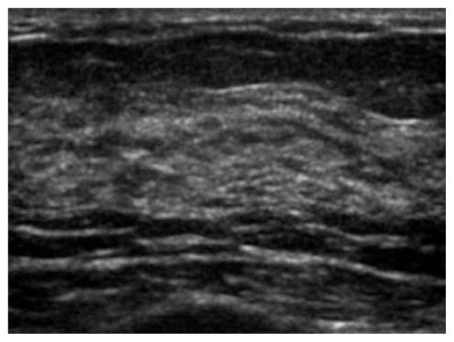
non-mass, diffuse increment of the glandular tissue (subareolar antero-posterior mean thickness > 1 cm);
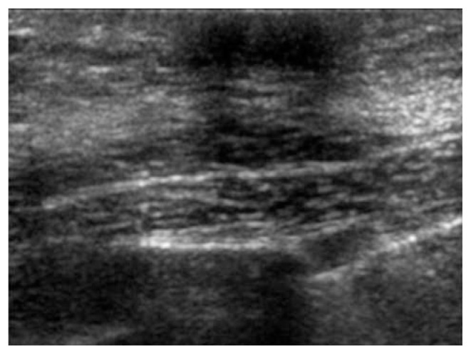
nodular-discrete round or oval hypoechoic area in the subareolar region;
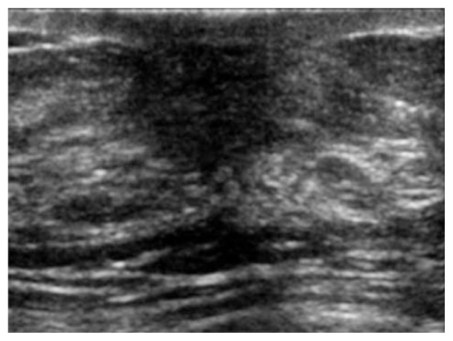
flame shaped-irregular hypoechoic area with extensions into the surrounding tissue.
Mammographic findings were categorized into 3 groups on the basis of the parenchymal pattern as described by Appelbaum et al. (20):
diffuse;
nodular;
dendritic.
The sonographic and mammographic category assignations were compared in order to find any difference of classification between the two imaging tools.
The reference standard for both the classification systems was represented by the cytological examination in 18 out of 44 cases (41%) and the six-month US follow-up in the remaining cases. Fine needle aspiration cytology (FNAC) was performed free hand, under US guidance, using a 21 G needle and the sampled material was treated with spray fixative solution.
Results
There were 48 patients included in our study population, who presented with pain (n=8), breast lump (n=26), pain and lump (n=16). The age range was 24 to 74 years (mean age, 52±3 years). The sonographic examination revealed pseudogynecomastia in four out of 48 (8%) and true gynecomastia in the remaining 44 patients (92%).
In our study population, the etiology of gynecomastia was represented by endocrine dysfunctions in 8 (18%) cases, non-endocrine disease in 16 (36%), tumors in 2 (5%), drug therapy in 13 (30%), idiopathic in the remaining 5 (11%) patients. Gynecomastia was bilateral in 25 (57%) out of 44 patients and unilateral in the remaining 19 (43 %) patients.
The cases of true gynecomastia included non mass shape in 26 (59%) out of 44 patients, nodular shape in 12 (27%) and flame shape in 6 (14%).
The mammographic examination revealed the same results as compared with sonographic findings. In particular, pseudogynecomastia was found in four out of 48 (8%) and true gynecomastia in the remaining 44 patients (92%). The cases of true gynecomastia included diffuse shape in 26 (59%) out of 44 patients, nodular shape in 12 (27%) and dendritic shape in 6 (14%).
18 out of 44 (41%) patients affected by nodular or dendritic gynecomastia as detected by sonography and mammography underwent cytological examination, confirming the presence of glandular tissue and the benign nature of the clinical condition.
Discussion
The abnormal increase in the stromal and ductal components of the male breast can be caused by physiological factors (puberty or aging), alterations in the testosterone-to-estrogen ratio, which can arise from endocrine tumors (testicular, adrenocortical tumors or ectopic hCG-secretions), endocrine dysfunctions (hypogonadism, hyperthyroidism, obesity, diabetes), non-endocrine disease (chronic liver disease, renal failure or HIV). It is also associated with numerous drug therapies such as antidepressants, anti-hypertensives, glucocorticoids, chemotherapeutic agents and illicit drugs. Many cases of gynecomastia are idiopathic accounting for 25% especially in the prepuberal forms (6, 9).
In our series, the etiology of gynecomastia was represented by endocrine dysfunctions in 18% of cases, non-endocrine disease in 36%, tumors in 5%, drug therapy in 30%, idiopathic in the remaining 11%. Therefore, our results confirm this data, except for the idiopathic forms accounting for 11% in our series, probably due to the higher mean age of the examined patients. Besides, gynecomastia is bilateral in approximately half of the patients as also occurred in our series.
Diagnostic imaging of gynecomastia has already been reported in the medical literature and unanimous agreement about mammographic diagnosis and classification already exists. In particular, Appelbaum et al. described three mammographic patterns: diffuse, nodular, dendritic. Diffuse glandular gynecomastia has a mammographic appearance similar to that of a heterogeneously dense female breast (18, 20, 21). Nodular gynecomastia appears as a fan-shaped density radiating from the nipple, which may be symmetric or more prominent in the upper outer quadrant. The density usually blends gradually into the surrounding fat, but it may be more spherical. This pattern correlates with the pathological classification of florid gynecomastia, which is the early phase of this clinical condition. Dendritic gynecomastia appears as a retroareolar soft-tissue density with prominent extension which radiates into the deeper adipose tissue. This pattern correlates with fibrous gynecomastia. Basing on these diagnostic information, mammography is reported to be the primary imaging modality for diagnosing gynecomastia with a negative predictive value of 100% and also a sensitivity and specificity values of 100% and 90%, respectively, for cancer detection (8, 22).
However, mammography presents several limitations. First, the X-ray exposure in case of prepuberal gynecomastia and young patients as compare with ultrasound; several men also refuse mammography being a more invasive and less comfortable tool. Moreover, Dialani also reported that it could be insufficient when there is asymmetric nodular gynecomastia or in the case of a cluster of subareolar ducts with a convex margin mimicking a nodular breast lesion. Finally, mammography is usually more accurate for detecting microcalcifications as compared with sonography; however, the pattern of microcalcifications in male breast cancer are not so classic as women cancer and could also present benign features (2).
Therefore, many authors propose the use of sonography combined with mammography (3). In particular, a negative predictive value close to 100% has been demonstrated for both mammography and sonography. Munoz Carrasco et al. (5) reported that the combined use of the two techniques makes it possible to avoid many unnecessary surgical procedures in men, while Chen et al. (3) suggested that targeted sonography is of limited value in symptomatic male patients where mammography is negative or reveals gynecomastia and that sonography is useful only in the case of suspected mammographic findings. Therefore, the precise role of sonography in this field is still debated, as also if it could be used alone or in combination with mammography in all cases, and the related findings not yet standardized.
In fact, different sonography classification systems have been reported. Wigley et al. (23) reported two patterns represented by focal and diffuse. Dialani et al. (2) described four patterns on sonography: nodular, poorly defined, flame-shaped and non-mass. Nodular consists of round or oval hypoechoic area in the retroareolar region, surrounded by normal fatty tissue; poorly defined of vague hypoechoic area in the retroareolar region; flame-shaped of a subareolar hypoechoic area with an anechoic star-shaped posterior border and with finger like projections; non-mass of increased AP depth at the nipple defined as greater than 1 cm depth of breast parenchyma at the nipple, with isoechoic, hypoechoic or hyperechoic shape. The most commonly described sonographic pattern of gynecomastia in adults is the flame-shaped or triangular retroareolar density (18, 21).
In our series, in order to compare mammographic and sonographic diagnosis and classification systems and to standardize the relative findings, we considered three sonographic patterns of gynecomastia (diffuse, nodular and flame-shaped), corresponding to the three mammographic patterns already described in the medical literature (diffuse, nodular and dendritic). In our series pseudogynecomastia was found in 8% of cases and true gynecomastia in the remaining 92%. True gynecomastia appeared as diffuse in 59%, nodular in 27% and dendritic in 14% of patients on both mammography and ultrasound. Basing on the obtained results, no difference between the two imaging tools in terms of categorization was found.
In particular, all the nodular and dendritic lesions were recognized at sonography and confirmed at the following cytological examination in all cases. Therefore, in our experience, sonography appeared as able as mammography for both diagnosing and classifying gynecomastia and the two classification systems appeared superimposable in all cases with sonography being able to be proposed as the primary X-ray free imaging modality for gynecomastia.
Our study has some limitations, mainly represented by the retrospective setting of the study, the relative small number of the examined patients, the lack of an inter-observer agreement, the lack of an histologic control in all cases and also the lack of a long-term sonography follow-up of more than six months.
Conclusions
Sonography could be proposed as the primary imaging tool for diagnosing and classifying gynecomastia, avoiding unnecessary X-ray examinations or invasive procedures in case of diffuse gynecomastia. In case of nodular or dendritic patterns, biopsy remains mandatory for a definitive diagnosis and for excluding breast cancer. Mammography could be reserved only in the case of suspected sonographic malignant findings to confirm diagnosis before interventional procedures.
Fig. 1.
Non-mass US pattern of gynecomastia in a 54-years-old male patient represented by a diffuse increment of the glandular tissue.
Fig. 2.
Nodular US pattern of gynecomastia in a 36-years-old male patient represented by an oval hypoechoic area in the subareolar region with regular edges. The cytological examination confirmed the US diagnosis of gynecomastia.
Fig. 3.
Flame shaped US pattern of gynecomastia in a 61-years-old male patient represented by an irregular hypoechoic area with extensions into the surrounding tissue. The cytological examination confirmed the US diagnosis of gynecomastia.
Footnotes
Conflict of interest
All authors have no conflicts of interest nor financial or personal relationships regarding this paper.
References
- 1.Iuanow E, Kettler M, Slanetz PJ. Spectrum of disease in the male breast. AJR Am J Roentgenol. 2011;196(3):W247–59. doi: 10.2214/AJR.09.3994. [DOI] [PubMed] [Google Scholar]
- 2.Dialani V, Baum J, Mehta TS. Sonographic Features of Gynecomastia. J Ultrasound Med. 2010;29:539–547. doi: 10.7863/jum.2010.29.4.539. [DOI] [PubMed] [Google Scholar]
- 3.Chen Po-Hao, Slanetz Priscilla J. Incremental clinical value of ultrasound in men with mammographically confirmed gynecomastia. European Journal of Radiology. 2014;83:123–129. doi: 10.1016/j.ejrad.2013.09.021. [DOI] [PubMed] [Google Scholar]
- 4.Johnson RE, Hassan Murad M. Gynecomastia: Pathophysiology, Evaluation, and Management. Mayo Clin Proc. 2009;84(11):1010–1015. doi: 10.1016/S0025-6196(11)60671-X. [DOI] [PMC free article] [PubMed] [Google Scholar]
- 5.Munoz Carrasco R, Alvarez Benito M, Munoz, et al. Mammography and ultrasound in the evaluation of male breast disease. Eur Radiol. 2010;20:2797. doi: 10.1007/s00330-010-1867-7. [DOI] [PubMed] [Google Scholar]
- 6.Simões Dornellas de BarrosI Alfredo Carlos, de Castro Moura Sampaio Marcelo. Gynecomastia: physiopathology, evaluation and treatment. Sao Paulo Med J. 2012;130(3):187–97. doi: 10.1590/S1516-31802012000300009. [DOI] [PMC free article] [PubMed] [Google Scholar]
- 7.Giordano SH, Cohen DS, Buzdar AU, Perkins G, Hortobagyi G. Breast carcinoma in men: a population-based study. Cancer. 2004;101:51–57. doi: 10.1002/cncr.20312. [DOI] [PubMed] [Google Scholar]
- 8.Patterson SK, Helvie MA, Aziz K, Nees AV. Outcome of men presenting with clinical breast problems: the role of mammography and ultrasound. Breast J. 2006;12:418–23. doi: 10.1111/j.1075-122X.2006.00298.x. [DOI] [PubMed] [Google Scholar]
- 9.Athwal Ruvinder Kaur, Donovan Rosamund, Mirza Mehboob. Clinical Examination Allied to Ultrasonography in the Assessment of New Onset Gynaecomastia: An Observational Study. Journal of Clinical and Diagnostic Research. 2014;8(6):NC09–NC11. doi: 10.7860/JCDR/2014/7920.4507. [DOI] [PMC free article] [PubMed] [Google Scholar]
- 10.Mathew J, Perkins GH, Stephens T, Middleton LP, Yang W-T. Primary breast cancer in men: clinical, imaging, and pathologic findings in 57 patients. AJR Am J Roentgenol. 2008;191(6):1631–9. doi: 10.2214/AJR.08.1076. [DOI] [PubMed] [Google Scholar]
- 11.Telegrafo M, Rella L, Stabile Ianora AA, Angelelli G, Moschetta M. Unenhanced breast MRI (STIR, T2-weighted TSE, DWIBS): An accurate and alternative strategy for detecting and differentiating breast lesions. Magn Reson Imaging. 2015;33:951–5. doi: 10.1016/j.mri.2015.06.002. [DOI] [PubMed] [Google Scholar]
- 12.Moschetta M, Telegrafo M, Carluccio DA, Jablonska JP, Rella L, Serio G, Carrozzo M, Stabile Ianora AA, Angelelli G. Comparison between fine needle aspiration cytology (FNAC) and core needle biopsy (CNB) in the diagnosis of breast lesions. G Chir. 2014;35:171–6. [PMC free article] [PubMed] [Google Scholar]
- 13.Moschetta M, Telegrafo M, Rella L, Capolongo A, Stabile Ianora AA, Angelelli G. MR evaluation of breast lesions obtained by diffusion-weighted imaging with background body signal suppression (DWIBS) and correlations with histological findings. Magn Reson Imaging. 2014 Jul;32(6):605–9. doi: 10.1016/j.mri.2014.03.009. [DOI] [PubMed] [Google Scholar]
- 14.Moschetta M, Scardapane A, Lorusso V, Rella L, Telegrafo M, Serio G, Angelelli G, Ianora AA. Role of multidetector computed tomography in evaluating incidentally detected breast lesions. Tumori. 2015;101(4):455–60. doi: 10.5301/tj.5000291. [DOI] [PubMed] [Google Scholar]
- 15.Telegrafo M, Rella L, Stabile Ianora AA, Angelelli G, Moschetta M. Supine breast US: how to correlate breast lesions from prone MRI. Br J Radiol. 2016;89:20150497. doi: 10.1259/bjr.20150497. [DOI] [PMC free article] [PubMed] [Google Scholar]
- 16.Yang WT, Whitman GJ, Yuen EH, Tse GM, Stelling CB. Sonographic features of primary breast cancer in men. AJR Am J Roentgenol. 2001;176(2):413–6. doi: 10.2214/ajr.176.2.1760413. [DOI] [PubMed] [Google Scholar]
- 17.American College of Radiology. Breast Imaging Reporting and Data System: Ultrasound. Reston, VA: American College of Radiology; 2003. [Google Scholar]
- 18.Gunhan-Bilgen I, Bozkaya H, Ustun EE, Memis A. Male breast disease: clinical, mammographic, and ultrasonographic features. European Journal of Radiology. 2002;43:246–55. doi: 10.1016/s0720-048x(01)00483-1. [DOI] [PubMed] [Google Scholar]
- 19.Munoz Carrasco R, Álvarez Benito M, Rivin del Campo E. Value of mammography and breast ultrasound in male patients with nipple discharge. European Journal of Radiology. 2013;82(3):478–84. doi: 10.1016/j.ejrad.2012.10.019. [DOI] [PubMed] [Google Scholar]
- 20.Appelbaum AH, Evans GFF, Levy KR, Amirkhan RH, Schumpert TD. Mammographic appearances of male breast disease. Radiographics. 1999;19:559–68. doi: 10.1148/radiographics.19.3.g99ma01559. [DOI] [PubMed] [Google Scholar]
- 21.Adibelli ZH, Oztekin O, Gunhan-Bilgen I, Postaci H, Uslu A, Ilhan E. Imaging characteristics of male breast disease. Breast Journal. 2010;16(5):510–8. doi: 10.1111/j.1524-4741.2010.00951.x. [DOI] [PubMed] [Google Scholar]
- 22.Evans GFF, Anthony T, Appelbaum AH, et al. The diagnostic accuracy of mammography in the evaluation of male breast disease. AJR Am J Surg. 2001;181:96–100. doi: 10.1016/s0002-9610(00)00571-7. [DOI] [PubMed] [Google Scholar]
- 23.Wigley KD, Thomas JL, Bernardino ME, Rosenbaum JL. Sonography of gynecomastia. AJR Am J Roentgenol. 1981;136:927–930. doi: 10.2214/ajr.136.5.927. [DOI] [PubMed] [Google Scholar]


