
Figure 8. Immunofluorescence analysis of GFAP expression (green) at time 0, after 6 and 15 days of differentiation in medium containing 10% FBS.
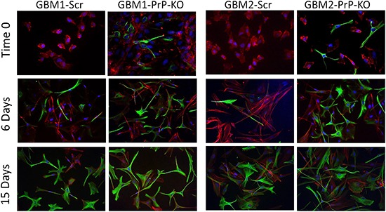
Nuclei were counterstained with DAPI (blue) and cells morphology was evidenced by staining with Dil, a lipophilic red-fluorescent dye. Magnification 40X.