Abstract
We investigated the effect of ethnicity and gender on optic nerve head morphology in healthy subjects using spectral-domain optical coherence tomography (SD-OCT). Thirty-five Indian (i.e. Indian subcontinent) females, 34 Caucasian females, 32 Indian males, and 32 Caucasian males were examined using SD-OCT (Copernicus, Optopol Technology). Disc and rim areas were larger in Caucasian males compared with females but smaller in Indians males compared with females. Indian participants had significantly larger cup areas and volumes without significant differences in retinal nerve fibre layer (RNFL) thicknesses between groups. Gender and ethnicity differences should be considered in assessment of patients.
Keywords: Gender and ethnicity, healthy optic nerve, OCT
Introduction
Pathology of the optic nerve including glaucoma and optic nerve atrophy is one of the main causes of visual impairment and blindness across the world. The development of optical coherence tomography (OCT) gives access to high-resolution three-dimensional images of optic nerve head (ONH) structure with high reproducibility, giving the potential for ONH pathologies to be detected at earlier stages.1
ONH parameters in healthy eyes measured by spectral-domain OCT (SD-OCT) are generally published by the equipment manufacturer for detection of ONH pathology. Although these measurements are provided on the majority of commercially available OCT machines, surprisingly little data have been published in the literature on whether these control data are applicable across different ethnicities and for both genders. This decreases the chance of providing accurate diagnosis and timely treatment for the different ONH pathologies.
Two-dimensional (2D) fundus analysis2,3 and more recently three-dimensional (3D) OCT imaging have been used to show that clear differences exist in ONH morphology between ethnicities in parameters such as of optic disc, cup, and rim areas and volumes.4–8 Individuals originating from the Indian subcontinent show particularly distinctive features in ONH morphology compared with other ethnicities, with the most obvious differences apparent in comparison with individuals of Caucasian ethnicity.4
The effect of gender on ONH morphology is more controversial, especially in relation to different ethnic groups. Varma et al.3 and Ramrattan et al.2 used 2D fundus analysis to suggest that larger discs exist in males from both white European and African American ethnic groups based in the USA. Stereo-photography optic disc assessment by Arvind et al. also found a similar tendency in Indian population.9 However, Knight et al. in a large, recent OCT study that compared European, Chinese, and Hispanic ethnic groups describe no gender-related difference in any optic nerve head parameters.5 Also, a recent OCT study of subjects of Indian nationals has not found any gender-related difference in optic nerve morphology.8 One possibility may be that gender differences in ONH morphology may show contrasting patterns in various ethnic groups, which could be investigated by analysing the statistical interaction (different pattern between males and females in different ethnicities) of ONH parameters between gender and ethnicity.
The current study has been performed in the city of Leicester, UK, which has one of the most ethnically diverse populations in the UK, with the largest Indian (Indian subcontinent) population of any local authority area, accounting for about 30% of the population. The Caucasian and Indian populations in Leicester also share similar socioeconomic status and access to health care.10 In the present study we aim to determine the effect of ethnicity and gender on ONH morphology and peripapillary retinal nerve fibre layer (RNFL) thickness in healthy subjects using SD-OCT, in order to enable better diagnostic criteria of normal and abnormal optic nerve morphology in different ethnicities.
Participants and Methods
Participants
The population cohort for this study included 133 clinically healthy subjects (Indian female: n = 35, Caucasian female: n = 34, Indian male: n = 32, and Caucasian male: n = 32) consecutively selected from the university and National Health Service (NHS) staff, their relatives and friends (82.7%), and relatives and partners of the patients of general clinics (17.3%). The mean age was 32.7 years (SD: 14.5 years). All participants had best-corrected visual acuity of 0.2 logMAR or better, and normal visual field test results. All refractive errors were between −3.0 and +3.0 dioptres and there were no differences in mean refractive error between the groups. Participants and their close relatives did not suffer from any eye pathology including glaucoma or systemic disease, did not use any medication, and had no previous intraocular operations on the analysed eye. All patients underwent ophthalmic examination, which included measurement of best-corrected visual acuity, measurement of intraocular pressure, automated perimetry, and slit-lamp and fundus examination. As a result of these examinations, we excluded one Caucasian female who had an intraocular pressure of 26 mm on the day of the test. Gender, ethnicity, and age were self-reported by the participants.
Informed consent was obtained from all participants of this study. The study adhered to the tenets of the Declaration of Helsinki and was approved by the local ethics committee.
Optical Coherence Tomography Imaging
Ultra-high-resolution spectral-domain OCT (Copernicus; Optopol Technology S.A., Zawiercie, Poland) was used to acquire tomograms with a theoretical axial resolution of 3 μm and a transverse resolution of 12 μm. This study included data from one eye for each participant using the image with the best quality. ONH analysis was performed on a 3D scan profile (75 B-scans, 7 × 7 × 2 mm volume, each B-scan consisted of 743 A-scans) with a fixation target set eccentrically to image the ONH.
The ONH measurements including cup, disc, and rim diameters, areas, and volumes and the thickness of the RNFL were estimated using an automated algorithm provided in the Copernicus OCT software that detects the internal limiting membrane and the edges of the retinal pigment epithelium. Rim and cup dimensions were measured relative to a fixed reference point of the plane of the disc (defined from retinal pigment epithelium [RPE] edges). A default offset of 150 µm was used, since this avoids zero measurements of the cup and rim volume (none were present in the current data set). To minimise any inaccurate measurements by the software the disc margins, position of the internal limiting membrane and RNFL could be adjusted manually by the user (by A.V.P.).
RNFL thickness was measured within the temporal, superior, nasal, and inferior quadrants of an annulus with internal diameter of 2.4 mm and width of 0.4 mm (default settings).
Statistical Methods
The sample size was based on pilot data recorded from 40 participants (10 in each group: Indian male, Indian female, Caucasian male, and Caucasian female) in which the power calculation (power = 95%, p < 0.05) showed that 32 participants were required in each group in order to detect a significantly different pattern between males and females in different ethnicities.
Statistical analysis was carried out using SPSS software version 16.0 (SPSS, Inc., Chicago, IL, USA). Parameters of ONH and RNLF thickness were analysed using general linear model including ethnicity and gender as fixed factors and age as a covariate with interaction (coeffect if gender and ethnicity) terms. Original data were normally distributed only for rim area, inferior distance between disc and cup edges, and cup/disc vertical ratio. Consequently, ONH and RNFL thickness parameters were transformed so that data were more closely approximated to a normal distribution as tested using a Shapiro-Wilk test. Square route was applied for the area measurements, cube route for the volume measurements and log for distance measurements. p ≤ 0.05 was considered statistically significant.
Results
Optic Nerve Head Measurements
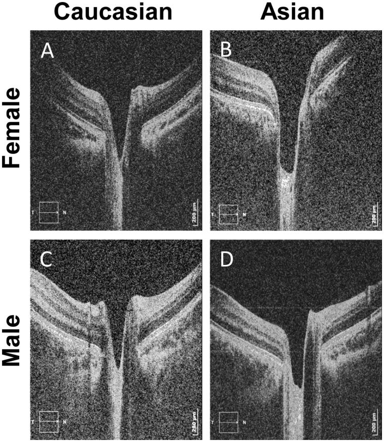
Visual observation of the optic disc scans (representative examples shown for the four participant groups in Figure 1) suggested that mean cup volume was larger in Indian participants, especially in females.
FIGURE 1.
Tomograms of the optic nerve head. Right eyes of (A) a 31-year-old Caucasian female, (B) a 34-year-old Indian female, (C) a 35-year-old Caucasian male and (D) a 27-year-old Indian female participant. Refer to the scale of the scan in the right bottom corner of each tomogram where the horizontal axis is 106 pixels/mm and the vertical axis is 513 pixels/mm.
Disk and Rim Dimensions
Mean values (with standard deviations) and statistical comparisons for the most important parameters are shown in Table 1.
TABLE 1.
Means and standard deviations and statistical comparison for different ethnicity and gender groups for optic nerve parameters.
| Asian |
Caucasian |
||||||||||
|---|---|---|---|---|---|---|---|---|---|---|---|
| Male (n = 32) |
Female (n = 35) |
Male (n = 32) |
Female (n = 34) |
p Value |
|||||||
| Parameter | Mean | SD | Mean | SD | Mean | SD | Mean | SD | Gender | Ethnicity | Different pattern between males and females in two ethnicities |
| Age | 32.19 | 15.25 | 31.49 | 14.92 | 33.53 | 13.75 | 33.59 | 14.92 | 0.90 | 0.50 | 0.88 |
| Disc area (mm2) | 1.58 | 0.33 | 1.80 | 0.32 | 1.63 | 0.26 | 1.53 | 0.30 | 0.21 | 0.13 | 0.009 |
| Cup area (mm2) | 0.60 | 0.34 | 0.71 | 0.41 | 0.45 | 0.32 | 0.51 | 0.31 | 0.066 | 0.001 | 0.81 |
| Rim area (mm2) | 0.98 | 0.25 | 1.06 | 0.35 | 1.13 | 0.33 | 1.00 | 0.29 | 0.59 | 0.33 | 0.049 |
| Cup volume (mm3) | 0.12 | 0.09 | 0.17 | 0.16 | 0.08 | 0.07 | 0.11 | 0.12 | 0.086 | 0.001 | 0.98 |
| Rim volume (mm3) | 0.14 | 0.07 | 0.12 | 0.09 | 0.18 | 0.11 | 0.11 | 0.05 | 0.002 | 0.17 | 0.36 |
| Cup disc ratio | 0.37 | 0.16 | 0.40 | 0.20 | 0.28 | 0.16 | 0.31 | 0.16 | 0.29 | 0.008 | 0.68 |
| Retinal nerve fibre layer thickness (µm) | |||||||||||
| Mean | 118.0 | 14.41 | 115.0 | 16.45 | 117.0 | 13.83 | 119.8 | 12.30 | 0.83 | 0.58 | 0.19 |
| Temporal | 74.2 | 13.37 | 73.1 | 13.36 | 68.9 | 10.24 | 70.1 | 13.90 | 0.98 | 0.07 | 0.59 |
| Superior | 135.2 | 19.75 | 132.0 | 23.57 | 131.2 | 15.97 | 135.8 | 14.97 | 0.80 | 0.96 | 0.23 |
| Nasal | 95.4 | 21.47 | 92.8 | 21.41 | 99.0 | 19.58 | 97.3 | 20.42 | 0.56 | 0.29 | 0.91 |
| Inferior | 135.1 | 15.93 | 133.4 | 19.74 | 134.8 | 17.17 | 137.21 | 18.25 | 0.92 | 0.56 | 0.50 |
Rim volume was significantly larger in males, whereas cup volume and area and cup/disc ratio was significantly larger in Indians. The interaction term (gender × ethnicity) indicates a different pattern between males and females in the two ethnic groups and was statistically significant for disc and rim areas.
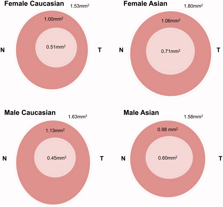
The mean values are also illustrated schematically in Figures 2 and 3. Disc and rim areas (Table 1) showed a similar significant interaction between gender and ethnicity (i.e. different pattern between males and females in different ethnicities). This cross-difference was due to Indian females demonstrating significantly larger disc and rim areas compared with males, whereas in Caucasians an opposite pattern was observed, with females demonstrating smaller disc and rim areas than males (Figure 2).
FIGURE 2.
Schematic diagram of optic disc (outer circle), cup (light pink inner circle), and rim (dark pink area) dimensions for each of the four groups. Cup and disc horizontal and vertical dimensions are represented as an ellipse and the location of the cup within the disc is also represented. Dotted lines indicate SE around the mean (solid lines). T = temporal; N = nasal.
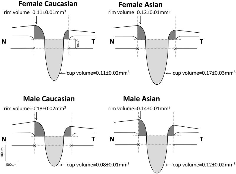
FIGURE 3.
Cross-sectional schematic diagram of optic nerve head (mean ± SE). Top dotted lines represent cup horizontal diameters, bottom dotted lines indicate disc horizontal diameters, and vertical dotted lines show margins of rim areas. T = temporal; N = nasal.
Disc areas had a different dimensions in Indian and Caucasian groups due to horizontal elongation in Indian individuals (disc horizontal diameter was statistically larger in Indian group, p = 0.003, vertical/horizontal disc ratio, p = 0.001).
Cup Dimensions
Figure 2 summarises the measurements of the disc and cup in the four groups, illustrating also significant ethnicity-related differences in cup measurements. Indian participants had significantly larger cup diameters (p = 0.03 and p = 0.002 for vertical and horizontal, respectively), resulting in the larger areas of the cup (p = 0.001) than Caucasian participants (Figure 2).
Moreover, cup volume was also larger in Indian participants compared with Caucasians (p = 0.001), as illustrated in Figure 3. Cup volumes also appeared larger in the female individuals, although this was not statistically significant (p = 0.086).
Ethnicity still had a significant effect on both cup area (p = 0.01) and cup volume (p = 0.01) after adjusting for disc area.
Investigating age-related changes in ONH morphology were not the main purpose of this study. However, there was a significant increase in cup volume (p = 0.04) and cup/disk ratio (p = 0.02) with the decreasing in rim area (p = 0.01) with age.
Peripapillary RNFL
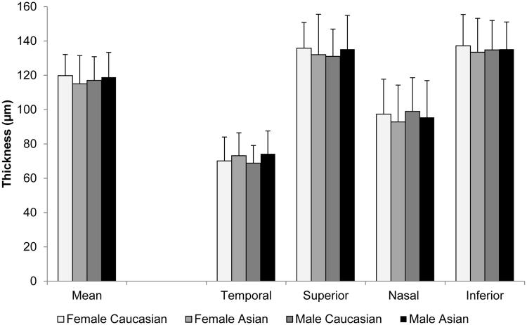
Figure 4 clearly demonstrates that despite a variety of differences in disc, cup, and rim measurements across ethnicity and gender, all individuals showed remarkable similar patterns in RNFL thickness for the various segments (Table 1, Figure 4). The only one factor in the statistical model that showed a significant effect was age (p = 0.02). As expected, decrease of the mean RNFL thickness with age corresponded to the decrease in the rim area (p = 0.01) and increase in cup volume (p = 0.04) and cup/disc ratio (p = 0.02).
FIGURE 4.
Average thickness of the peripapillary retinal nerve fibre layer across gender and ethnicity in different quadrants. Error bars are standard deviations.
The RNFL thickness was significantly larger in superior and inferior quadrants compared with the other two quadrants, with no significant difference between them. Moreover, the RNFL thickness in the temporal quadrant was significantly lower value than in the nasal quadrant (p = 0.001).
Discussion
In this study we describe for the first time a significant interaction between gender and ethnicity for optic nerve head morphology. In particular, disc and rim areas were significantly larger in Caucasian males compared with females but smaller in Indians (i.e. Indian subcontinent) males compared with females (p = 0.009 and p = 0.049, respectively). We also found “physiological cupping” of the optic nerve head in Indian compared with Caucasian individuals that was particularly obvious in Indian females. The cup areas were 36.5% larger and cup volumes 52.6% larger in Indians compared with Caucasians. This was not associated with changes in the RNFL integrity where there were no significant differences between the groups. The study cohort was derived from populations in the same UK city with similar socioeconomic status and access to health care, with identical retinal imaging methodology and analysis applied to both groups.
Different Pattern between Males and Females in Different Ethnicities
In agreement with our findings, larger disc and rim areas have been described in males compared with females in Americans originating from white European ethnic groups based on fundus photography.2,3 A similar pattern was also observed in African Americans in these studies. This is the first time an interaction has been described between ethnicity and gender for optic nerve head structure where disc and rim area show contrasting patterns between males and females for two different ethnicities. A recent large OCT study described no gender-related differences for individuals of European, African, Chinese, and Hispanic5 descent. A group originating from the Indian subcontinent was not included in this study.
Another recent study has investigated ONH morphology within an Indian (Indian subcontinent) population and found no gender-related differences.8 However, disc area was not compared but included as a predictor variable in the statistical model. Also, the mean age of the study cohort was older (mean age = 47 years). Our findings highlight the importance of determining normal parameters not only for different ethnicities but also for male and female groups within each ethnicity.
It is possible that different pattern between males and females may be specific to Indian (Indian subcontinent) compared with European ethnicities, since these are two ethnicities that show some of the most marked differences in ONH morphology.4 For example, Girkin et al.4 find that in comparing five ONH parameters (disc area, cup/disc ratio, vertical cup/disc ratio, cup area, and rim area) between five ethnic groups (African, European, Indian, Hispanic, Japanese) that European and Indian ethnicities are the only comparison that differ for all five ONH measurements. It would be useful to determine whether the ethnicity/gender interaction exists for other comparisons of ethnic groups, especially Indian populations compared with other groups.
In contrast to the recent findings of Girkin et al. who described significantly smaller rim areas in Indian compared with Caucasian individuals and no significant difference in vertical cup/disc ratio,4 we observed no significant difference in rim area between Indian and Caucasian individuals but a highly significant difference in cup/disk ratio (cup area to disc area, p = 0.008). This could be explained by the findings we observe between gender and ethnicity for rim area, since Girkin et al. do not report the proportion of males and females or explore gender for significant effects on ONH morphology. We also find that elongation of the ONH is mainly horizontal in the Indian group, resulting in larger cup areas and cup/disc ratio (cup area/disc area); therefore, the area measurements are more sensitive for cup/disc ratio. Our findings that peripapillary RNFL thickness show similar patterns and thicknesses regardless of ethnicity agree with the Marsh et al.7 and Girkin et al.4 This substantiates our postulation that optic nerve cupping in Indian populations compared with Caucasian populations is physiological with no associated thinning of the peripapillary RNFL.
Physiological Cupping in Indian Individuals
We observed significantly larger cup areas and volumes in Indian participants. The pattern was particular obvious in Indian females, although the difference did not reach statistical significance (p = 0.066 for cup area and p = 0.086 for cup volume). Girkin et al. have recently described individuals of Indian origin having particularly large cups compared with individuals of other ethnic origins. They showed that Indians demonstrated significant differences in cup area to individuals of African and Hispanic descent (p < 0.05 to >0.005), and highly significant differences to individuals of European descent (p < 0.005 to >0.0001). Although Girkin et al. did not describe differences in cup volume, our findings demonstrate that larger cup areas are also associated with larger cup volumes in Indian individuals.4 In a recent study, Knight et al. have suggested that there are no differences for ethnicity in ONH parameters such as cup/disc ratios, cup area, and volumes and rim areas if these values are adjusted for disc area.5 However, Knight et al. did not include an Indian population. Here, we show that differences in cup area and volume between ethnicities are still apparent after adjusting for disc area and cannot simply be explained by the larger disc size in this ethnic group.
The larger cup volumes we have observed in Indian individuals are possibly the most important parameter that gives the clinical impression of enlarged cups and raises suspicion of ONH damage. Enlarged cup in glaucoma patients indicates pathological changes in the rim area and RNFL thickness. However, in the current study mean RNFL thicknesses were remarkably similar regardless of ethnicity or gender, with the pattern of RNFL thickness in quadrants around the ONH paralleled by each group.
Overall rim area was not significantly different between Indian and Caucasian groups, although a statistically significant interaction between gender and ethnicity was caused by rim area being larger in Caucasian males compared with females but smaller in Indians males compared with females.
Disc area is an ONH parameter that demonstrates some of the clearest differences between ethnic groups, with European populations demonstrating smaller disc sizes compared with most other ethnic groups.4 Previously, Dandona et al.11 have attempted to explain the larger optic disc sizes in African individuals compared with white Europeans as due to the larger total lamina cribrosa area in African individuals. However, they also observed that the connective tissue proportion and pore size were almost identical in the two groups. Consequently, there is still little known about the mechanism by which increased intraocular pressure preferentially leads to ONH damage in individuals with larger disc and cup areas. The risk of glaucoma in patients with different ethnicities is highly debated in literature. Recent publications claim that prevalence of glaucoma in Indian group is considerably higher than in Caucasians.12,13 If this is true, it is unclear whether the larger cup sizes in Indian individuals are physiological, leading to overdiagnosis of glaucoma, or whether larger cupping increases susceptibility of the ONH to damage from increased intraocular pressure.
Although age-related changes in ONH morphology were not the main purpose of this study, we found a significant increase in cup volume and cup/disk ratio as well as the decrease in rim area with age, which confirm the findings of previous studies.7,14–16
Strengths and Weaknesses of the Study
We observed disc sizes that were smaller than those reported in most earlier OCT studies.4,5,17 The Copernicus OCT device used in the present study was able to resolve up to 3 µm in the axial plane, which is significantly better than most other commercially available OCT devices. Improvements in axial resolution are likely to lead to more accurate measurement of disk margins in the future due to less ambiguity in resolving the edges of the RPE. Interestingly, the cup sizes we observed were comparable to earlier OCT studies, possibly due to the internal limiting membrane being much easier to define at lower imaging resolutions.
Other strengths of the current study include using a population derived from the same city with similar socioeconomic status and access to health care. We also used the same imaging device, image analysis, and statistical analysis. Participants in all groups were matched for age and refractive errors, which have both been shown to have a significant effect on ONH morphology.4,5
Although the Indian population of Leicester UK is diverse, with participants derived from many regions across Indian, one limitation of the study is that since the patients were all recruited from a specific location in the UK, the study sample may not reflect the entire European and Indian ethnic group.
We believe that future studies could include using enhanced depth imaging to explore difference between ethnicities and gender in deeper structures such as the lamina cribrosa.
In summary, this study provides quantitative evidence that the diagnostic potential of OCT for clinical practice could be improved by taking into account both ethnicity and gender, in addition to other factors such as age to establish normative data sets on ONH morphology. Our results specifically show an interaction between gender and ethnicity for disc and rim areas. These parameters were larger in Caucasian males compared with females but smaller in Indian males compared with females. Indian participants, especially females, have larger cup area, cup/disc ratio, and cup volume. However, these are not accompanied by ethnicity and/or gender-related changes in RNFL thickness. These observations may also lead to a greater understanding of the biomechanics behind the link between increased intraocular pressure and pathology of the optic nerve or RNFL.
Declaration of interest: The authors report no conflicts of interest. The authors alone are responsible for the content and writing of the paper.
Note: Figure 2 of this article is available in colour online at http://www.informahealthcare.com/oph
References
- 1.Budenz DL, Anderson DR, Varma R, Schuman J, Cantor L, Savell J, Greenfield DS, Patella VM, Quigley HA, Tielsch J. Determinants of normal retinal nerve fiber layer thickness measured by Stratus OCT. Ophthalmology 2007;114:1046–1052 [DOI] [PMC free article] [PubMed] [Google Scholar]
- 2.Ramrattan RS, Wolfs RC, Jonas JB, Hofman A, de Jong PT. Determinants of optic disc characteristics in a general population: The Rotterdam Study. Ophthalmology 1999;106:1588–1596 [DOI] [PubMed] [Google Scholar]
- 3.Varma R, Tielsch JM, Quigley HA, Hilton SC, Katz J, Spaeth GL, Sommer A. Race-, age-, gender-, and refractive error-related differences in the normal optic disc. Arch Ophthalmol 1994;112:1068–1076 [DOI] [PubMed] [Google Scholar]
- 4.Girkin CA, McGwin G, Jr, Sinai MJ, Sekhar GC, Fingeret M, Wollstein G, Varma R, Greenfield D, Liebmann J, Araie M, Tomita G, Maeda N, Garway-Heath DF. Variation in optic nerve and macular structure with age and race with spectral-domain optical coherence tomography. Ophthalmology 2011;118:2403–2408 [DOI] [PubMed] [Google Scholar]
- 5.Knight OJ, Girkin CA, Budenz DL, Durbin MK, Feuer WJ. Effect of race, age, and axial length on optic nerve head parameters and retinal nerve fiber layer thickness measured by Cirrus HD-OCT. Arch Ophthalmol 2012;130:312–318 [DOI] [PMC free article] [PubMed] [Google Scholar]
- 6.Mansoori T, Viswanath K, Balakrishna N. Optic disc topography in normal Indian eyes using spectral domain optical coherence tomography. Indian J Ophthalmol 2011;59:23–27 [DOI] [PMC free article] [PubMed] [Google Scholar]
- 7.Marsh BC, Cantor LB, WuDunn D, Hoop J, Lipyanik J, Patella VM, Budenz DL, Greenfield DS, Savell J, Schuman JS, Varma R. Optic nerve head (ONH) topographic analysis by Stratus OCT in normal subjects: correlation to disc size, age, and ethnicity. J Glaucoma 2010;19:310–318 [DOI] [PMC free article] [PubMed] [Google Scholar]
- 8.Rao HL, Kumar AU, Babu JG, Kumar A, Senthil S, Garudadri CS. Predictors of normal optic nerve head, retinal nerve fiber layer, and macular parameters measured by spectral domain optical coherence tomography. Invest Ophthalmol Vis Sci 2011;52:1103–1110 [DOI] [PubMed] [Google Scholar]
- 9.Arvind H, George R, Raju P, Ve RS, Mani B, Kannan P, Vijaya L. Optic disc dimensions and cup-disc ratios among healthy South Indians: The Chennai Glaucoma Study. Ophthalmic Epidemiol 2011;18:189–197 [DOI] [PubMed] [Google Scholar]
- 10.Leicester City Council. (2012). Area profile for the City of Leicester: demographic and cultural. http://www.leicester.gov.uk/your-council-services/council-and-democracy/city-statistics/demographic-and-cultural/
- 11.Dandona L, Quigley HA, Brown AE, Enger C. Quantitative regional structure of the normal human lamina cribrosa. A racial comparison. Arch Ophthalmol 1990;108:393–398 [DOI] [PubMed] [Google Scholar]
- 12.Stein JD, Kim DS, Niziol LM, Talwar N, Nan B, Musch DC, Richards JE. Differences in rates of glaucoma among Asian Americans and other racial groups, and among various Asian ethnic groups. Ophthalmology 2011;118:1031–1037 [DOI] [PMC free article] [PubMed] [Google Scholar]
- 13.Cedrone C, Mancino R, Cerulli A, Cesareo M, Nucci C. Epidemiology of primary glaucoma: prevalence, incidence, and blinding effects. Prog Brain Res 2008;173:3–14 [DOI] [PubMed] [Google Scholar]
- 14.Medeiros FA, Zangwill LM, Bowd C, Vessani RM, Susanna R, Jr, Weinreb RN. Evaluation of retinal nerve fiber layer, optic nerve head, and macular thickness measurements for glaucoma detection using optical coherence tomography. Am J Ophthalmol 2005;139:44–55 [DOI] [PubMed] [Google Scholar]
- 15.Parikh RS, Parikh SR, Sekhar GC, Prabakaran S, Babu JG, Thomas R. Normal age-related decay of retinal nerve fiber layer thickness. Ophthalmology 2007;114:921–926 [DOI] [PubMed] [Google Scholar]
- 16.Savini G, Zanini M, Carelli V, Sadun AA, Ross-Cisneros FN, Barboni P. Correlation between retinal nerve fibre layer thickness and optic nerve head size: an optical coherence tomography study. Br J Ophthalmol 2005;89:489–492 [DOI] [PMC free article] [PubMed] [Google Scholar]
- 17.Moghimi S, Hosseini H, Riddle J, Lee GY, Bitrian E, Giaconi J, Caprioli J, Nouri-Mahdavi K. Measurement of optic disc size and rim area with spectral-domain OCT and scanning laser ophthalmoscopy. Invest Ophthalmol Vis Sci 2012;53:4519–4530 [DOI] [PubMed] [Google Scholar]


