

ABSTRACT
We report 7 cases of syphilitic optic neuropathy during a 2-year period. All patients were newly diagnosed with human immunodeficiency virus (HIV) infection. Six cases (86%) initially presented with swollen optic disc either unilaterally or bilaterally. Blind spot enlargement was the most common type of visual field defect. Final visual acuity of at least 20/25 was achieved together with visual field improvement and resolution of swollen optic disc. Optic nerve involvement can be the first manifestation of syphilis and HIV co-infection. Syphilitic optic neuropathy has an excellent prognosis if the disease diagnosed promptly and treated properly.
KEYWORDS: Enlarged blind spot, human immunodeficiency virus, optic neuropathy, syphilis
Introduction
Syphilis is a sexually transmitted disease caused by spirochete, Treponema pallidum. Syphilitic infection is commonly associated with human immunodeficiency virus (HIV) infection.1 One retrospective study reported 49.3% of syphilitic cases were known to be HIV positive.1 Ocular involvement in acquired syphilis is rare and occurs mainly in secondary and tertiary syphilis.2 Although the ocular manifestation of syphilis can affect any structures of the eye, including keratitis, scleritis, episcleritis, anterior and posterior uveitis, optic nerve involvement is not a common presentation and can be found in only 20%.3
During the 2003–2012 period, only 1 case of syphilitic optic neuropathy had been diagnosed in our institution, a tertiary medical centre in Bangkok, Thailand. However, in the last 2 years, the number of patients had significantly increased. We report a series of 7 new cases who had syphilitic optic neuropathy and HIV co-infection.
Material and methods
The medical records of all patients with syphilitic optic neuropathy seen in the Neuro-Ophthalmology Clinic at Ramathibodi Hospital, Bangkok, Thailand, between 2013 and 2014 were reviewed. Ethical approval for this study was obtained from the institutional review board of Faculty of Medicine Ramathibodi Hospital, Mahidol University. Informed consent was obtained from all participants. The study adhered to the tenets of the Declaration of Helsinki. The diagnosis of syphilitic optic neuropathy was based on the following criteria: (1) a history of acute or sub-acute visual loss secondary to an optic neuropathy; (2) positive serologic testing with Venereal Disease Research Laboratory (VDRL) and Treponema pallidum hemagglutination (TPHA); (3) elevated cerebrospinal fluid (CSF) protein and/or elevated CSF leukocyte count and/or positive VDRL or TPHA tests in CSF; and (4) no evidence of other known causes of CSF abnormalities or optic neuropathies.
All patients underwent complete history taking and ocular examination, including visual acuity measurement, ocular tonometry, slit-lamp bio-microscopy, and complete fundus examination using non-contact lens. Fundus photography, computerized static visual field test using Humphrey SITA 30-2 program, colour vision test using Ishihara pseudoisochromatic plate, and Farnsworth panel D-15 were performed. Fluorescein angiogram was obtained in 1 patient.
All patients underwent imaging of brain and orbits using magnetic resonance imaging (MRI) with gadolinium injection prior to the lumbar puncture. CSF fluid was sent for routine laboratory tests, including cell count and differential cell count, protein and glucose levels, bacterial culture, and VDRL and TPHA tests.
Results
Demographics
During a 2-year period, 10 eyes of 7 patients were diagnosed with syphilitic optic neuropathy by serologic and cerebrospinal fluid tests (Table 1). All patients were male, aged between 35 and 45 years (mean 40.1 years). Six of 7 patients were men who have sex with men (MSM).
Table 1.
Demographics, clinical presentations, treatments and visual outcomes.
| No. | Age (years) | Laterality | Disc oedema | AC and vitreous cells | Duration of visual loss (days) | Presenting VA | Final VA | Presenting HVF 30-2 | Final HVF 30-2 | Antibiotics | Steroid | Follow-up period (months) |
|---|---|---|---|---|---|---|---|---|---|---|---|---|
| 1 | 35 | Unilateral | Yes | Present | 14 | 20/25 | 20/20 | Enlarged blind spot | Normal | IV PGS | Oral | 10 |
| 2 | 45 | Unilateral | No | Absent | 8 | 20/400 | 20/20 | Central scotoma | Central scotoma (Improved) | IV PGS | IV + oral | 2 |
| 3 | 37 | Unilateral | Yes | Absent | 7 | 20/150 | 20/25 | Enlarged blind spot | Normal | IV PGS | IV + oral | 18 |
| 4 | 44 | Unilateral | Yes | Absent | 21 | 20/400 | 20/25 | Enlarged blind spot | Enlarged blind spot (Improved) | IV Ceftriaxone | IV + oral | 5 |
| 5 | 44 | Bilateral | Yes | Absent | 14 | 20/25 | 20/20 | Normal | Normal | IV PGS | No | 10 |
| 20/50 | 20/25 | Enlarged blind spot | Normal | |||||||||
| 6 | 38 | Bilateral | Yes | Present | 7 | 20/40 | 20/20 | Enlarged blind spot | Normal | IV PGS | No | 4 |
| 20/20 | 20/20 | Normal | Normal | |||||||||
| 7 | 38 | Bilateral | Yes | Present | 7 | 20/50 | 20/20 | Enlarged blind spot | Normal | IV PGS | No | 6 |
| 20/40 | 20/20 | Enlarged blind spot | Normal |
Note. AC = anterior chamber; VA = visual acuity; HVF = Humphrey visual field; IV = intravenous; PGS = benzylpenicillin (penicillin G).
Clinical presentation
The presenting symptoms included blurry vision (6 patients), eye pain or headache (2 patients), and floater (1 patient). Presenting visual acuity ranged from 20/20 to 20/400. Most of patients (4 patients, 57%) had mild visual loss (≥20/50), 1 patient (14%) had moderate visual loss (20/70–20/200), and 2 patients (29%) had severe visual loss (<20/200). Blind spot enlargement was the most common type of visual field defect (7 eyes, 70%) (Figures 1 and 2). Central scotoma and normal visual field were found in 1 and 2 eyes, respectively.
Figure 1.

Left optic disc photograph of patient 1 showing diffuse disc oedema (A) and Humphrey visual field 30-2 at initial presentation (B), and 1 month (C) and 3 months (D) after treatment.
Figure 2.
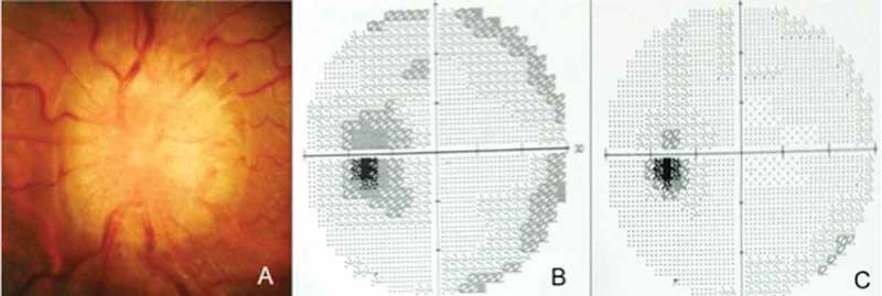
Left optic disc photograph of patient 3 (A) and visual field at initial presentation (B) and 2 months after treatment (C).
Three cases (43%) presented with bilateral disc oedema, 3 (43%) with unilateral disc oedema, and 1 (14%) with unilateral retrobulbar optic neuropathy. Anterior chamber and vitreous reactions were found in 3 patients. The appearance of disc swelling in all patients typically showed diffuse 360-degree oedema with profound elevation and blurring of disc margins (Figures 1 and 2). No peripapillary haemorrhages, exudates, or cotton wool spots were observed. Macula and retinal background were normal in all cases; however, fluorescein angiogram demonstrated peripheral retinal venous leakage in 1 patient. Neurological examination was normal in 6 of 7 patients. Left facial palsy, lower motor neuron type was found in 1 case.
Laboratory investigations
All cases were newly diagnosed with HIV infection. The CD4 T-cell count ranged from 116 to 613 cells/μL. Erythrocyte sedimentation rate ranged from 39 to 103 (mean 80.5) mm/h. The CSF profile of all patients showed common features: elevated protein level, normal glucose level, and mild leukocytosis with mononuclear cell predominance. All patients had non-reactive CSF VDRL, and 5 patients had reactive CSF TPHA.
Treatments
Six patients received intravenous benzylpenicillin 14.4 g (penicillin G 24 million international units) per day for 14 days, and 1 patient received intravenous ceftriaxone 2 g per day for 14 days. Intravenous methylprednisolone 1 g per day for 3 days, followed by oral prednisolone 1 mg/kg/day for 2 weeks and then rapidly tapered off was given to 3 patients who had moderate to severe visual loss. Oral prednisolone alone was given to 1 patient who had severe disc oedema with markedly enlarged blind spot. Corticosteroid was administered at least 7 days after the initiation of antibiotics. Three patients did not receive any adjunctive corticosteroid treatment because they initially had mild visual loss and responded rapidly to intravenous benzylpenicillin (penicillin G). The disc oedema in all eyes resolved within 2 months after treatment.
Visual outcome
Follow-up period ranged from 2 to 18 months. All patients had favorable visual outcome. Final visual acuity ranged from 20/20 to 20/25. Visual field was improved in 8 eyes presented with visual field defects. Six of them were completely normal, and 2 had significantly smaller defect (Figures 1 and 2).
Discussion
Syphilitic optic neuropathy is considered a manifestation of late syphilis.2 The incidence rate was rare among patients who have ocular involvement.4 In a case series of 8 patients with ocular syphilis over a 2.5-year period, only 2 patients had isolated optic neuritis, whereas most cases presented with either anterior or posterior uveitis.2
In our institution, we had 7 patients with syphilitic optic neuropathy visited our neuro-ophthalmologic clinic over a short period of 2 years. All patients were middle-aged, sexually active male with positive HIV antibody test. Six of the seven patients were known to be men who have sex with men (MSM). This high incidence might suggest an outbreak of syphilitic optic neuropathy, which is probably due to increasing HIV and syphilis epidemics among men who have sex with men (MSM) in Bangkok, Thailand.5
Optic nerve involvement in patients with syphilis can be unilateral or bilateral and have variable presentations, including anterior optic neuritis, retrobulbar optic neuritis, and optic perineuritis. Patients with optic perineuritis usually have reasonably good central vision associated with constricted peripheral visual field, whereas patients with optic neuritis have poor central vision.6
Our patients had similar demographic data and clinical feature as previous literature.2,7–11 All patients in previous reports were male, aged between 37 and 65 years, and most of them had HIV co-infection. Presenting visual acuity ranged from 20/15 to counting finger, but most of them were in normal and subnormal range. Most patients had some degree of visual field defects.
Optic disc oedema in syphilitic infection, as shown in our series, is usually diffuse, elevated, and not associated with haemorrhages or exudates. However, this finding is non-specific; thus, the diagnosis could not be based solely on the optic disc appearance.
Treatment of syphilitic optic neuropathy should be considered as the same regimen given in neurosyphilis, which is intravenous administration of benzylpenicillin 14.4 g (penicillin G 24 million international units) per day for 10–14 days. Adjunctive corticosteroid treatment previously has been given in both oral and intravenous forms without any proven benefit.4
In our series, we prescribed intravenous methylprednisolone and/or oral prednisolone to 4 patients in order to decrease the optic nerve inflammation and disc oedema. Although all patients had recovery of visual acuity to 20/25, the benefit of adjunctive corticosteroid administration is still inconclusive, due to the limitation of our retrospective study.
In summary, syphilitic optic neuropathy has re-emerged in our institution in the past 2 years. Visual loss from optic nerve involvement can be the first manifestation of syphilis and HIV co-infection. Patients usually have optic disc oedema and mild visual dysfunction. Syphilitic optic neuropathy has an excellent prognosis if the disease is diagnosed promptly and treated properly.
Declaration of interest
The authors report no conflicts of interest. The authors alone are responsible for the content and writing of the paper.
References
- [1].González-López JJ, Guerrero ML, Luján R, Tostado SF, de Górgolas M, Requena L.. Factors determining serologic response to treatment in patients with syphilis. Clin Infect Dis 2009;49:1505–1511. [DOI] [PubMed] [Google Scholar]
- [2].Puech C, Gennai S, Pavese P, Pelloux I, Maurin M, Romanet JP, Chiquet C.. Ocular manifestations of syphilis: recent cases over a 2.5-year period. Graefes Arch Clin Exp Ophthalmol 2010;248:1623–1629. [DOI] [PubMed] [Google Scholar]
- [3].Tucker JD, Li JZ, Robbins GK, Davis BT, Lobo AM, Kunkel J, Papaliodis GN, Durand ML, Felsenstein D.. Ocular syphilis among HIV-infected patients: a systematic analysis of the literature. Sex Transm Infect 2011;87:4–8. [DOI] [PMC free article] [PubMed] [Google Scholar]
- [4].Smith GT, Goldmeier D, Migdal C.. Neurosyphilis with optic neuritis: an update. Postgrad Med J 2006;82:36–39. [DOI] [PMC free article] [PubMed] [Google Scholar]
- [5].Griensven FV. HIV and syphilis infection among men who have sex with men-Bangkok, Thailand, 2005–2011. Centers for Disease Control and Prevention Web site Available at: http://www.cdc.gov/mmwr/preview/mmwrhtml/mm6225a2.htm/. Published June 28, 2013. Accessed July 14, 2015. [Google Scholar]
- [6].Sadiq SB, Corbett JJ, Abubakr A.. Idiopathic optic perineuritis: disguised as recurrent optic neuritis. Clin Neurol Neurosurg 2015;132:12–15. [DOI] [PubMed] [Google Scholar]
- [7].JM Weinstein, Lexow SS, Ho P, Spickards A.. Acute syphilitic optic neuritis. Arch Ophthalmol 1981;99:1392–1395. [DOI] [PubMed] [Google Scholar]
- [8].JB Carter, Hamill RJ, Matoba AY.. Bilateral syphilitic optic neuritis in a patient with a positive test for HIV. Arch Ophthalmol 1987;105:1485–1486. [DOI] [PubMed] [Google Scholar]
- [9].Rodríguez-Uña I, Serrador-García M, Santos-Bueso E, Díaz-Valle D, García-Feijóo J.. Simultaneous optic and vestibulocochlear syphilitic neuropathy in a patient with HIV infection. J Ophthalmic Inflamm Infect 2013;3:1–4. [DOI] [PMC free article] [PubMed] [Google Scholar]
- [10].Bandettini di Poggio M, Primavera A, Capello E, Bandini F, Mazzarello G, Viscoli C, Schenone A.. A case of secondary syphilis presenting as optic neuritis. Neurol Sci 2010;31:365–367. [DOI] [PubMed] [Google Scholar]
- [11].McBurney J, Rosenberg ML.. Unilateral syphilitic optic perineuritis presenting as the big blind spot syndrome. J Clin Neuroophthalmol 1987;7:167–169. [PubMed] [Google Scholar]