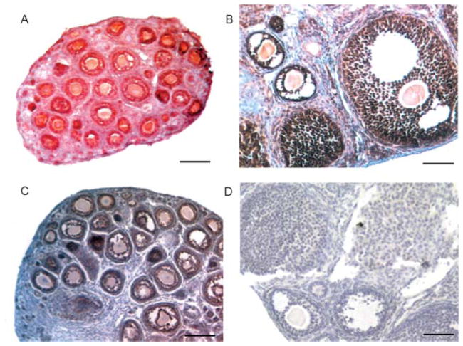
Figure 7.
Detection of SSB-1 and SSB-4 by immunohistochemistry. Ovaries were fixed in 4% para-formaldehyde, embedded in paraffin and sectioned at 5 μm thickness. Sections were subjected to an antigen retrieval technique. (A and B) SSB-1 is detected in granulosa cells, including both mural and cumulus cells of antral follicles, but not in the thecal cells or interstitial tissue. (C) SSB-4 is detected in granulosa cells in follicles at different stages of growth. (D) Sections processed as above but using the same concentration of rabbit IgG as the primary antibody show no specific staining. Non-specific staining of oocytes is sometimes observed using this immunodetection procedure.

