Figure 4.
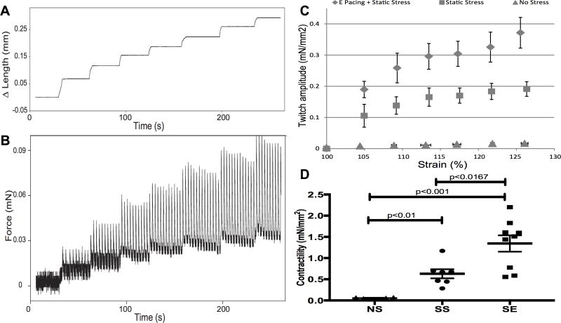
Stress conditioning and electrical stimulation increase contractility. Representative length (A) and force (B) traces demonstrate the response of a spontaneously contracting cardiac tissue construct to a series of stretches up to 125% of slack length. The amplitude of the isometric twitch force increases with increasing preparation length, in accordance with the Frank-Starling mechanism. C, Isometric twitch force amplitude measured at different preparation lengths is enhanced by 2 weeks of SS conditioning (triangles) in comparison to NS conditioning (triangles). Addition of electrical stimulation (diamonds) further increases contractility as show in (D). D, Contractility of constructs from the 3 stimulation conditions. Contractility is measured from the slope of the twitch force-strain curve, which is the active force development. The contractility of no stress constructs is 0.055±0.009 mN/mm2. Stress conditioning promotes the contractility 10-fold (0.63±0.10 mN/mm2) and addition of electrical pacing further enhances force development another 2-fold (1.34±0.19 mN/mm2). NS vs SS: p<0.01; SS vs SE: p<0.01. n=6 for NS, n=7 for SS, and n=9 for SE.

