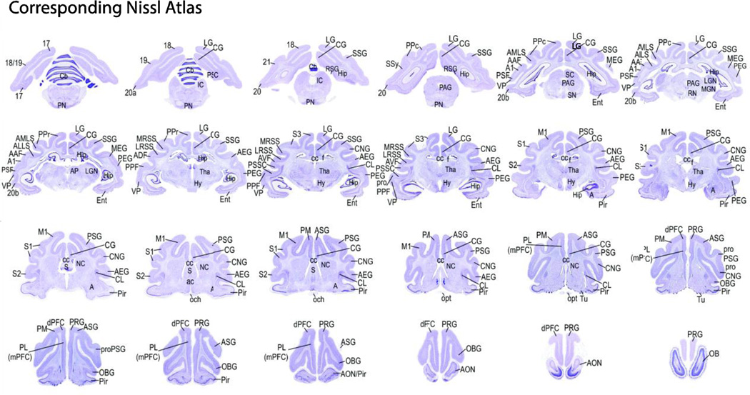
Figure 4.
Nissl stain sections corresponding to MRI slices. Nissl stained sections of the ferret brain atlas (50 µm thick cryostat sections; Radtke-Schuller et al., unpubl.) corresponding to the MR images from this study are shown with relevant anatomical and functional labels for the main foci found in the gICA maps and further anatomical labels for orientation. The labeling of anatomical structures used in this atlas was based on currently published data on the ferret and/or other carnivores (mainly cat and dog). Functional and structural nomenclatures are indicated on the left and right hemisphere, respectively.

