Abstract
Objective:
Radiopaque markers are commonly deployed following breast biopsies to indicate the location of the targeted lesion. A frequently encountered complication is the displacement of these markers. This study compared the degree of displacement among four newer generation markers after stereotactic core needle biopsy.
Methods:
80 consecutive biopsies were performed at three breast centre sites. The markers included: HydroMARK® (Mammotome, Cincinnati, OH), MammoMARK™ (Mammotome, Cincinnati, OH), MammoStar™ (Mammotome, Cincinnati, OH) and SecurMark® (Hologic, Bedford, MA). Each marker was composed of a radiopaque core with a unique polymeric encasing component. Post-procedure mammograms were obtained and the degree of marker displacement was measured.
Results:
MammoMARK™ exhibited the greatest mean net displacement, followed by HydroMARK®, SecurMark® and MammoStar™ (13.9, 7.7, 5.8 and 4.7 mm, respectively), although these differences did not reach statistical significance (p = 0.398). 73% of the markers did not displace at all. However, in the 19 of 22 markers in which displacement occurred, the distance from the biopsy cavity was >10 mm. No statistically significant contributing factors to predict displacement were found.
Conclusion:
Newer generation biopsy markers perform comparably with one another. However, clinically significant and unpredictable marker displacement persists. Compared with multiple similar studies of older generation bare metallic markers, the overall displacement rate of newer generation markers seems to be lower, possibly owing to the use of polymeric embedding agents that self-expand within the biopsy cavity.
Advances in knowledge:
This article compares the post-procedure displacement of breast biopsy markers, which have not been evaluated or discussed in detail since markers with polymeric embedding agents gained widespread use.
INTRODUCTION
Stereotactic vacuum-assisted biopsy (SVAB) has become the standard of care for the work-up of suspicious breast calcifications and other sonographically occult lesions. The procedure is minimally invasive, more convenient and significantly less expensive than surgical excisional biopsy. However, when there is a histologic diagnosis of malignancy, it is often necessary to proceed to lumpectomy. Frequently, all radiographic evidence of the lesion can be completely removed after SVAB, reportedly in up to 50–72% of cases.1 This can make pre-operative localization challenging, forcing the radiologist to have to rely on mammographic landmarks and/or the presence of a haematoma for targeting. In the early to mid-1990s, the placement of a radiopaque metallic post-biopsy clip began gaining popularity in cases where the mammographic abnormality was determined to be no longer visible.2 Since then, the use of post-procedure markers after SVAB has become widespread, with most institutions, including ours, routinely placing the markers after all breast biopsies. Studies have shown that clear margins are obtained in 90–92% of excisional specimens when lesions are localized using biopsy markers vs 31–62% when wire localization is used alone.2,3
A well-known complication of marker use is the immediate displacement from the biopsied lesion. Most cases of displacement are believed to be caused by the so-called “accordion effect”.2,4–9 During the SVAB procedure and subsequent marker placement, the breast is in compression. After the procedure, when released from compression, the breast resumes its normal shape and the marker can displace along the axis of compression (the z-axis). Causes for delayed marker migration, which occurs any time in the non-immediate interval following biopsy, include displacement by a haematoma, simple migration within the fatty tissue and changes secondary to neoadjuvant chemotherapy.10,11 Any instance in which a marker is distant from the targeted lesion poses a risk that a malignancy may be incompletely excised at surgery, particularly when displacement of the marker from the biopsy cavity goes unrecognized.
The first generation of breast biopsy markers were bare metallic clips, usually composed of either titanium or stainless steel. Many of these markers were designed to physically attach to the biopsy cavity wall, similar to vascular ligating clips. Others were simply deposited within or near the biopsy cavity.2,4 Use of bare metallic biopsy markers continues today at many institutions. Performance of these markers is variable, with reported rates of displacement <10 mm ranging from 54 to 99% in the literature.1,4–6,12–14 However, some of these higher published numbers are thought to be inflated, as displacement was measured with the breast still under compression on the stereotactic table, before the accordion effect is thought to take place.6,12,14
In 2003, the first marker embedded within a biocompatible polymeric material was introduced to the market, the MammoMARK™ (Mammotome, Cincinnati, OH). In a comparative analysis, Rosen et al4 showed that the MammoMARK™ resulted in a statistically significant decrease in the number of occurrences of marker-to-biopsy site displacement >10 mm vs older generation bare metallic markers (16 vs 44%, respectively). However, this study was somewhat limited by the relatively low number of markers with embedding agents (n = 31) and bare metallic markers (n = 43). Moreover, markers were not deployed following all biopsy cases—only cases in which the radiologist determined that the lesion was no longer mammographically visible, and the patients were not randomized, potentially introducing a selection bias. The embedding agent used in the MammoMARK™ is collagen. Markers employing various other kinds of polymeric embedding materials have since come to market and are now widely used. These embedding materials attempt to limit post-biopsy displacement by a common mechanism: expansion within the biopsy cavity immediately after deployment.
While several authors have studied the displacement of breast biopsy clips, after performing a systematic literature review, we found no published journal articles comparing newer generation markers with polymeric embedding agents with each other. We prospectively evaluated the performance of four newer generation breast biopsy site markers immediately following SVAB using post-procedure mammograms.
METHODS AND MATERIALS
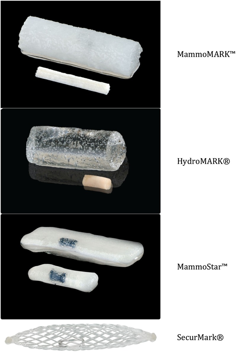
Upon obtaining approval from our institutional review board, four commercially available biopsy site markers composed of a central metallic or ceramic radiopaque core encased in unique polymeric coatings were evaluated to determine the degree of displacement following SVAB. The institutional review board waived the requirement to obtain informed consent from the patients because the study protocol did not differ from the standard of care in the routine work-up of suspicious breast lesions at our institution. The markers included the MammoMARK™, HydroMARK® (Mammotome, Cincinnati, OH), MammoStar™ (Mammotome, Cincinnati, OH) and SecurMark® (Hologic, Bedford, MA) (Table 1, Figure 1). The markers were selected based on those we currently held in stock. Bare metallic clips were not evaluated because they are no longer considered the standard of care at our institution and are not stocked.
Table 1.
List of breast biopsy site markers included in study and material properties
| Brand | Manufacturer | Radiopaque marker material | Embedding material |
|---|---|---|---|
| MammoMARK™ | Mammotome, Cincinnati, OH | Titanium | Collagen |
| HydroMARK® | Mammotome, Cincinnati, OH | Stainless steel or titanium | Polyethylene glycol-based hydrogel |
| MammoStar™ | Mammotome, Cincinnati, OH | Ceramic | Lyophilized (freeze-dried) beta-glucan gel |
| SecurMark® | Hologic, Bedford, MA | Stainless steel or titanium | Bioabsorbable suture-like netting (glycoprene) |
Figure 1.
Photographs of the four markers evaluated in the study. The MammoMARK™ (Mammotome, Cincinnati, OH), HydroMARK® (Mammotome, Cincinnati, OH) and MammoStar™ (Mammotome, Cincinnati, OH) are depicted in both their hydrated and non-hydrated states, courtesy of Mammotome. The SecurMark® (Hologic, Bedford, MA) was provided courtesy of Hologic.
Between 15 May 2014 and 19 February 2015, 74 consecutive patients underwent SVAB for a suspicious mammographic abnormality by a single fellowship-trained breast radiologist at one of three breast centre campuses. One of the campuses was located in an urban setting in Detroit, Michigan, USA proper, and the other two in suburban settings in the greater Detroit Metropolitan Area. Inclusion criteria were any female with a breast imaging reporting and data system® category 4 or 5 assessment with recommendation for SVAB. Patients were excluded if SVAB was contraindicated for any reason, including the inability to remain in the position required for the duration of the procedure, weight greater than the weight limit of the biopsy table (300 lbs), inability to visualize the breast lesion mammographically or inaccessible position of the lesion within the breast. All SVABs were performed in the prone position using the Hologic Eviva® nine-gauge system and Hologic MultiCare Platinum® stereotactic biopsy table (Bedford, MA). 6–12 samples were taken per lesion depending on the performing radiologist level of confidence that the lesion was adequately sampled.
Patients were randomly assigned to receive one of the four biopsy markers using a randomization schedule created by SAS v. 9.4 (SAS Institute Inc., Cary, NC). Five of the patients underwent biopsies of two lesions on the same day immediately following one another, and different biopsy markers were used in these instances. One patient underwent two biopsies on the same breast separated by a time interval of 1 week. A total of 80 biopsy markers were deployed, with 20 markers from each marker group. The markers were placed according to the in-service training provided by each marker vendor representative. Per recommended guidelines, all markers were deployed through the end of the introducer cannula after vacuum assistance was turned off. Following each biopsy case, the breast was slowly decompressed and manual pressure was applied for several minutes to obtain haemostasis.
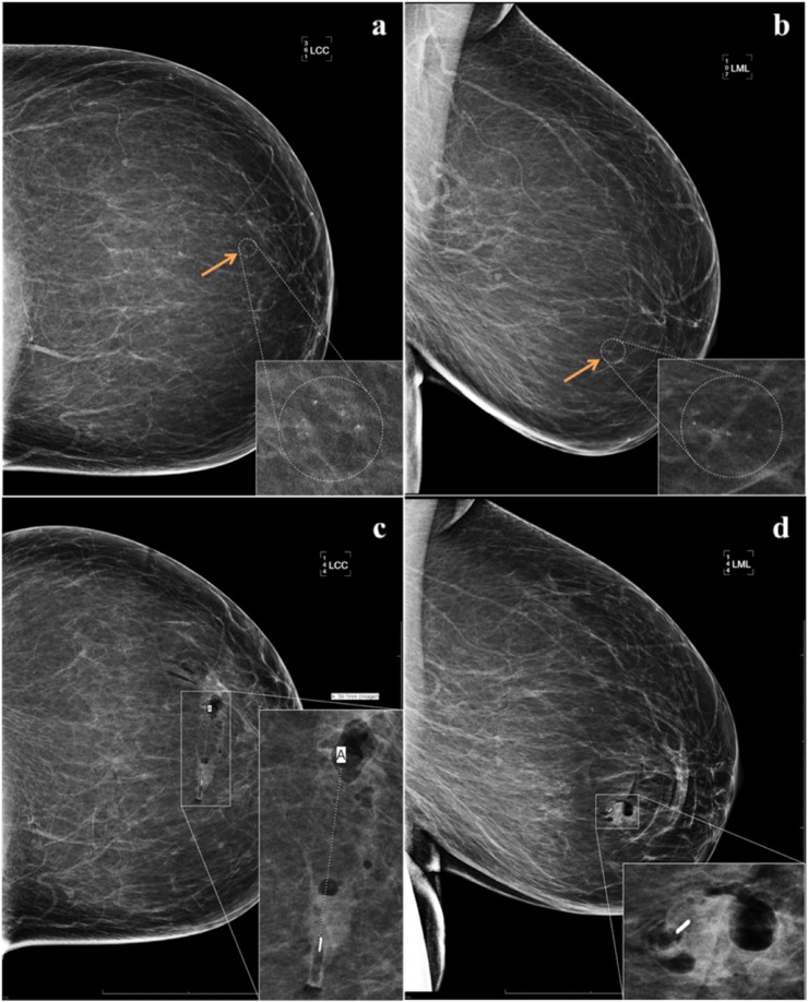
All patients received post-biopsy 90° true lateral and craniocaudal mammograms immediately following the procedure (within 15 min) per our institution standard operating procedures to verify and measure marker location in relationship to the targeted biopsy cavity. These images were compared with true lateral and craniocaudal images that were obtained during the previous diagnostic mammography work-up. The marker-to-biopsy site distance was determined using the direct measurement method in which a line was drawn from the edge of the biopsy cavity to the proximal edge of the marker on both craniocaudal and true lateral views (Figure 2). Prior to measuring, the biopsy cavity was confirmed to be located within the targeted lesion. If the lesion was no longer mammographically visible, soft-tissue landmarks were used to confirm that the appropriate lesion was biopsied. The distance was measured by the same radiologist who performed the procedure. In addition, patient age and lesion characteristics including the mammographic description of the lesion, size of lesion, assigned breast imaging reporting and data system® category and final pathology were recorded (Table 2). Variables related to biopsy technique including breast tissue composition, thickness of the breast under compression on the stereotactic table, biopsy approach, number of core samples obtained, whether a second biopsy was performed on the same day and the presence of a post-procedure haematoma were also documented (Table 3).
Figure 2.
63-year-old female with new grouped amorphous calcifications (arrows) at 4 o'clock within the left breast anteriorly for which stereotactic vacuum-assisted biopsy was recommended. (a, b) The calcifications were biopsied and a MammoMARK™ (Mammotome, Cincinnati, OH) was deployed. Immediate post-procedure mammogram images reveal 40 mm of medial displacement from the biopsy cavity (c, d). LCC, left craniocaudal view; LML, left mediolateral view.
Table 2.
Summary of descriptive data related to patient demographics and lesion characteristics
| Factor | Variable | Result | p-value |
|---|---|---|---|
| Age (years) | Mean (SD) | 57.9 (12.4) | 0.122 |
| Mammographic description | Calcifications | 60 (75.0%) | 0.436 |
| Focal asymmetry | 5 (6.3%) | ||
| Mass | 11 (13.8%) | ||
| Calcifications with focal asymmetry or mass | 4 (5.0%) | ||
| Size of lesion (mm) | Mean (SD) | 16.6 (18.6) | 0.585 |
| BI-RADS® category | 3 | 2 (2.5%) | 0.822 |
| 4A | 27 (33.8%) | ||
| 4B | 38 (47.5%) | ||
| 4C | 10 (12.5%) | ||
| 5 | 1 (1.3%) | ||
| Nonea | 2 (2.5%) | ||
| Pathology | Benign | 49 (61.3%) | 0.726 |
| Malignant | 22 (27.5%) | ||
| High risk | 8 (10.0%) | ||
| Discordant results without follow-up | 1 (1.3%) |
BI-RADS, breast imaging reporting and data system; SD, standard deviation.
Numbers in the result column represent the n-value, unless otherwise indicated. Correlation between variables and displacement was analyzed (statistical significance was set at p < 0.05).
Two cases were referrals from outside hospitals in which reports were not available prior to biopsy.
Table 3.
Summary of variables related to stereotactic biopsy technique
| Factor | Variable | Result | p-value |
|---|---|---|---|
| Tissue composition | Fatty | 9 (11.3%) | 0.657 |
| Scattered | 42 (52.5%) | ||
| Heterogeneous | 27 (33.8%) | ||
| Extremely | 2 (2.5%) | ||
| Thickness in compression on biopsy table (mm) | ≤40 | 12 (15.0%) | 0.858 |
| >40 | 68 (85.0%) | ||
| Approach | Superior | 27 (33.8%) | 0.067 |
| Inferior | 19 (23.8%) | ||
| Lateral | 27 (33.8%) | ||
| Medial | 7 (8.8%) | ||
| Number of core samples | Mean (SD) | 6.6 (1.8) | 0.119 |
| Single vs second same-day biopsy performed (n = 75) | Single | 70 (93.3%) | 0.311 |
| Double | 5 (6.7%) | ||
| Haematoma | No | 76 (95.0%) | 0.340 |
| Yes | 4 (5.0%) |
SD, standard deviation.
Numbers in the result column represent the n-value, unless otherwise indicated. Correlation between variables and displacement was analyzed (statistical significance was set at p < 0.05).
Measurements of displacement were broken down into x–y-axis (orthogonal to compression), z-axis (plane of compression) and net displacement, which were calculated using the Pythagorean theorem. The markers were compared using the Kruskal–Wallis test to evaluate for statistically significant differences in biopsy cavity-to-marker distances. Spearman's rank tests were used to analyze whether any of the aforementioned variables listed above had a correlation with net displacement of the markers as a group. Analysis was performed using SAS v. 9.4. Statistical significance was defined as a p-value <0.05.
RESULTS
22 (28%) of the 80 markers were displaced from the biopsy site on post-procedure mammograms. The median marker-to-biopsy site displacement for all four groups was 0 mm. MammoMARK™ exhibited the greatest mean net displacement (13.9 mm), followed in order by HydroMARK®, SecurMark® and MammoStar™ (Table 4). However, the differences in displacement among the four tested markers did not reach statistical significance (p = 0.398). Of the four markers, MammoMARK™ had the greatest maximal net displacement (73.4 mm).
Table 4.
Marker-to-biopsy site displacement of the four markers
| Biopsy site marker | n | Net marker-to-biopsy site displacement (mm) |
|||
|---|---|---|---|---|---|
| Median | Mean | SD | Range | ||
| MammoStar™ (Mammotome, Cincinnati, OH) | 20 | 0 | 4.7 | 11.5 | 0–42.0 |
| SecurMark® (Hologic, Bedford, MA) | 20 | 0 | 5.8 | 15.0 | 0–62.0 |
| HydroMARK® (Mammotome, Cincinnati, OH) | 20 | 0 | 7.7 | 15.9 | 0–56.0 |
| MammoMARK™ (Mammotome, Cincinnati, OH) | 20 | 0 | 13.9 | 23.6 | 0–73.4 |
SD, standard deviation.
The most cases of marker displacement occurred with MammoMARK™ [8/20 (40%)], with the majority displacing >10 mm [7/20 (35%)]. MammoStar™ and SecurMark® both had the least number of marker displacement [4/20 (20%) each]; but again, most of the instances of displacement of these markers were >10 mm (Table 5). In fact, 19 of the 22 markers that demonstrated positive displacement were found to be at a distance >10 mm from the biopsy cavity, indicating that when there was displacement, it was substantial. This is reflected by a mean distance of 29.2 mm from the biopsy cavity among the 22 markers in which displacement occurred. When displacement occurred in just 1 axis (17 of the 22 cases), it was exclusively in the z-axis.
Table 5.
Absolute number and percentage of cases of marker displacement by marker group
| Biopsy site marker | n | Cases of post-procedure displacement |
Markers displaced (net) (%) |
||||
|---|---|---|---|---|---|---|---|
| z-axis only | x–y-axis only | z-axis and x–y-axis | Total | >0 cm | ≥1 cm | ||
| MammoStar™ (Mammotome, Cincinnati, OH) | 20 | 4 | 0 | 0 | 4 | 20 | 15 |
| SecurMark® (Hologic, Bedford, MA) | 20 | 3 | 0 | 1 | 4 | 20 | 20 |
| HydroMARK® (Mammotome, Cincinnati, OH) | 20 | 4 | 0 | 2 | 6 | 30 | 25 |
| MammoMARK™ (Mammotome, Cincinnati, OH) | 20 | 6 | 0 | 2 | 8 | 40 | 35 |
| Total | 80 | 17 | 0 | 5 | 22 | 27.5 | 23.8 |
We found no statistically significant correlation between patient and lesion characteristics and the incidence of displacement when data from all four types of markers were aggregated (Table 2). 80% of the biopsied lesions involved calcifications and 22 biopsies yielded malignant results. Including diagnoses with high-risk pathology, 24 cases proceeded to surgery at our institution. Of the eight high-risk lesions, one was upgraded to malignancy at surgical excision. Positive-predictive value was 29.1%. We also found that factors related to biopsy technique were not associated with displacement (Table 3). It should be noted that some lesion characteristics and variables related to technique had low n-values, which limits their interpretability. For instance, just two females in the study had extremely dense breast tissue composition. Medial approach was also uncommonly utilized (n = 7). However, when a biopsy was performed using a medial approach, it is interesting to note that four out of seven cases exhibited displacement in both the x–y and z-axes. Only one other instance of x–y displacement occurred during the study (in which lateral approach was used) and this marker also displaced along the z-axis. Although the “approach” category did not technically achieve statistical significance (p = 0.067), it appears there is a trend towards correlation. Four cases (involving MammoMARK™, HydroMARK® and two MammoStar™ markers) were complicated by haematomas measuring 9.0, 2.7, 7.3 and 1.5 cm, respectively. In all of these cases, no displacement from the biopsy cavity was observed.
DISCUSSION
In general, no single marker performed significantly better or worse than the others evaluated in this study, with most [58/80 (73%)] markers showing no displacement on two-view mammography immediately following SVAB. This implies that the presence of a biocompatible polymeric embedding agent is more important than the specific type of polymer used when it comes to minimizing the occurrence of post-biopsy displacement. Our results reveal that 19/80 (24%) of the markers displaced >10 mm, which roughly approximates Rosen et al's study of the MammoMARK™ marker in which 5/31 (16%) markers were located >10 mm from the target. Additional studies on the immediate post-procedure displacement of newer generation breast biopsy markers with embedding agents were not found after performing a literature review. There are more robust data available regarding the displacement of the older generation bare metallic markers. In similar study protocols to our own, Rosen et al,4 Chaveron et al5 and Kass et al1 found immediate post-SVAB displacement rates of bare metallic markers measuring >10 mm of 44, 32 and 46%, respectively. However, of these three studies, Rosen et al were the only group of investigators to utilize a prospective technique like we did in our study.
Three of the four markers investigated in this study (MammoMARK™, HydroMARK® and MammoStar™) have embedding agents that expand via osmotic water absorption and the fourth, the SecurMark®, utilizes a self-expanding net. The mechanism by which embedding agents minimize displacement is thought to relate to the fact that these types of markers are deployed directly into the biopsy cavity, exerting force on the walls as they expand and anchoring them within the cavity.4 Self-expansion continues over the ensuing minutes and hours, which secures them within the cavity further and helps to decrease the incidence of long-term migration. Delayed migration can occur from days to over 1 year after the initial biopsy and is a clinically relevant issue itself.15 However, it should be noted that our results do not apply to long-term migration, which was not evaluated in this study in order to avoid additional radiation exposure to the patients.
Besides reducing the incidence of displacement, polymeric embedding agents offer additional benefits compared with bare metallic markers. They are highly visible sonographically, especially when hydrogel is used (e.g. HydroMARK®), as we have analyzed in prior research.16 This can be useful for ultrasound-guided pre-operative localization procedures and even for intraoperative ultrasound-guided excisional biopsies and lumpectomies. Furthermore, the pressure of the polymeric embedding agent against the biopsy cavity wall reduces the chance of post-procedure haematoma formation. Collagen (used in the MammoMARK™) has the added benefit of activating the coagulation cascade, which confers enhanced haemostasis.2,4,17
One area of concern was that when the markers did displace in our study, the distance from the biopsy cavity was considerable—10 mm or more in 86% of the displaced cases. This is speculated to relate, at least in part, to the slippery surface of the gel-like polymeric component of some of the markers. Consequently, if the marker is not deployed exactly within the biopsy cavity, the self-expanding nature of the markers could result in extrusion along the biopsy tract. In fact, in a retrospective review by Klein et al,18 breast surgeons who were attempting to perform excisional biopsies using intraoperative ultrasound alone (without a separate pre-operative localization procedure) reported extrusion of the HydroMARK® in 14 of 31 cases when the biopsy tract was traversed during surgery.
Our results are in agreement with previous literature, suggesting that the accordion effect is the primary means of immediate post-biopsy displacement, given that all markers displacing in one dimension did so along the z-axis. Uematsu et al8 examined variables that could predict when markers will be more likely to displace via the accordion effect and found that breast thickness was the only statistically significant predictor, with thinner compressed breasts tending to exhibit greater degrees of displacement. Other variables, including age of the patient, breast tissue composition and number of specimens were not useful in predicting marker displacement. We also evaluated multiple variables to see whether marker displacement could be predicted in our study group. We did not find a statistically significant correlation with any of these variables, including breast thickness. The average breast thickness in our study population was 53 mm, while in Uematsu et al's study, it was 37 mm. Perhaps this may have contributed to our discrepant results.
Aside from using markers with polymeric embedding agents, other strategies for reducing displacement include gently releasing the breast from compression and closely following manufacturer instructions when deploying the markers. A study by Chaveron et al5 also suggested that when feasible, using mediolateral rather than craniocaudal compression tended to decrease the incidence of displacement, although the reasoning behind this was uncertain. They postulate that breast ptosis or gravity effects may have played a role. We did not see this same effect in our study. In fact, more cases of displacement occurred under craniocaudal compression [13/22 (59%)] in comparison with mediolateral compression [9/22 (41%)]. Notably, medial approach seemed to predispose the markers to biaxial displacement in our study. We have no clear explanation as to why this occurred, but this apparent trend could be investigated in future research.
With only 4 (5%) cases of post-procedure haematomas, it is not possible to infer whether this complication influenced displacement of the newer generation markers, despite there being no associated marker displacement in these cases. The use of smaller 11-gauge SVAB devices may decrease post-procedure bleeding and haematoma formation, although evidence suggests that this effect is device dependent. Schaefer et al19 showed that the 11-gauge Mammotome® system had a significantly lower haematoma rate than the 8-gauge Mammotome®. At our institution, the Hologic Eviva nine-gauge SVAB system is used for all breast biopsies and is preferred over smaller gauge devices in an attempt to limit sampling error. In addition, the decrease in the number of required core samples has itself been correlated with lower incidences of haematoma formation and thus can counterbalance the risks of taking larger cores.20 Our haematoma rate is lower than comparable studies, which reached as high as 45%, and may relate at least in part to our study protocol's short interval from biopsy to post-procedure mammogram.21 The haemostatic effect of the polymeric embedding agents may have also played a role.
Our study was underpowered owing to financial constraints and limited access to the various biopsy markers, which made it difficult to detect small differences in displacement. However, repeating this study with a higher n-value may be of limited clinical yield, given that all biopsy markers had a median displacement of 0 mm and as a group, marker displacement was unpredictable. Since the study was performed at three clinical sites, different technologists and nurses participated in the biopsy and post-procedure care, which could have potentially introduced a confounding variable if different techniques were used. For example, the duration of post-biopsy manual compression may have varied among sites. Five of the patients in our study underwent two separate biopsies immediately following one another. The effects of placing the breast back into compression to perform the second biopsy and the delayed post-procedure mammogram with respect to the first biopsy may have influenced displacement.
In conclusion, newer generation markers with biocompatible polymeric embedding agents perform relatively similarly to one another on immediate post-SVAB mammograms with no statistically significant difference in mean displacement among the four markers that were tested. Furthermore, there was no definitive evidence to suggest an ability to predict when this displacement may occur. The percentage of markers displacing >10 mm was in line with the only other known research study measuring displacement of newer generation breast biopsy markers, and both are seemingly lower than the reported rate of displacement of bare metallic markers according to studies utilizing comparable methodological protocols. More research is necessary to discover methods to further reduce the incidence of displacement, possibly through markers that self-expand at a faster rate or are made of a substance that better adheres to the biopsy cavity wall once deployed.
Contributor Information
David M Pinkney, Email: davidpi@rad.hfh.edu.
Mirek Mychajlowycz, Email: mmychajl@med.wayne.edu.
Biren A Shah, Email: birens@rad.hfh.edu.
REFERENCES
- 1.Kass R, Kumar G, Klimberg V, Kass L, Henry-Tillman R, Johnson A, et al. Clip migration in stereotactic biopsy. Am J Surg 2002; 184: 325–31. doi: 10.1016/S0002-9610(02)00952-2 [DOI] [PubMed] [Google Scholar]
- 2.Thomassin-Naggara I, Lalonde L, David J, Darai E, Uzan S, Trop I. A plea for the biopsy marker: how, why and why not clipping after breast biopsy? Breast Cancer Res Treat 2011; 132: 881–93. doi: 10.1007/s10549-011-1847-x [DOI] [PubMed] [Google Scholar]
- 3.Corsi F, Sorrentino L, Bossi D, Sartani A, Foschi D. Preoperative localization and surgical margins in conservative breast surgery. Int J Surg Oncol 2013; 2013: 1–9. doi: 10.1155/2013/793819 [DOI] [PMC free article] [PubMed] [Google Scholar]
- 4.Rosen E, Baker J, Soo M. Accuracy of a collagen-plug biopsy site marking device deployed after stereotactic core needle breast biopsy. AJR Am J Roentgenol 2003; 181: 1295–9. doi: 10.2214/ajr.181.5.1811295 [DOI] [PubMed] [Google Scholar]
- 5.Chaveron C, Bachelle F, Fauquet I, Rocourt N, Faivre-Pierret M, Ceugnart L. Clip migration after stereotactic macrobiopsy and presurgical localization: technical considerations and tricks. J Radiol 2009; 90: 31–6. doi: 10.1016/S0221-0363(09)70075-7 [DOI] [PubMed] [Google Scholar]
- 6.Liberman L, Dershaw D, Morris E, Abramson A, Thornton C, Rosen P. Clip placement after stereotactic vacuum-assisted breast biopsy. [In French.] Radiology 1997; 205: 417–22. doi: 10.1148/radiology.205.2.9356622 [DOI] [PubMed] [Google Scholar]
- 7.Burbank F, Forcier N. Tissue marking clip for stereotactic breast biopsy: initial placement accuracy, long-term stability, and usefulness as a guide for wire localization. Radiology 1997; 205: 407–15. [DOI] [PubMed] [Google Scholar]
- 8.Uematsu T, Kasami M, Takahashi K, Junichiro W, Yamasaki S, Tanaka K, et al. Clip placement after an 11-gauge vacuum-assisted stereotactic breast biopsy: correlation between breast thickness and clip movement. Breast Cancer 2011; 19: 30–6. doi: 10.1007/s12282-011-0252-y [DOI] [PubMed] [Google Scholar]
- 9.Philpotts L, Lee C. Clip migration after 11-gauge vacuum-assisted stereotactic biopsy: case report. Radiology 2002; 222: 794–6. doi: 10.1148/radiol.2223011263 [DOI] [PubMed] [Google Scholar]
- 10.Esserman L, Cura M, Dacosta D. Recognizing pitfalls in early and late migration of clip markers after imaging-guided directional vacuum-assisted biopsy. Radiographics 2004; 24: 147–56. doi: 10.1148/rg.241035052 [DOI] [PubMed] [Google Scholar]
- 11.Birdwell R, Jackman R. Clip or marker migration 5–10 weeks after stereotactic 11-gauge vacuum-assisted breast biopsy: report of two cases. Radiology 2003; 229: 541–4. doi: 10.1148/radiol.2292021594 [DOI] [PubMed] [Google Scholar]
- 12.Kruger B, Burrowes P, MacGregor J. Accuracy of marker clip placement after mammotome breast biopsy. Can Assoc Radiol J 2002; 53: 137–40. [PubMed] [Google Scholar]
- 13.Rosen E, Vo T. Metallic clip deployment during stereotactic breast biopsy: retrospective analysis. Radiology 2001; 218: 510–16. doi: 10.1148/radiology.218.2.r01fe39510 [DOI] [PubMed] [Google Scholar]
- 14.Margolin F, Kaufman L, Denny S, Jacobs R, Schrumpf J. Metallic marker placement after stereotactic core biopsy of breast calcifications: comparison of two clips and deployment techniques. AJR Am J Roentgenol 2003; 181: 1685–90. doi: 10.2214/ajr.181.6.1811685 [DOI] [PubMed] [Google Scholar]
- 15.Parikh J. Delayed migration of Gel Mark Ultra clip within 15 days of 11-gauge vacuum-assisted stereotactic breast biopsy. AJR Am J Roentgenol 2005; 185: 203–6. doi: 10.2214/ajr.185.1.01850203 [DOI] [PubMed] [Google Scholar]
- 16.Pinkney D, Shah B. Prospective comparative study to evaluate the sonographic visibility of five commercially available breast biopsy markers. J Diagn Med Sonog 2013; 29: 151–8. doi: 10.1177/8756479313486962 [DOI] [Google Scholar]
- 17.Pachence J. Collagen-based devices for soft tissue repair. J Biomed Mater Res 1996; 33: 35–40. doi: [DOI] [PubMed] [Google Scholar]
- 18.Klein R, Mook J, Euhus D, Rao R, Wynn R, Eastman A, et al. Evaluation of a hydrogel based breast biopsy marker (HydroMARK®) as an alternative to wire and radioactive seed localization for non-palpable breast lesions. J Surg Oncol 2011; 105: 591–4. doi: 10.1002/jso.22146 [DOI] [PubMed] [Google Scholar]
- 19.Schaefer F, Order B, Echmann-Scholz C, Strauss A, Hilpert F, Kroj K, et al. Interventional bleeding, hematoma and scar-formation after vacuum-biopsy under stereotactic guidance: mammotome®-system 11g/8g vs ATEC®-system 12g/9g. Eur J Radiol 2012; 81: 739–45. [DOI] [PubMed] [Google Scholar]
- 20.Zagouri F, Gounaris A, Liakou P, Dimosthenis C, Flessas I, Bletsa G, et al. Vacuum-assisted breast biopsy: more cores, more hematomas? In Vivo 2011; 25: 703–5. [PubMed] [Google Scholar]
- 21.Melotti M, Berg W. Core needle breast biopsy in patients undergoing anticoagulation therapy: primary results. AJR Am J Roentgenol 2000; 174: 245–9. doi: 10.2214/ajr.174.1.1740245 [DOI] [PubMed] [Google Scholar]


