Fig. 4.
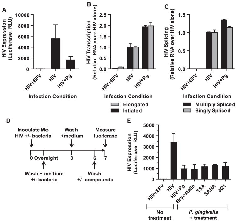
P. gingivalis mediated repression of HIV is post-transcriptional. MDMs were infected with HIV-NL4-3ΔEnv-Luc(VSV-G) for 4 h in the presence or absence P. gingivalis at an MOI of 30. (A) The cells were collected 3 days post-infection to measure HIV expression by luciferase. Error bars represent the standard deviation of 3 infections. In a parallel infections, the cells were harvested to measure the levels of (B) initiated and elongated HIV transcripts and (C) multiply- and singly-spliced HIV transcripts by RT-PCR. Error bars represent the standard deviation of measurements done in triplicate. The data shown is representative of 2 independent experiments. (D) Latency reversing compounds were used to reactivate repressed HIV in MDMs exposed to P. gingivalis. After removing the bacteria and culturing MDMs in the absence of bacteria for 3 days, the cells were exposed to a panel of stimulating agents for 24 h and followed by analysis of luciferase expression (E): bryostatin (10 nM), trichostatin A (100 nM), SAHA (10 μM), and JQ1 (10 μM). Error bars represent the standard deviation of measurements done in triplicate. The data is representative of more than 3 experiments.

