Fig. 5.
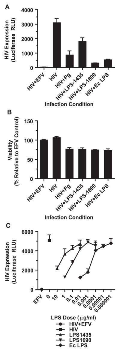
LPS from P. gingivalis is sufficient to repress HIV expression. (A) MDMs were infected with HIV-NL4-3ΔEnv-Luc(VSV-G) with or without P. gingivalis (MOI 30), purified P. gingivalis LPS1435 (1 μg/ml), purified P. gingivalis LPS1690 (1 μg/ml) or E. coli LPS (10 ng/ml) following the outline in Fig. 2A and monitored luciferase expression at 3 days post-infection. Cultures treated with 1 μM efavirenz (EFV) served as a negative control. Error bars represent the standard deviation of triplicate measurements. Data is representative of more than 3 individual experiments. (B) Cell viability was monitored using CellTiter-Glo and represents the viability relative to the EFV-treated cells. Graph was generated from a parallel infections as the experiment in (A). Error bars represent the standard deviation from triplicate measurements. The data is representative of 2 individual experiments. (C) MDMs were infected with HIV-NL4-3ΔEnv-Luc(VSV-G) with or without serial dilutions of P. gingivalis and E. coli LPS. Error bars represent the standard deviation of measurements done in triplicate. The data is representative of 2 independent experiments.

