. 2016 Nov 29;5:e14707. doi: 10.7554/eLife.14707

Figure 4. Structure of the RBM5 OCRE/SmN peptide complex.

(A,B) Side and top views in a cartoon presentation of the RBM5 OCRE/SmN peptide complex.The secondary structure and loops in the RBM5 OCRE domain are colored in green and grey, respectively, the proline-rich motif (PRM) peptide corresponding to SmN residues 221–229 is shown in yellow. (C–E) Zoomed views of key interaction sites. (C) The SmN PRM adopts a proline-type II helical conformation and is recognized by stacking with key tyrosine residues from the OCRE domain. The side chain of SmN Ile227 packs against the hydrophobic surface of the PPII helix. (D) Recognition of SmN Arg221 by interactions with hydroxyl groups of Tyr472, Tyr479 and Ser490 (E) Arg228 forms electrostatic contacts with the side chains of Asp481 and Ser484.

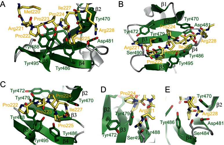
Figure 4.

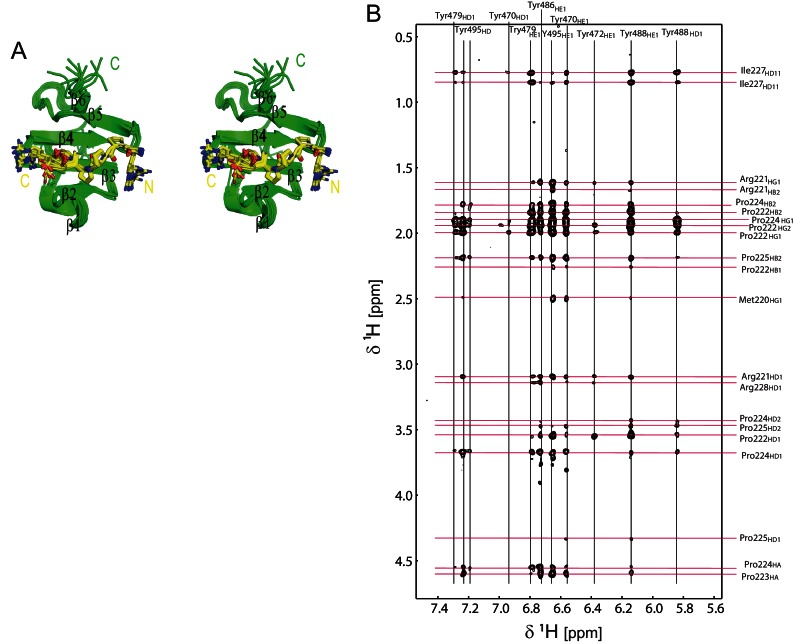
Figure 4—figure supplement 1. Structural analysis of the OCRE/SmN complex.

Figure 4—figure supplement 1.

(A) Stereo view of the ensemble of 10 lowest energy NMR structures of the RBM5 OCRE domain in complex with SmN peptide, same view as in Figure 1D. (B) Intermolecular NOEs between the RBM5 OCRE domain and the SmN peptide. A zoomed view of a 2D ω1-filtered NOESY spectrum recorded on a 2 mM 15N,13C-labeled OCRE domain bound to 14 mM unlabelled SmN (residues 219–229). Assignments of the OCRE domain and the unlabelled SmN peptide are indicated on top and on the right, respectively.
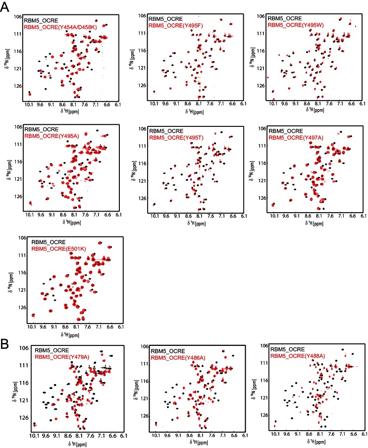
Figure 4—figure supplement 2. NMR spectra and ITC data for OCRE domain mutants.

Figure 4—figure supplement 2.

Comparison of NMR spectra of the RBM5 OCRE domain wild type (black) and different mutants (red, as indicated).Spectra in (A) indicate that the overall fold is intact, with some potential local effects near the residue mutated. (B) These mutants suggest local unfolding of the β-strand, which comprises the altered residue, but are also consistent with an overall folded domain.