Abstract
Childhood cataract (CC) has an incidence of 3.5 per 10,000 by age 15 years. Diagnosis of any underlying cause is important to ensure effective and prompt management of multisystem complications, to facilitate accurate genetic counselling and to streamline multidisciplinary care. Next generation sequencing (NGS) has been shown to be effective in providing an underlying diagnosis in 70% of patients with CC in a research setting. This project aimed to integrate NGS testing in CC within six months of presentation and increase the rate of diagnosis.
A retrospective case note review was undertaken to define the baseline efficacy of current care in providing a precise diagnosis. Quality improvement methods were used to integrate and optimize NGS testing in clinical care and measure the improvements made.
The percentage of children receiving an NGS result within six months increased from 26% to 71% during the project period. The mean time to NGS testing and receiving a report decreased and there was a reduction in variation over the study period. Several patients and families had a change in management or genetic counselling as a direct result of the diagnosis given by the NGS test.
The current recommended investigation of patients with bilateral CC is ineffective in identifying a diagnosis. Quality Improvement methods have facilitated successful integration of NGS testing into clinical care, improving time to diagnosis and leading to development of a new care pathway.
Problem
Childhood cataract (CC) has a reported incidence of 3.5 per 10,000 by age 15 years (Rahi et al) and is an important cause of lifelong visual loss. Early identification and diagnosis facilitate timely and appropriate surgical intervention, which is critical to ensure optimal visual outcomes. Diagnosis of any underlying cause of CC is necessary in order to ensure effective and prompt management of multisystemic complications, to facilitate accurate genetic counselling and to streamline multidisciplinary care. Despite the publication of care pathways guiding management and investigation of congenital cataracts (Lloyd et al, Wirth et al), the proportion of cases of congenital cataracts in which the underlying cause is established remains low.
Here, we report a Health Services quality improvement project. This defined the baseline efficacy of current mainstream, multidisciplinary care in providing a precise diagnosis for patients with CC. The aim of the programme was then to transfer into mainstream service the NGS technologies described by Gillespie et al. and through a process of PDSA cycles, to assess the improvements for patients who were prospectively ascertained with bilateral CC. We aimed to improve the proportion of children diagnosed with CC receiving a result of genomic testing, from 26% to over 50% within six months of diagnosis of CC. Through routine genomic testing of such patients, we hypothesize the proportion of patients with a known underlying cause of CC will improve. This project was undertaken as part of a Quality Improvement (QI) Project within Haelo and the Manchester Academic Health Science Centre at Saint Mary's Hospital and the Manchester Royal Eye Hospital. The paediatric ophthalmology unit at Manchester Royal Eye Hospital is a tertiary referral centre for paediatric cataracts with three consultants and four registrars.
Background
The heterogeneous nature of congenital cataract results in significant delay in diagnosis. Rahi et al (2000) reported that no cause of cataract could be identified in 61% of bilateral and 47% of unilateral cataract. Traditionally, numerous clinical, imaging, biochemical, and genetic tests are pursued sequentially in an attempt to delineate the precise cause at significant cost to health services; a process that is complicated, inefficient, and frequently unsuccessful (Gillespie et al). Where a diagnosis is made, often the time taken to achieve this is very long with multiple professionals involved, many appointments, and investigations required.
Bilateral CC is highly heterogeneous and as many as 50% of cases have an underlying genetic aetiology (Francis and Moore). These are most commonly transmitted as an autosomal dominant trait although autosomal recessive cases are frequently reported (Francis and Moore, Aldamesh et al). Many syndromic forms of CC are genetic including Stickler, Cockayne, Nance-Horan Warburg-MICRO syndromes (reviewed in Gillespie et al). Importantly a small group results from biochemical defects that are amenable to therapeutic intervention, such as galactosaemia, cerebrotendinous xanthomatosis, stomatin-deficient cryohydrocytosis (Flatt et al; Klepper; Leen et al; Luyckx et al).
Advances in DNA sequencing technologies, so-called Next Generation Sequencing (NGS) have revolutionized the throughput and diagnosis rate of genomic testing, in particular for genetically heterogeneous conditions. The application of such tests in a diagnostic setting has extended eligibility to greater numbers of patients (e.g. O'Sullivan et al) and this has recently been demonstrated for CC, which has shown that precise genetic diagnosis can positively impact clinical management and diagnostic outcomes (Gillespie et al).
Baseline measurement
A retrospective case note review was undertaken of 29 consecutive paediatric patients aged 10 years and under identified through the paediatric ophthalmic surgical list at the Manchester Royal Eye Hospital (MREH) from Jan 2011, under the care of the same consultant for bilateral congenital and developmental cataracts. Unilateral cases of congenital cataract were excluded. The outcomes of clinical investigations were assessed up until April 1st 2014. Patients were categorized into groups based on investigations received. The paediatric work up group received the investigations used to specifically determine a cause of the patient's cataract. These investigations are TORCH screen (including plasma Ig, culture, or PCR investigations), karyotype, urine for reducing substances, and organic amino acids. The extensive investigations group received investigations in addition to the basic paediatric work-up. This list includes DNA probe microarray, gene sequencing, fluorescent in situ hybridization (FISH), and cholesterol studies. The time awaiting a diagnosis would be routinely measured throughout the project to confirm whether NGS has the potential to improve this.
Over this time period, 29 new patients were identified with bilateral congenital cataracts. Of these 29 patients, 18 were male (62%), with an age range at presentation between 3 and 3875 days (10.6 years/127 months). The average age at time of first presentation to the MREH was 1130 days (3.1 years/37 months). 9 patients (31%) had a known family history of congenital cataracts and 5 (17%) had a relevant history of consanguinity. These patients presented from locations across the north of England, referred from primary and specialist care as well as one case of self-referral. In total, 8 patients (28%) were referred from primary care services including both general practice and community orthoptists. 20 patients (69%) were tertiary referrals. The majority of patients (23/29, 79.3%) received care from clinical specialities in addition to ophthalmology. Ophthalmic examination revealed diverse cataract morphology demonstrated considerable clinical diversity and of the 29 patients, 11 (38.0%) had systemic features, developmental delay, and learning disability being the most frequent. Patients with systemic features tended to be investigated more extensively than other patients, with 6/11 receiving extensive investigations (55%) and four (36.4%) a full paediatric work up.
By April 1st 2014, only 1/29 (3.4%) patients had a diagnosis identified using current, routine investigative techniques. The patient receiving a diagnosis received extensive investigation, made by microarray. This patient (patient 17) was diagnosed with 17q21.31 microdeletion syndrome before the development of cataracts. Investigations in all remaining 28 patients failed to yield a diagnosis, i.e. 96.5% of cataracts remained of undetermined origin. The mean time awaiting diagnosis was 54 months/4.5 years (SD + 42 months/3.5 years). Of these 29 patients, ten (34.5%) received no investigations to determine the cause of their cataracts, of whom 50% (5/10) had a family history of congenital or developmental cataract. 13 (44.9%) received the full paediatric work-up. Six (20.7%) received extensive investigations, of whom only one (14.3%) had a family history of congenital cataract.
As well as extensive investigations that were costly and ineffective, there was no uniformity in care pathway and families spent many years attending multiple hospital appointments without a diagnosis.
To assess the completion of our original aim, we routinely measured the time taken for genomic testing to be initiated following the diagnosis of congenital cataract, as well as the outcome of these investigations (See supplementary files).
bmjqir.u211094.w4602supp1.pptx (254.7KB, pptx)
Design
NGS testing for CC had been carried out since 2013 as part of an externally funded research project. This research project had demonstrated that NGS testing gave a positive diagnosis in 70% of cases (Gillepsie et al 2015). We wished to determine if NGS testing could be integrated into the clinical care of patients with CC to optimise the speed and rate of diagnosis. The participants of this quality improvement project were Mohammud Musleh (MRes student), Jane Ashworth (consultant paediatric ophthalmologist), Georgina Hall (geneticist), and Graeme Black (honorary consultant in genetics and ophthalmology). A range of methods were employed for this project including data collection from case note sampling to review baseline current care and small tests of change (PDSA cycles) to develop and test changes to the current system. These tests were designed iteratively to inform the key drivers for change (Supplement Figure 1). Patient surveys provided qualitative data on satisfaction with the NGS process.
Early PDSA cycles (Supplement Figure 2) tested new ways of working between teams in paediatric ophthalmology, clinical genetics, and genetic scientists. Communication, variation in practices, and unfamiliarity with NHS genetic tests were identified as barriers. Improvements to working included improved integration with monthly MDT meetings involving clinicians and scientists for education and discussion of NGS cataract test results. Overcoming barriers for phlebotomy involved improving availability of laboratory request cards, consent forms in the paediatric eye clinics and agreement for genetic phlebotomy services to collect samples. Closer working with the genetic laboratory also enabled a PDSA for urgent testing during which results were received in six weeks (compared to standard reporting times of 80 days).
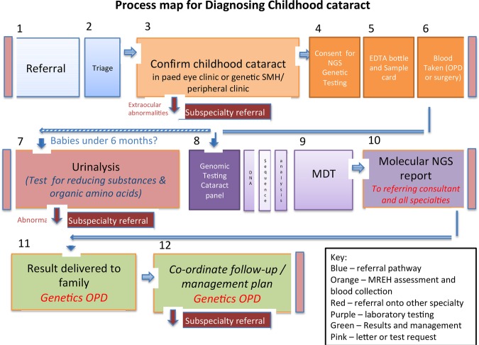
Evidence from PDSA cycles were used to engage the team in a shared goal for a single care pathway to improve care. In November 2014, a proposed pathway was agreed and tested with further refinements in May 2015 (Figure 1). In addition to a defined care pathway, the financial arrangements for NGS genetic testing within MREH was also agreed.
Figure 1.
Process map of new care pathway
Strategy
Implementation of NGS testing in clinical care of patients at Manchester Royal Eye Hospital and Saint Mary's hospital with paediatric cataract was made using a series of PDSA cycles based on a driver diagram.
PDSA cycle 1 aimed to improve time to blood taking for the NGS test. We hypothesized the delay in blood sampling for NGS contributed to the prolonged time awaiting diagnosis. To address this, the paediatric ophthalmology team would alert the genetic team at the time of the first referral or appointment so that arrangements for consent for testing and blood taking could be made as soon as possible. This was implemented from June 2014. Delays were identified due to limitations in access to phlebotomy, which was addressed in later addressed in cycle 3.
PDSA cycle 2 aimed to improve the turnaround time for NGS test results when needed for an urgent case. We predicted the testing time could fall through the genetics department contacting the laboratory on referral of a baby with congenital cataracts and asking for urgent processing of the sample. Through this, an urgent sample could be analysed in three weeks when prioritized over all other samples. We identified communication delays between ophthalmology and genetics requesting tests and the laboratory further contributed to delays. This was addressed in cycle 3.
PDSA cycle 3 aimed to engage the wider team in the project to improve time to blood testing and NGS result. We predicted that different clinicians and different care pathways will make it difficult to agree a single care pathway for the child with cataract, further contributing to delays. An interdisciplinary meeting was held in November 2014 with the Quality Improvement team, administrative and management team, and the paediatric ophthalmology and ophthalmic genetics and laboratory team to improve knowledge of the project and share ideas. Following this, distribution of posters and leaflets providing information to patients and clinicians about NGS testing, changes to the phlebotomy arrangements so that blood for NGS testing can be taken at the time of the first ophthalmology clinic visit if appropriate, and changes to the consent process and the referral pathway to genetics were implemented. Through these actions, the engagement of the paediatric ophthalmology team with the genetics team was greatly improved. However, the process was inconsistent. Some patients underwent blood testing in ophthalmology clinic, others were referred to genetics, and others were sent by email to the genetic counsellor for information and consent. There was also confusion from the lab regarding funding for testing. These issues were addressed in a single, defined care pathway that was agreed for a standard of genetic testing in the clinical pathway. Financial proposals were also approved to fund the test within the eye hospital, enabling ophthalmologists to deliver the test, rather than refer to the genetics team.
PDSA cycles tested new ways of working between teams in paediatric ophthalmology, clinical genetics, and genetic scientists. Communication, variation in practices, and unfamiliarity with NHS genetic tests were identified as barriers. Improvements to working included improved integration with monthly MDT meetings involving clinicians and scientists for education and discussion of NGS cataract test results. Overcoming barriers for phlebotomy involved improving availability of laboratory request cards, consent forms in the paediatric eye clinics, and agreement for genetic phlebotomy services to collect samples. Closer working with the genetic laboratory also enabled a PDSA for urgent testing during which results were received in six weeks (compared to standard reporting times of 80 days).
Evidence from PDSA cycles were then used to engage the team in a shared goal for a single care pathway to improve care. In November 2014, a proposed pathway was agreed and tested with further refinements in May 2015. In addition to a defined care pathway, the financial arrangements for NGS genetic testing within MREH was also agreed.
Results
51 patients were identified prospectively from January 2014 as having paediatric or congenital cataracts. Of these 31 were new patients and 20 patients were already under the care of MREH or Saint Mary's genetic clinic but who had not had previous NGS testing.
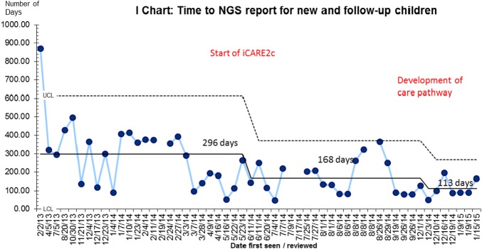
Data was retrieved to record time to NGS testing and time to NGS report across patients seen in either MREH or MCGM across the project period. As a set of individual data points across time (date referred or reviewed in clinic), a run chart is used to display time to testing or reporting for each patient. An Individual Chart (I Chart) was chosen as the data points are quantitative and each patient has a single data value (Provost et al 2011). The I chart calculates both the mean and the standard variation around the mean. Special cause variation can be seen when data points fall outside the standard variation and this can be helpful in exploring and understanding cases that fall outside the expected system.
Chart were analysed across 3 phases as the QI project progressed. A range of samples prior to the project start date (June 2014) “Phase 1” illustrate previous care. “Phase 2” is the period where small PDSA cycles were testing different working pathways and in November 2014, we held our first Engagement meeting across the ophthalmology and genetics department to begin agreeing a new care pathway which continued to be developed and agreed in May 2015. “Phase 3” shows the time after this engagement meeting.
The I charts show both a reduction in the average (mean) time to testing and reporting and a reduction in the variation (a narrowing between the upper and lower limits).
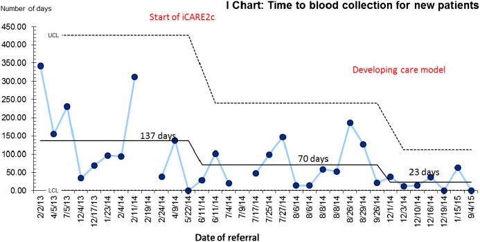
The time to blood collection reduced from a mean of 137 days to 70 days following the start of the project in June 2014 (Chart 1) and to 23 days following the improvements implemented in November 2014.
Chart 1:
Time (in days) to blood collection for genetic testing for new patients referred to CMFT.
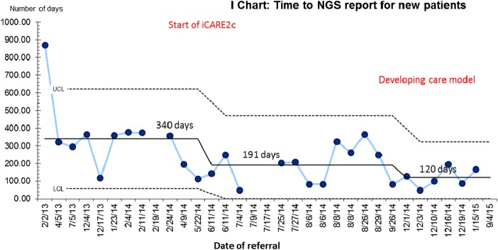
The time to NGS result also reduced from a mean of 340 days to 191 days following the start of the project in June 2014 (Chart 2) and to 120 days following the improvements implemented in November 2014.
Chart 2:
Time (in days) to a genetic NGS report for new patients
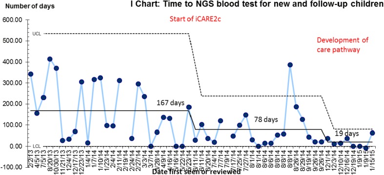
Data was also analysed across the new and follow-up patients (Charts 3 and 4). This data was investigated separately as the referral pathways across the different groups varied more significantly and it was anticipated that we would have achieved a lower success in the follow-up patients. However, a similar level of improvement was seen across the whole sample set.
Chart 3:
Time (in days) to genetic NGS testing for new and follow-up patients
Chart 4:
Time (in days) to NGS report in new and follow-up patients.
One special cause* case is highlighted in the time to testing. Examination of this case shows a patient who was not tested at their initial eye hospital appointment but referred to genetics. The family then did not attend their genetic appointment and the nature of the waiting lists meant they waited another six months for an appointment. Within the new care pathway, the family should be tested at the first appointment but patients who do not attend appointments will increase the time to testing and this cannot be controlled .
Diagnostic success rates from NGS testing:
After the introduction of NGS in April 2014 and until the 1st of April 2015, 15 of the 29 patients who had been followed as part of the base line evaluation underwent genomic testing as part of their routine care. In 66.7% patients (10/15) a diagnosis was found. No mutation was identified in the remaining five patients, including the one patient who had already been diagnosed with the 17q microdeletion Syndrome. The genes identified were associated with both syndromic (n=6) and non-syndromic (n=4) CC.
Of 51 children later identified, 11 had a confirmed diagnosis from NGS testing, 1 had a confirmed diagnosis from an array, 5 had a genetic diagnosis of uncertain significance, 5 are still waiting for results, and 31 (%) had ‘no pathogenic change’ on mutation report (Figure 2). These results suggest around a 20% diagnostic yield in this cohort of patients. Exploring the impact of these diagnosis was outside the scope of this project, however anecdotal experience from these families demonstrates the importance and relevance of a genetic diagnosis for families (see Appendix).
Figure 2.
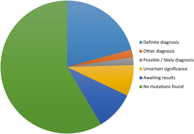
Pie chart to show diagnostic outcome from NGS testing
Percentage receiving a result within six months:
The percentage of children receiving a result within six months (180 days) increased from 26% to 71% during the project period (Table 1).
Table 1.
Percentage of patients receiving a result within 6 months
| Time period | Number of patients | Number with result within 6 months | Percentage of patients |
|---|---|---|---|
| Before June 2014 | 26 (12 new, 14 follow up) | 7 | 26 |
| June 2014-November 2014 | 18 (14 new, 4 follow-up) | 7 | 39 |
| December 2014-?March 2015 | 7 (6 new, 1 follow-up) | 5 | 71 |
Patient experience of NGS cataract testing:
Questionnaires were distributed to 21 parents/carers of patients who had undergone NGS testing for CC, to measure its impact. 11 completed questionnaires were returned (Table 3). These participants had been offered NGS testing during 2014 during the testing and development phase of the care pathway. Half the patients reported being satisfied with the service, and the remaining half highlighted difficulties with the existing pathway. In particular, patients mentioned the time waiting for results and the method of receiving the results. The comments were incorporated into the new process in which patients are all seen back in clinic for the results.
In terms of integrating the test into clinical care, the majority of patients felt that NGS testing was very important and they highlighted the benefits of getting a diagnosis, such as an explanation for the cataracts and genetic risk information.
Lessons and limitations
Data collection on prospective cases relied on existing databases including laboratory databases, Medisec, and a paper diary of surgical patients. This limited the scope of data collection and the sample in this project is unlikely to represent full ascertainment of patients attending the Trust over this time period. However, the sample was collected across a range of databases and is therefore likely to be an unbiased group. In addition, funding restrictions and inequality in access to NGS testing may limit the application of the proposed care pathway.
The low diagnostic rate during this period compared to previous research studies (Gillespie et al 2014) may reflect the case mix and will be improved with further refinement of the test and new improving technology.
Conclusion
The utility of NGS testing will further increase with the completion of the 100,000 Genome Project in 2017. This project aims to sequence 100,000 genomes, initially focusing on cancer, infectious, and rare diseases. The completion of this project will further enable NGS to detect mutations associated with even rarer conditions. Overall, we report similar findings to previous studies that demonstrate current recommended investigation for patients with bilateral paediatric cataract is ineffective in identifying a diagnosis. As demonstrated in previous studies, NGS enabled identification of underlying causes of CC in the majority of patients. This QI project enabled a substantial increase in the proportion of patients undergoing genomic testing and receiving a diagnosis within six months, as was originally aimed. NGS may confirm the presence of isolated cataract and exclude systemic associations, as well as provide information on hereditability within a few months. The adoption of such novel investigative techniques is required to be incorporated into clinical care if future care pathways are to improve the rate, efficiency, and speed of diagnosis.
The Quality Improvement approach has demonstrated the methods and evidence for successful translation of these technologies into patient care, improving the time to diagnosis and developing and integrating a new care pathway across hospital specialties. Embedding this integrated care will enable rapid translation of new genomic technology to patients not only with congenital cataracts but all inherited eye conditions. The next challenge from this project will be extending this care pathway to patients with inherited eye conditions seen in other hospitals across the region. It is also hoped that this care pathway and the learning from this project will help to inform other inherited conditions such as neurological or cardiac conditions.
Acknowledgments
This work was funded by Fight for Sight, grant number: 1831. The authors would also like to acknowledge the support of the Manchester Academic Health Science Centre and the Manchester National Institute for Health Research Biomedical Research Centre.
The funding organization had no role on the design or conduct of this research.
Footnotes
Declaration of interests: No conflicting relationship exists for any author
Ethical approval: Ethics committee approval was obtained from the NW Research Ethics Committee (11/NW/0421).
References
- 1.Rahi JS, Dezateux C. Measuring and interpreting the incidence of congenital ocular anomalies: lessons from a national study of congenital cataract in the UK. Investigative ophthalmology & visual science 2001;42:1444–8. [PubMed] [Google Scholar]
- 2.Francis PJ, Moore AT. Genetics of childhood cataract. Current opinion in ophthalmology 2004;15:10–5. [DOI] [PubMed] [Google Scholar]
- 3.Aldahmesh MA, Khan AO, Mohamed JY, Hijazi H, Al-Owain M, Alswaid A et al. Genomic analysis of pediatric cataract in Saudi Arabia reveals novel candidate disease genes. Genetics in medicine : official journal of the American College of Medical Genetics 2012;14:955–62. [DOI] [PubMed] [Google Scholar]
- 4.Gillespie RL, O'Sullivan J, Ashworth J, Bhaskar S, Williams S, Biswas S et al. Personalized diagnosis and management of congenital cataract by next-generation sequencing. Ophthalmology 2014;121:2124–37.e1-2. [DOI] [PubMed] [Google Scholar]
- 5.Flatt JF, Guizouarn H, Burton NM, Borgese F, Tomlinson RJ, Forsyth RJ et al. Stomatin-deficient cryohydrocytosis results from mutations in SLC2A1: a novel form of GLUT1 deficiency syndrome. Blood 2011;118:5267–77. [DOI] [PubMed] [Google Scholar]
- 6.Klepper J. Glucose transporter deficiency syndrome (GLUT1DS) and the ketogenic diet. Epilepsia 2008;49:46–9. [DOI] [PubMed] [Google Scholar]
- 7.Leen WG, Klepper J, Verbeek MM, Leferink M, Hofste T, van Engelen BG et al. Glucose transporter-1 deficiency syndrome: the expanding clinical and genetic spectrum of a treatable disorder. Brain : a journal of neurology 2010;133:655–70. [DOI] [PubMed] [Google Scholar]
- 8.Luyckx E, Eyskens F, Simons A, Beckx K, Van West D, Dhar M. Long-term follow-up on the effect of combined therapy of bile acids and statins in the treatment of cerebrotendinous xanthomatosis: a case report. Clinical neurology and neurosurgery 2014;118:9–111. [DOI] [PubMed] [Google Scholar]
- 9.Lloyd IC, Goss-Sampson M, Jeffrey BG, Kriss A, Russell-Eggitt I, Taylor D. Neonatal cataract: aetiology, pathogenesis and management. Eye (London, England) 1992;6:184–96. [DOI] [PubMed] [Google Scholar]
- 10.Wirth MG, Russell-Eggitt IM, Craig JE, Elder JE, Mackey DA. Aetiology of congenital and paediatric cataract in an Australian population. The British journal of ophthalmology 2002;86: 782–6. [DOI] [PMC free article] [PubMed] [Google Scholar]
- 11.Rahi JS, Dezateux C. Congenital and infantile cataract in the United Kingdom: underlying or associated factors. British Congenital Cataract Interest Group. Investigative ophthalmology & visual science 2000;41:2108–14. [PubMed] [Google Scholar]
- 12.O'Sullivan J, Mullaney BG, Bhaskar SS, Dickerson JE, Hall G, O'Grady A et al. A paradigm shift in the delivery of services for diagnosis of inherited retinal disease. Journal of medical genetics 2012;49:322–6. [DOI] [PubMed] [Google Scholar]
- 13.Haargaard B, Wohlfahrt J, Fledelius HC, Rosenberg T, Melbye M. A nationwide Danish study of 1027 cases of congenital/infantile cataracts: etiological and clinical classifications. Ophthalmology 2004;111:2292–8. [DOI] [PubMed] [Google Scholar]
- 14.Provost L P, Murray S K. The Health Care Data Guide. Learning from data for improvement. 2011. Wiley and Sons; p.149–151 [Google Scholar]
- 15.Moen R D, Nolan T W, Provost L P. Quality Improvement through planned experiment. 2012. McGraw-Hill Companies Inc. [Google Scholar]
Associated Data
This section collects any data citations, data availability statements, or supplementary materials included in this article.
Supplementary Materials
bmjqir.u211094.w4602supp1.pptx (254.7KB, pptx)


