Abstract
The ankylosing spondylitis (AS) is a systemic, multi-factorial, chronic rheumatic disease. Patients are highly susceptible to vertebral fractures with or without spinal cord injury (AS-SCI), even after a minor trauma. The study is a retrospective descriptive survey of post-acute, traumatic AS-SCI patients, transferred from the neurosurgical department and admitted in a Romanian Neurorehabilitation Clinic, during 2010–2014. There were 11 males associating AS-SCI (0.90% of all consecutive SCI admitted cases), with an average age of 54.6 years (median 56, limits 42–73 years). The average duration between the medically diagnosed AS and the actual associated spinal fracture(-s) moment was 21.4 years (median 23; limits 10–34 years). Low-energy trauma was incriminated in 54.5% cases. The spinal level of fracture was: cervical (four cases), thoracic (three), lumbar (four), assessed at admission as: American Spinal Injury Association (ASIA) Impairment Scale (AIS) A (four subjects), C (five) and D (two). By the time of discharge, neither patient has neurologically deteriorated; five patients (45.5%) improved of at least grade 1 (AIS). The overall complications were mainly infections: symptomatic urinary tract infections (seven patients; 63.6%), pulmonary (three subjects; 27.3%) and spondylodiscitis (one case; 9%). The average follow-up period was 15.3 months (median 12; limits 1–48 months) after discharge; three subjects gained functional improvement to AIS-E. The clinical profile (different risk factors, mechanisms, types and levels of spinal fractures, additional encephalic and/or cord lesions, co-morbidities), different post-surgical and/or general complications acquired during admission in our rehabilitation ward, served us for future prevention strategies and a better therapeutic management.
Subject terms: Fracture repair, Ankylosing spondylitis
Introduction
The spondyloarthropathies—which includes ankylosing spondylitis (AS) as the most known condition—are chronic, often progressive inflammatory diseases, ‘localized’ at some main ‘target tissue structures’: entheses, bone, articular mesenchyme, skin and nails, mucosae (oral, bowel, urogenital, eyes—including anterior uvea) and possibly muscles, endocardium, lung and kidney parenchyma. Their characteristic immune auto-aggression lesional phenotype is the enthesitis.1
Regarding the AS axial lesions (synthetically: sacroiliitis, spondylitis, spondylodiscitis and spondyloarthritis2), their morphopathological characteristics have been described over 40 years ago: chronic osteolytic foci, local bone edema and inflammatory reactive fibrosis, which lead to subsequent bone formation (mainly on endochondral way). Thereby, it would result in the vertebral bodies’ squaring (‘anterior spondylitis’/‘Romanus lesion’)3 and/or discitis (‘Andersson lesion’),4 as expressions of the erosive process and syndesmophytes formation.
As for the posterior zygapophyseal joints, the classical enthesitis sequence (erosion, fibrosis, ossification) merges with that of arthritis, thus resulting in a ‘capsular enthesitis’, with later ossification in a ‘bony shell’ enclosing pattern.5,6
Decreased bone mineral density—with a prevalence between 19 and 62%7,8—parallels the long disease evolution; literature pointed unexpected high prevalence of osteoporosis (13–16%) even within 10 years of onset of AS, at relatively young males.7
The intimate/molecular basis of the underlying physiopathological process is the genetic, immune-reactive stigmatization of the affected patients, connected to the presence of the HLA B27 antigen, especially to its (class I) defective allelic subtype.9 The axial forms of AS have the strongest association with the presence of the HLA B27 antigen, thus sustaining a genetic topographic determination.1,2,9,10
The AS has a quite stable prevalence (0.1–1.4%),11 and can be diagnosed in people of any age, gender or race; the onset occurs most commonly between the ages of 17 and 35, typically affecting males. Epidemiological studies have estimated the AS prevalence per 10 000 persons to be 18.6 (in Europe), 18.0 (in Asia) and 12.2 (in Latin America).12
The progressive biomechanical skeletal degradation augments the susceptibility of the spine for fractures in AS patients, even after low-energy impacts.13,14 Two decades of old paradigm postulates that a stiff (ankylosed) and osteoporotic spine is prone to fractures, even after a trivial trauma.14 Patients with AS have a fourfold fracture risk during their lifetime, compared with unaffected individuals,13 the prevalence of vertebral fractures being estimated at 10%.12 Spinal fractures in AS are usually associated with advanced age and a longstanding disease, impaired back mobility, syndesmophyte formation, lower bone mineral density and deteriorated microarchitecture in both the axial and the peripheral skeleton.14–17
In about two third of the cases, the main etiology consists of falls from standing or sitting position, the cervical region being the most frequently injured (in about 81% cases).18,19
Subjects and methods
This observational study aims to depict the clinical profile and functional outcomes of patients with AS who suffered a spinal cord injury (AS-SCI), and to evaluate the incidence of AS-SCI among the cohort of all consecutive subjects with traumatic SCI, admitted in a Romanian rehabilitation clinic.
Our hospital’s Bioethics Commission approval was obtained for this survey. This is a retrospective analysis of 11 consecutive patients with spinal trauma and AS (AS-SCI), first time admitted in our Neuromuscular Rehabilitation clinic during 2010–2014, after surgical intervention. The data were collected from the medical files (from the neurosurgical and rehabilitation departments in our hospital). All the patients had been diagnosed with AS, previous to their admission in the neurosurgical ward.
The variables studied were: demographic items (age and gender), time elapsed since the onset of AS, predisposing and pre-morbid factors, mechanism of the spinal trauma, involved vertebral level and the fracture characteristics, the neurological status—evaluated through the American Spinal Injury Association (ASIA) Impairment Scale (AIS) for spinal cord injuries, neurosurgical interventions, evolution and functional outcomes, complications during the hospital stay and individual follow-up.
Neuroimaging diagnosis and evaluation consist of conventional radiography, computed tomography and magnetic resonance imaging (MRI).
Results
There were 11 males with AS-SCI transferred from the neurosurgery spine department. The incidence of AS-SCI in our hospital was 0.90% from the cohort of all SCI patients, transferred to the rehabilitation clinic.
The clinical data are summarized in Table 1. The patients are grouped in the descendant order of the AS oldness. The average duration between the medically diagnosed AS and the actual associated spinal fracture(-s) moment was 21.4 years (median 23; limits 10–34 years). All patients were male, with an average age of 54.6 years (median 56, limits 42–73 years).
Table 1. Summarized clinical data of 11 consecutive patients with AS-SCI admitted in our rehabilitation clinic, during 2010–2014.
| Case no. | Age years | Gap AS-SCI (years) | Previous spinal fracture | Co-morbidities | Etiology | Severity of trauma | Fracture level/complications | AIS add. | AIS disch. | Surgical treatment | Post surgical complications | Reintervention | General medical complications | Follow-up (months) AIS |
|---|---|---|---|---|---|---|---|---|---|---|---|---|---|---|
| 1 | 66 | 34 | — | TB | Fall (on stairs) | Medium energy | L2 transvertebral | C (T12) | D (T12) | PLF | Dehiscence | Suture, per secundam healing | N.bladder; N. bowel; anemia; | 1; D |
| 2 | 61 | 32 | Old SCI (odontoid & C6-7 dislocation) | Stroke sequela; T2D; HBP | Fall (0.5 m; wheelchair) | Low energy | C5; C5-6 dislocation | C (C5) | D (C5) | ACCF+Halo | — | — | N.bladder; UTI; depression | 15; D |
| 3 | 73 | 31 | Old SCI (C5-6, operated) | HBP; hip prosthesis; CHC | Back fall; body level | Low energy | L2 transvertebral; retroperitoneal hematoma | C (L2) | D (L2) | PLF | Minor bleeding, per secundam healing | — | N.bladder; UTI; N.Pain; HBP | 2; D |
| 4 | 57 | 27 | — | IHD; LBB; AF (pace maker); HBP; T2D; Depression; | Back fall; body level | Low energy | T7; epidural hematoma (C4-T9) | A (C7) | A (T1) | DL (C5, C6-7; T1,3, 5,7) | Spondylodiscitis (staphylococcus) | PLF | N.bladder; UTI; N. bowel; N.Pain; spasticity; postural hypotension: depression; anemia | 12; A |
| 5 | 59 | 25 | — | Obesity II; [2]Cox; T2D; SMPN | Back fall; body level | Low energy | T11 (+T2 lamina) | C (T7) | D (T11) | DL (T10) | T10 non- compressive meningocele | DL (T2-3) | N.bladder; N.Pain; spasticity; depression | 48; E |
| 6 | 51 | 23 | Old SCI (C1,C2 odontoid 1989) | T2D; obesity; polytrauma: mild TBI; bilat.pleurisy | Car accident (passenger) | High energy | L1 transvertebral; T12-L1 dislocation | D (T10) | D (T10) | ALIF+PLF | Dehiscence | Suture, per secundam healing | N.bladder; UTI | 36; E |
| 7 | 42 | 15 | — | HBP; alcoholism | Fall (1;5 m) | Medium energy | C3-4 transvertebral; epidural hematoma | D (C4) | D (C4) | DL (C3-4; C6-7) | — | — | Spasticity; anemia | 1; D |
| 8 | 43 | 14 | — | Obesity III | Body level fall | Low energy | C5-6 dislocation | A (C5) | A (C5) | ACCF | — | — | N.bladder; N. bowel; UTI; spasticity; diabetes insipidus; respiratory infection; OcI | 21; A |
| 9 | 45 | 14 | — | Polytrauma: mild TBI; bilat. pleurisy Heavy smoker; alcoholism | Fall (height) work accident | High energy | T10/11 dislocation | C (T11) | D (L2) | PLF+PMMA | — | — | N.bladder; UTI; N. bowel; respiratory infection; enterocolitis | 6; E |
| 10 | 48 | 11 | — | T2D; obesity II; stroke sequela | Body level fall | Low energy | C6 | A (C5) | A (C5) | ACCF | — | — | N.bladder; UTI; N. bowel; N.Pain; pressure sore; spasticity; postural hypotension; autonomous dysreflexia (HBP+epileptic seizure); depression; respiratory infection | 24; A |
| 11 | 56 | 10 | — | polytrauma:TBI;Heavy smoker | Cyclist (rode accident) | High energy | L1 | A (L1) | A (L1) | PLF +PMMA | Implant loosening, migration | Repositioning | N.bladder; N. bowel; anemia; | 2; A |
Abbreviations: [2]Cox, [bilateral] coxarthrosis; ACCF, anterior cervical corpectomy and fusion (cervical decompression, anterior bone graft, metallic instrumentation); AF, atrial fibrillation; ALIF, (anterior lumbar interbody fusion, with bone); bilat., bilaterally; CHC, chronic hepatitis C; DL, decompressive laminectomy; Gap AS-SCI, time (measured in years) elapsed since the onset of AS and the associated spinal fracture (-s) moment; Halo, halo-vest instrumentation; HBP, high blood pressure; IHD, ischemic heart disease; LBB, left bundle branch block; N.bladder, neurogenic bladder; N.bowel, Neurogenic bowel; N.Pain, neuropathic pain; OcI, sub-occlusive intestinal syndrome; PLF, posterior lumbar fusion (decompressive laminectomy and unilateral/bilateral posterior metallic instrumentation with screws and rods); PMMA, polymethylmethacrylate vertebroplasty; SCI, spinal cord injury; SMPN, sensory-motor polyneuropathy; T2D, type 2 diabetes; TB, pulmonary tuberculosis; TBI, traumatic brain injury; UTI, symptomatic urinary tract infection.
Patients had the following co-morbidities: stroke sequelae (2 subjects), type 2 diabetes (4), arterial hypertension (4), obesity (4), sensory-motor polyneuropathy (1), ischemic heart disease (1), left bundle branch block (1), atrial fibrillation (1 patient, who after his first admission in our clinic received a cardiac pace maker in a specialized unit, outside our hospital), bilateral coxarthrosis (1), hip prosthesis (1), chronic hepatitis C (1), chronic alcoholism (2), polytrauma (3 patients), pulmonary tuberculosis (1); 2 were heavy smokers (defined by a daily cigarettes consumption of >20 pieces, or ⩾20 pack-years, according to Fagerström nicotine dependence test) (http://ndri.curtin.edu.au/btitp/documents/Fagerstrom_test.pdf).
Two patients had (an old) cervical and (a new) thoracolumbar fracture, acquired at different times, and one subject suffered an additional cervical spine fracture.
Some cases, which need a special emphasis, are depicted below.
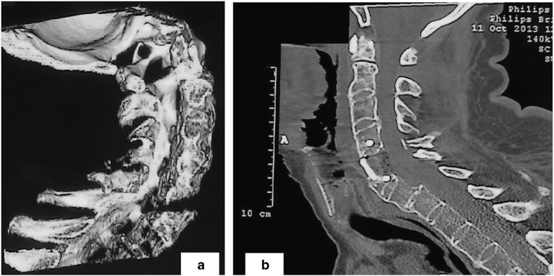
Patient no. 2 (Figures 1a and b) was previously disabled and restricted to wheelchair, because of a left hemiparesis (hemorrhagic stroke sequela, which occurred 14 years ago); he also had some old multilevel cervical fractures (the odontoid process, associated with a C6-7 dislocation, operated in 2006). This vulnerable patient was admitted in our rehabilitation department in an early stage, after the surgical instrumentation of a recent hyperextension fracture through the C5 vertebral body.
Figure 1.
Patient no. 2. Male, 61 years old: left hemiparesis (hemorrhagic stroke sequela) and cervical hyperextension fracture through the C5 vertebral body (after a minor trauma, falling from the wheelchair). Computed tomography sagittal-reconstruction: preoperative (a) and post-surgical aspects (b): anterior cervical corpectomy and fusion. Neurological status: C5 AIS-C tetraplegia (at admission) and C5 AIS-D (at discharge).
Half of the subjects (54.5%) experienced a low-energy trauma, spinal injuries resulting from a fall to the ground-level (patient no. 2 has fallen from the wheelchair).
Two cases underwent a medium energy impact, falling from stairs (~1–1.5 m height). Other three persons submitted a polytrauma (two subjects were victims of a traffic accident, the third fell from height, during a work accident) followed by instable transvertebral fractures and dislocations of the thoracolumbar junction.
The infra-axis cervical spine was predominantly affected (in four patients): in three subjects the C5-6 vertebrae, the C3-4 level, resulting in different AIS (Frankel) grades of tetraplegia.
Patient no. 5 was admitted from another hospital after a T11 vertebral fracture, operated and complicated with a non-compressive meningocele. The subject and the surgeon noticed any symptom, in the context of co-existence of a concomitant T2 occult (undiagnosed) compressive lamina fracture, detected after a delayed neurological deterioration. The diagnosis of the missed associated fracture and of the (mentioned) post-surgical complication were established in our rehabilitation clinic, after the patient’s abrupt neurological aggravation; after MRI confirmation, the patient was directed to the neurosurgical department in our hospital for a successful T2 decompression operation, followed by a gradual improvement.
All of our 11 AS-SCI cases were transferred after surgical management, indicated due to their posttraumatic spinal instability and/or the presence of a compressive epidural hematoma (2 cases).
All of the cervical patients underwent decompression and anterior stabilization (ACCF—anterior cervical corpectomy, fusion with autologous bone graft and metallic instrumentation), associating in one case halo-vest instrumentation. None of them required both anterior and posterior fixation.
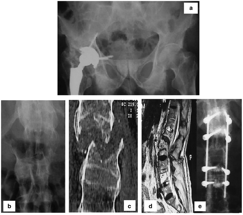
The thoracolumbar-injured patients underwent posterior lumbar fusion (PLF—decompressive laminectomy, unilateral or bilateral posterior metallic instrumentation with screws and rods); in two cases polymethylmethacrylate vertebroplasty was needed, to ‘anchor’ the screws in the fragile, osteoporotic bone (Figures 2a–e). One subject imposed ALIF (associated anterior lumbar interbody fusion with autologous bone) and posterior spondylodesis (PLF).
Figure 2.
Patient no.3: Male, 73 years old, instable L2 transvertebral fracture and retroperitoneal hematoma after a trivial trauma. Radiographs of the pelvis—old hip arthroplasty (a), and of the lumbar spine (b, c). T2-weighted sagittal MRI, documents the transvertebral fracture (d). Postoperative radiograph (e) after realignment and posterior fusion (T12-L4 spondylodesis with titanium instrumentation and polymethylmethacrylate vertebroplasty). Neurological status: L2 AIS-C paraplegia (at admission) and L2 AIS-D (at discharge).
Post surgical complications were noticed in six cases (54.5%); the most prominent were: metallic implantant migration (one subject, who needed surgical reintervention), infectious spondylodiscitis (one case), a non-compressive meningocele (conservatively followed-up for 48 months), some minor bleeding and per secundam healing (three patients).
The cervical SCI cases (noticed in 36.4%) were associated with AIS-A (complete) tetraplegia in two of the four subjects. After the surgical evacuation of a compressive hematoma (encountered in two of the cervical cases), the neurological status was: C7 AIS-A tetraplegia (in patient no.4) and C4 AIS-D tetraplegia (in patient no. 7).
Four of the seven patients with thoracolumbar SCI improved from AIS-C paraplegia (no ambulatory status) at admission, to AIS-D (ambulatory status) at discharge. The other three remained stationary: one functional AIS-D paraplegia, one complete paraplegia and the other an AIS-A tetraplegia.
By the time of discharge neither patient has neurologically deteriorated. Five patients (45.5%) improved of at least grade 1(AIS). Six patients (54.5%) had a stationary neurological evolution, regardless the topographical region or the specific feature (completeness or incompleteness) of the SCI.
The medical complications acquired during admission in our rehabilitation department are listed below: neurogenic bladder (10 cases), symptomatic urinary tract infections (7), neurogenic bowel (6), neuropathic pain (4), postural transient hypotension at mobilization in wheelchair (2), pressure sores (1), spasticity (5), respiratory tract infections (3), reactive anxious depressive neurotic syndrome (4), anemia (4), autonomous dysreflexia (with a sole generalized epileptic seizure, triggered by an abnormally high blood pressure spurt (1)), hypertension (2), (transient) diabetes insipidus (1), enterocolitis (with Clostridium difficile, 1). The most severe complication was noticed in patient no. 8, who was transferred to the general surgery department, with an acute sub-occlusive intestinal syndrome.
The average follow-up period after discharge was 15.3 months (median 12; limits between 1 and 48 months). More than half of the studied group (54.5%) had a long-term individual follow-up; the others were admitted during 2014, so it has been difficult to establish a pertinent appreciation about their long-term prognosis, due to their miscellaneous lesions and conditions.
Discussion
The incidence of AS-SCI in our clinic represented 0.90% from the cohort of all SCI patients, transferred from the neurosurgical department. Feldtkeller et al. 20 found an incidence of 1.3% vertebral fractures at AS patients, continuously increasing with the disease duration.
The authors performed a literature review, about this devastating pathological association: AS suffering spine fractures with/without cord injury (AS-SCI). After searching Pubmed were found 470 references (31 full text papers, between 1963 and October 2015) focused on AS-SCI. Very few papers depicted and analyzed cohorts of patients with AS-associating spine fractures, from a single medical center or region;17,21–23 other papers were systematic literature reviews.11,18 Most of these retrospective descriptive papers (about 98%) brought out—similarly with our study—only small series of AS subjects who suffered spine fractures: 18 patients,24 or 12 cases25 during a 10-year period and 12 subjects26 in 6 years. A plausible explanation for the scarcity of the cases consists of the epidemiological pattern of AS (mentioned earlier), making it difficult to establish a consistent cohort of patients in a single medical center. Grossly simplifying, according to the meta-analysis (describing 345 subjects cumulated in 76 papers) by Westervelds et al., 18 one can consider a mean of 4.5 AS-SCI cases per article. Another difficulty in gathering a large number of AS-SCI is represented by the remarkably high mortality rates in this redoubtable pathological association (17.7% deaths at 3 months, significantly higher compared with the usual SCI traumatic population18 (https://www.nscisc.uab.edu/public_content/pdf/Facts%202011%20Feb%20Final.pdf)—which is however, more than tenfold (9.3%)27 the general one (8.8‰ (http://www.medterms.com/script/main/art.asp?articlekey=2913)), particularly for those severely injured (https://www.nscisc.uab.edu/public_content/pdf/Facts%202011%20Feb%20Final.pdf). In our study group followed-up between 1 month and 4 years (average, as mentioned earlier, 15.3, and median 12 months) no deceased were registered.
In our group 54.5% of the subjects incriminated a trivial trauma (falling from the body level) as etiology for the spinal fractures. High-energy traumatic events (such as traffic accidents or fall from height) were responsible for 27.3% of injuries. Our data express quite a resemblance with the information provided by the literature (falls from standing or sitting position were noticed in 65.8% patients, and high energy impact trauma was incriminated in 31% of the spinal fractures18).
As a paradigm, most of the fractures in AS patients are caused by low-energy impacts, and the most vulnerable is the cervical segment (66.7%).18,24,28 Even in ‘usual’ traumatic conditions, the cervical segment is the most exposed region of the spinal column, due to its anatomical and biomechanical particularities (increased mobility, linear or translational kinetic momentum of the skull). Most of the AS cervical injuries involved the cervical spine between C5 and C7.18,25,28
The thoracolumbar junction was the second common topographic level of injury. Fractures of the thoracolumbar spine complicating AS were caused either by a major trauma (motor vehicle accident, fall from height—in three subjects), or even by a minor trauma (fall from the ground-level—in four cases).
The anatomical and biomechanical particularities of the cervical segment and thoracolumbar junction, expose these spinal regions to hyperextension or hyperflexion mechanisms of vertebral injury. Regarding the primary lesions pathways and topographic-related issues, ‘the critical velocity of tissue movement, which will lead to an axonal tear in a spinal cord contusion, is 0.5–1 m s−1, concentrating most of the stretching and shearing forces in the central part'.29,30
MRI control31 was mandatory in patient no. 5, with a symptom-free interval followed by neurological deterioration, and in patients no. 4 and 7, in whom MRI confirmed the suspicion of epidural hematomas (noticed in 18.2% of our cases). Post-traumatic compressive epidural hematoma is commonly reported in 23% of the patients with AS.26 Significant ossification of the ligaments increased the risk of epidural hematoma, favored bleeding from the epidural venous plexus and the fractured bone.32
Literature mentioned the possible co-existence of two33 or multiple spinal fractures34,35 in the advanced AS. In our lot, we depicted patients with associated multilevel SCIs (simultaneous—one case, or remote in time—three subjects).
Two patients had previous brain lesions (sequelae after hemorrhagic stroke, and lacunar strokes in the pontine and cerebellar regions). Traditional vascular risk factors seem to be more prevalent in patients with certain types of major rheumatic disorders, than in the general population.36,37 Many authors36–40 detected higher prevalence ratios for cardio-cerebrovascular pathology in AS patients, inflammation being incriminated in the pathogenesis and progression of atherosclerosis.38,39 Ischemic stroke has a higher prevalence in AS subjects (3.6%), compared with the general population (1.78%);39,40 increased risk of developing ischemic stroke was noticed even in young patients with AS.40 Prior (fewer) studies are in conflict with the ones aforementioned: no increased rate of acute myocardial infarction or stroke related to AS (mentioned by Brophy et al. 41 and Keller et al. 37 in their cohort studies) pointing to a higher prevalence of diabetes and hypertension,38,41 but not of hyperlipidemia/hypercholesterolemia.41
This peculiar association of triple severe disabling conditions (previous encephalic lesions, overlapped with AS and traumatic spinal cord injuries) represents a distinctive feature in our study group, not encountered in an advanced inquiry of the literature.
Diabetes insipidus secondary/in conjunction with a SCI, is an uncommon challenging complication.42,43 An interesting particularity noticed in one of our subjects was the triple association between AS-SCI and a transient diabetes insipidus.
Chronic alcoholic ingestion, noticed in 18.2% of the subjects in our group (less than that in the study by DeVivo et al. 27), represented also an important predisposing risk factor to fall and spine fracture in AS patients.
Post-surgery complications were noticed in 54.5% of our patients, but only two (18.2%) imposed reintervention: case no. 4 (an infectious osteodiscitis with Staphylococcus aureus) and case no. 11 (reposition of the metallic implant, loosened and migrated, due to the biological fragility of the bone).
Resembling categories (14% postoperative wound infections and 15% revision surgery due to insufficient stabilization)21 and quite similar rate of complications (51.1 or 41.7%)21 have been reported in the literature.
The most common complications cited in AS-SCI patients are pneumonia and urinary tract infections.44 The overall infectious complication rates in our patients were: symptomatic urinary tract infections (in 7 patients/63.6%, from the 10 subjects with neurogenic bladder) and pulmonary infections (3 cases/27.3%).
Absence of adequate follow-up data was considered19 the most significant weakness of the majority of the related papers. Hong45 followed up a group of eight subjects, from 4 up to 38 months (an average of 34 months) and noticed improvements of different degrees after operation, in five of them. From objective reasons (mentioned above), only half of our group (54.5%) benefited of an extended follow-up (between 12 and 48 months); they gained functional improvement (three of the six patients progressed to AIS-E). The other part of the lot was included only during the last year, and therefore, had a tiny—below the average—duration of follow-up.
Our data are consistent with most of the literature citations (with respect to the demographic items, risk factors, etiology and mechanisms of spinal fractures, diagnosis methodology, neurological, clinical, functional consequences, surgical and rehabilitation management, and follow-up).
This retrospective, descriptive person-centered approach, considered the multitude of factors that impact these two severe disabling conditions that merge and overlap (AS-SCI), and emphasized the individualized management in a collaborative integrated endeavor.
The presence of co-morbidities (concomitant or remote in time), different types and levels of spinal fractures, additional encephalic and/or cord lesions, and different (post-surgical and/or general) complications acquired during admission, all represented a supplementary challenge to our multidisciplinary rehabilitation team.
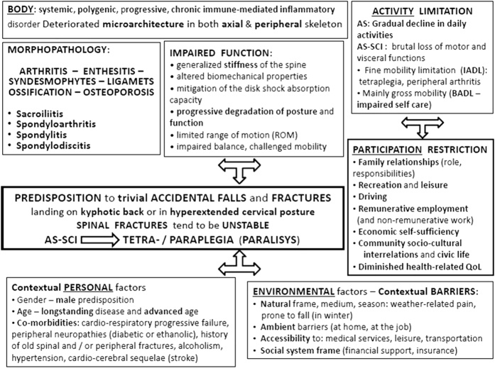
The complex evaluation of the risk factors for accidents, the clinical and morpho-physiopathological characteristics of our patients with AS and traumatic SCI (summarized in Table 1, and Figure 3) were used to implement prophylactic strategies in the patients’ community, and educational measures addressed to the patients, families and to the health professionals (general practitioners, rheumatologists and so on). The patients were instructed to avoid environmental circumstances predisposing to accidental falls (walking on slippery surfaces and loose carpets), to pay attention to doorsteps, to install preventive devices such as night lights and handrails and to sidestep activities involving risk of physical injury. They were encouraged to promote a healthy life style, to repel excessive use of alcohol,27,46,47 to diminish the metabolic and cardiovascular risk factors (behavioral changes, weight control, physical therapy and so on).
Figure 3.
Descriptive conceptual approach of patients with AS complicated with SCI, synthetically outlined in the general frame of the bio-psycho-social model of functioning, disability and health.
The authors declare no conflict of interest.
References
- Onose G , Stanescu-Rautzoiu L , Pompilian VM . Spondilartropatiile [The Spondyloarthropathies], Editura Academiei Române: Bucuresti (Bucharest), Romania, 2000. [Google Scholar]
- Colbert RA , DeLay ML , Layh-Schmitt G , Sowders DP . HLA-B27 misfolding and spondyloarthropathies. Prion 2009; 3: 15–26. [DOI] [PMC free article] [PubMed] [Google Scholar]
- Aufdermaur M . Pathogenesis of square bodies in ankylosing spondylitis. Ann Rheum Dis 1989; 48: 628–631. [DOI] [PMC free article] [PubMed] [Google Scholar]
- Naqi R , Ahsan H , Azeemuddin M . Spinal changes in patients with ankylosing spondylitis on MRI: case series. J Pak Med Assoc 2010; 60: 872–875. [PubMed] [Google Scholar]
- Ball J . Enthesopathy of rheumatoid and ankylosing spondylitis. Ann Rheum Dis 1971; 30: 213–223. [DOI] [PMC free article] [PubMed] [Google Scholar]
- Claudepierre P , Voisin MC . Les enthèses: histologie,anatomie pathologique et physiopathologie (The entheses: histology, pathology, and pathophysiology). Rev Rhum. 2005; 72: 34–41. [DOI] [PubMed] [Google Scholar]
- van der Weijden MA , Claushuis TA , Nazari T , Lems WF , Dijkmans BA , van der Horst-Bruinsma IE . High prevalence of low bone mineral density in patients within 10 years of onset of ankylosing spondylitis: a systematic review. Clin Rheumatol 2012; 31: 1529–1535. [DOI] [PMC free article] [PubMed] [Google Scholar]
- Davey-Ranasinghe N , Deodhar A . Osteoporosis and vertebral fractures in ankylosing spondylitis. Curr Opin Rheumatol 2013; 25: 509–516. [DOI] [PubMed] [Google Scholar]
- Kamanli A , Ardicoglu O , Godekmerdan A . HLA-B27 subtypes in patients with spondylarthropathies, IgE levels against some allergens and their relationship to the disease parameters. Bratil Lek Listy 2009; 110: 480–485. [PubMed] [Google Scholar]
- Goie The HS , Steven MM , van der Linden SM , Cats A . Evaluation of diagnostic criteria for ankylosing spondylitis: a comparison of the Rome, New York and modified New York criteria in patients with a positive clinical history screening test for ankylosing spondylitis. Br J Rheumatol 1985; 24: 242–249. [DOI] [PubMed] [Google Scholar]
- Braun J , Sieper J . Ankylosing spondylitis. Lancet 2007; 369: 1379–1390. [DOI] [PubMed] [Google Scholar]
- Dean LE , Jones GT , MacDonald AG , Downham C , Sturrock RD , Macfarlane GJ . Global prevalence of ankylosing spondylitis. Rheumatology (Oxford) 2014; 53: 650–657. [DOI] [PubMed] [Google Scholar]
- Taggard DA , Traynelis VC . Management of cervical spinal fractures in ankylosing spondylitis with posterior fixation. Spine 2000; 25: 2035–2039. [DOI] [PubMed] [Google Scholar]
- Hunter T , Forster B , Dvorak M . Ankylosed spines are prone to fracture. Can Fam Physician 1995; 41: 1213–1216. [PMC free article] [PubMed] [Google Scholar]
- Finkelstein JA , Chapman JR , Mirza S . Occult vertebral fractures in ankylosing spondylitis. Spinal Cord 1999; 37: 444–447. [DOI] [PubMed] [Google Scholar]
- Klingberg E , Geijer M , Göthlin J , Mellström D , Lorentzon M , Hilme E et al. Vertebral fractures in ankylosing spondylitis are associated with lower bone mineral density in both central and peripheral skeleton. J Rheumatol 2012; 39: 1987–1995. [DOI] [PubMed] [Google Scholar]
- Klingberg E , Lorentzon M , Göthlin J , Mellström D , Geijer M , Ohlsson C et al. Bone microarchitecture in ankylosing spondylitis and the association with bone mineral density, fractures, and syndesmophytes. Arthritis Res Ther 2013; 15: R179. [DOI] [PMC free article] [PubMed] [Google Scholar]
- Westerveld LA , Verlaan JJ , Oner FC . Spinal fractures in patients with ankylosing spinal disorders: a systematic review of the literature on treatment, neurological status and complications. Eur Spine J 2009; 18: 145–156. [DOI] [PMC free article] [PubMed] [Google Scholar]
- Westerveld LA , van Bemmel JC , Dhert WJ , Oner FC , Verlaan JJ . Clinical outcome after traumatic spinal fractures in patients with ankylosing spinal disorders compared with control patients. Spine J 2014; 14: 729–740. [DOI] [PubMed] [Google Scholar]
- Feldtkeller E , Vosse D , Geusens P , van der Linden S . Prevalence and annual incidence of vertebral fractures in patients with ankylosing spondylitis. Rheumatol Int 2006; 26: 234–239. [DOI] [PubMed] [Google Scholar]
- Backhaus M , Citak M , Kälicke T , Sobottke R , Russe O , Meindl R et al. Spine fractures in patients with ankylosing spondylitis: an analysis of 129 fractures after surgical treatment. Orthopade 2011; 40: 917–920, 922-4. [DOI] [PubMed] [Google Scholar]
- Mahajan R , Chhabra HS , Srivastava A , Venkatesh R , Kanagaraju V , Kaul R et al. Retrospective analysis of spinal trauma in patients with ankylosing spondylitis: a descriptive study in Indian population. Spinal Cord 2015; 53: 353–357. [DOI] [PubMed] [Google Scholar]
- Caron T , Bransford R , Nguyen Q , Agel J , Chapman J , Bellabarba C . Spine fractures in patients with ankylosing spinal disorders. Spine 2010; 35: E458–E464. [DOI] [PubMed] [Google Scholar]
- Thumbikat P , Hariharan RP , Ravichandran G , McClelland MR , Mathew KM . Spinal cord injury in patients with ankylosing spondylitis: a 10-year review. Spine 2007; 32: 2989–2995. [DOI] [PubMed] [Google Scholar]
- Seong-Bae A , Keung-Nyun K , Dong-Kyu C , Keun-Su K , Yong-Eun C , Sung-Uk K . Surgical outcomes after traumatic vertebral fractures in patients with ankylosing spondylitis. J Korean Neurosurg Soc 2014; 56: 108–113. [DOI] [PMC free article] [PubMed] [Google Scholar]
- Whang PG , Goldberg G , Lawrence JP , Hong J , Harrop JS , Anderson DG et al. The management of spinal injuries in patients with ankylosing spondylitis or diffuse idiopathic skeletal hyperostosis: a comparison of treatment methods and clinical outcomes. J Spinal Disord Tech 2009; 22: 77–85. [DOI] [PubMed] [Google Scholar]
- DeVivo MJ , Black KJ , Stover SL . Causes of death during the first 12 years after spinal cord injury. Arch Phys Med Rehabil 1993; 74: 248–254. [PubMed] [Google Scholar]
- Guo ZQ , Dang GD , Chen ZQ , Qi Q . [Treatment of spinal fractures complicating ankylosing spondylitis]. Zhonghua Wai Ke Za Zhi 2004; 42: 334–339. [PubMed] [Google Scholar]
- Blight AR , Decrescito V . Morphometric analysis of experimental spinal cord injury in the cat; the relation of injury intensity to survival of myelinated axons. Neuroscience 1986; 19: 321–341. [DOI] [PubMed] [Google Scholar]
- Onose G , Anghelescu A , Muresanu DF , Padure L , Haras MA et al. A review of published reports on neuroprotection in spinal cord injury. Spinal Cord 2009; 47: 716–726. [DOI] [PubMed] [Google Scholar]
- Pedrosa I , Jorquera M , Mendez R , Cabeza B . Cervical spine fractures in ankylosing spondylitis: MRI findings. Emerg Radiol 2002; 9: 38–42. [DOI] [PubMed] [Google Scholar]
- Elgafy H , Bransford RJ , Chapman JR . Epidural hematoma associated with occult fracture in ankylosing spondylitis patient: a case report and review of the literature. J Spinal Disord Tech 2011; 24: 469–473. [DOI] [PubMed] [Google Scholar]
- Foo D , Bignami A , Rossier AB . Two spinal cord lesions in a patient with ankylosing spondylitis and cervical spine injury. Neurology 1983; 33: 245–249. [DOI] [PubMed] [Google Scholar]
- Osgood CP , Abbasy M , Mathews T . Multiple spine fractures in ankylosing spondylitis. J Trauma 1975; 15: 163–166. [DOI] [PubMed] [Google Scholar]
- Samartzis D , Anderson DG , Shen FH . Multiple and simultaneous spine fractures in ankylosing spondylitis: case report. Spine 2005; 30: E711–E715. [DOI] [PubMed] [Google Scholar]
- Behrouz R . The risk of ischemic stroke in major rheumatic disorders. J Neuroimmunol 2014; 277: 1–5. [DOI] [PubMed] [Google Scholar]
- Keller JJ , Hsu JL , Lin SM , Chou CC , Wang LH , Wang J et al. Increased risk of stroke among patients with ankylosing spondylitis: a population-based matched-cohort study. Rheumatol Int 2014; 34: 255–263. [DOI] [PubMed] [Google Scholar]
- Nurmohamed MT , van der Horst-Bruinsma I , Maksymowych WP . Cardiovascular and cerebrovascular diseases in ankylosing spondylitis: current insights. Curr Rheumatol Rep 2012; 14: 415–421. [DOI] [PubMed] [Google Scholar]
- Lin CW , Huang YP , Chiu YH , Ho YT , Pan SL . Increased risk of ischemic stroke in young patients with ankylosing spondylitis: a population-based longitudinal follow-up study. PLoS ONE 2014; 9: e94027. [DOI] [PMC free article] [PubMed] [Google Scholar]
- Mathieu S , Pereira B , Soubrier M . Cardiovascular events in ankylosing spondylitis: an updated meta-analysis. Semin Arthritis Rheum 2015; 44: 551–555. [DOI] [PubMed] [Google Scholar]
- Brophy S , Cooksey R , Atkinson M , Zhou SM , Husain MJ , Macey S et al. No increased rate of acute myocardial infarction or stroke among patients with ankylosing spondylitis-a retrospective cohort study using routine data. Semin Arthritis Rheum 2012; 42: 140–145. [DOI] [PubMed] [Google Scholar]
- Closson BL , Beck LA , Swift MA . Diabetes insipidus and spinal cord injury: a challenging combination. Rehabil Nurs 1993; 18: 368–374. [DOI] [PubMed] [Google Scholar]
- Kuzeyli K , Cakir E , Baykal S , Karaarslan G . Diabetes insipidus secondary to penetrating spinal cord trauma: case report and literature review. Spine 2001; 26: E510–E511. [DOI] [PubMed] [Google Scholar]
- Mathews M , Bolesta MJ . Treatment of spinal fractures in ankylosing spondylitis. Orthopedics 2013; 36: e1203–e1208. [DOI] [PubMed] [Google Scholar]
- Hong F , Ni JP . [Retrospective study on the treatment of ankylosing spondylitis with cervical spine fracture: 8 cases report]. Zhongguo Gu Shang 2013; 26: 508–511. [PubMed] [Google Scholar]
- Anghelescu A , Onose LV , Magdoiu AM , Popescu C , Andone I , Onose G et al. Postacute evolution of traumatic spinal cord injury in patients with ankylosing spondylitis, in a romanian rehabilitation ward. Neurol Rehabil 2015; (Suppl 1): S78 (abstract 142). [DOI] [PMC free article] [PubMed]
- Alaranta H , Luoto S , Konttinen YT . Traumatic spinal cord injury as a complication to ankylosing spondylitis. An extended report. Clin Exp Rheumatol 2002; 20: 66–68. [PubMed] [Google Scholar]


