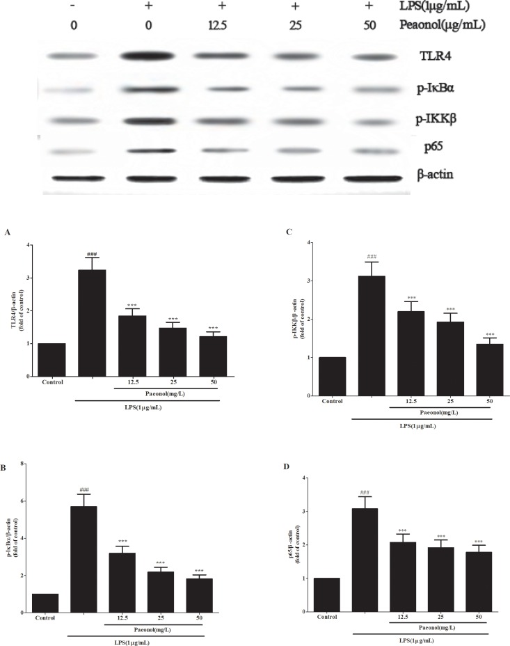
Figure 9. Paeonol modulates LPS-stimulated DCs by TLR4-NF-κB signaling.
DCs were incubated in presence or absence of different concentrations of paeonol (12.5, 25, 50 mg/L) for 24 h, then incubated with or without 1 μg/mL LPS for another 24 h. The expression levels of TLR4 (A), the phosphorylation of IKKβ (B), IκBα (C) and p65 (D) in cell lysates were determined by Western blot. β-actin was used as a standard control. Results are expressed as fold increase over control group. All data represent the means ± SD from three separate experiments. **p < 0.01, ***p < 0.001 compared to LPS group; ##p < 0.01 and ###p < 0.001 compared to control group.

