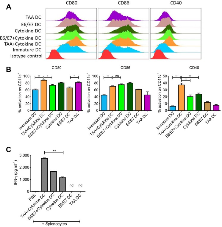
Figure 1. Total tumor lysate induces DC activation.
Bone marrow derived DCs were exposed to tumor associated antigen (TAA) or antigen specific E6/E7 peptides in presence or absence of proinflammatory cytokine cocktail. (A) Representative FACS analysis of costimulatory molecules on DCs after 18–24 hours of activation. (B) Statistical analysis of DC activation from A. (C) Levels of IFNγ induced after incubation of variously activated DCs with splenocytes for 3–4 days. Data are shown as + SEM. *p-value < 0.05, **p-value. < 0.01.

