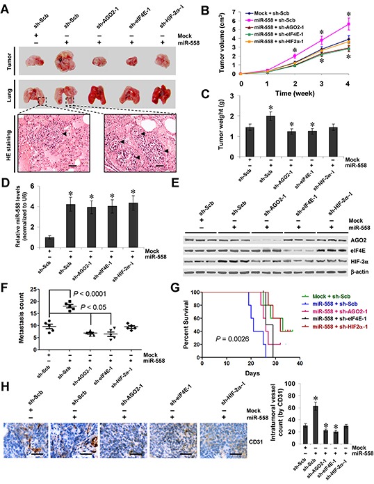
Figure 6. miR-558 promotes the growth, metastasis, and angiogenesis of NB cells through facilitating HIF-2α expression in vivo.

A. representative and HE staining images of xenograft tumors and lung metastatic colonies in athymic nude mice. B. tumor growth curve of SH-SY5Y (1×10⁶) stably transfected with empty vector (mock) or miR-558 precursor, and those co-transfected with sh-Scb, sh-AGO2, sh-eIF4E, or sh-HIF-2α in athymic nude mice (n=5 for each group), after hypodermic injection for 4 weeks. C. quantification of xenograft tumors formed by hypodermic injection of SH-SY5Y cells stably transfected with mock or miR-558 precursor, and those co-transfected with sh-Scb, sh-AGO2, sh-eIF4E, or sh-HIF-2α. D. and E. real-time qRT-PCR and western blot showing the expression of miR-558, AGO2, eIF4E, and HIF-2α in xenograft tumor tissues. F. and G. quantification of lung metastasis and Kaplan–Meier survival plots of nude mice with injection of SH-SY5Y cells (0.4×10⁶) stably transfected with mock or miR-558 precursor, and those co-transfected with sh-Scb, sh-AGO2, sh-eIF4E, or sh-HIF-2α via the tail vein (n=5 for each group). H. immunohistochemical staining (left) and quantification (right) of CD31 expression within tumors formed by hypodermic injection of SH-SY5Y cells stably transfected with mock or miR-558 precursor, and those co-transfected with sh-Scb, sh-AGO2, sh-eIF4E, or sh-HIF-2α. Scale bars: 100 μm. * P<0.001 vs. mock+sh-Scb.
