Abstract
In vitro Omics analysis (i.e. transcriptome) is suggested to predict in vivo toxicity and adverse effects in humans, although the causal link between high-throughput data and effects in vivo is not easily established. Indeed, the chemical-organism interaction can involve processes, such as adaptation, not established in cell cultures. Starting from this consideration we investigate the transcriptomic response of immortalized thyrocytes to ethylenthiourea and chlorpyrifos. In vitro data revealed specific and common genes/mechanisms of toxicity, controlling the proliferation/survival of the thyrocytes and unrelated hematopoietic cell lineages. These results were phenotypically confirmed in vivo by the reduction of circulating T4 hormone and the development of pancytopenia after long exposure. Our data imply that in vitro toxicogenomics is a powerful tool in predicting adverse effects in vivo, experimentally confirming the vision described as Tox21c (Toxicity Testing in the 21st century) although not fully recapitulating the biocomplexity of a living animal.
Toxicogenomics aims to define the molecular patterns predicting the in vivo toxicity, the adverse effects in humans, or the individual susceptibility to chemicals, allowing a better extrapolation of animal data to humans1. Since the publication of a report entitled “Toxicity Testing in the 21st Century: A Vision and a Strategy”, several researchers have been aiming to optimise the application of Omics technologies to in vitro cell systems, with a differentiated phenotype, for predictive toxicology2. Nowadays, there is a strong need for a rapid mechanism-based strategy in risk assessment, achievable in an easier manner in cell lines. This strategy could be used for the decision to opt out or to proceed with further animal tests, matching the need to apply the ‘3Rs’ concept (Replacement, Reduction and Refinement). At present, a practical application of the Tox21c vision is still far away because of the limited confidence in the new method as the causal link between in vitro data, obtained with new technologies, and adverse effects, tested in vivo, is difficult to establish. The crucial confidence can be reached by simultaneously evaluating phenotypic and molecular endpoints, the latter achievable through in vitro Omics analyses3,4, considering that the chemical-cell/organ interaction in vivo can be characterized by compensation and adaptive response not necessarily developed in differentiated cell cultures.
Hypothyroidism is increasing worldwide5 and several modifiable factors, i.e. diet and environmental pollutants, have been involved. Thyroid Disrupting Chemicals (THDCs) exert their effects on function and regulation of the thyroid tissue, especially during the early-life stages6,7.
Thyroid dysfunction has been associated to pesticides exposure in different epidemiological8,9 and experimental studies10,11, although a debate is heated. The traditional toxicology approach, based on long-lasting in vivo experiments, did not provide exhaustive information about the mechanism of action of THDCs7,12.
We have recently reported that THDCs toxicity of low-dose bisphenol-A (BPA) can be highlighted investigating directly the expression of thyroid specific genes in rat immortalized thyrocytes13 while unpredicted mechanisms of toxicity are evidenced by in vitro toxicogenomics14.
Here, we practically applied the Tox21c suggestions in the investigation of the dose-dependent effects of ethylenethiourea (ETU) and chlorpyrifos (CPF), both exerting THDCs activity10,11,15, starting from the transcriptomic analyses of exposed immortalized rat thyrocytes. We aimed to: a) consolidate the suggestion that in vitro toxicogenomics could draft the in vivo cell response; b) identify the thyroid signature and mechanisms of toxicity for in vivo validation; c) highlight in vitro cell type unrelated outcomes evaluable in vivo, as here shown for the haematopoiesis.
Results
Transcriptomic analyses in CPF and ETU treated cells
In vitro toxicogenomic experiments were performed to generate data to be verified in vivo. We assessed the low-dose and the mixture effects of CPF and ETU in PCCl3 cells, rat immortalized thyrocytes, considered a valuable model for studying thyroid cell function and transformation in vitro16. Our interest in ETU and CPF is due to their ability to impair thyroid physiology10,11, documented at morphological but not at molecular level. Considering the non-monotonic dose-response curves of the THDCs, we tested the transcriptomic effects of the pesticides at different concentrations.
Experimentally, PCCl3 cells were exposed for one week to different doses of both pesticides as reported in Table 1 and to their combinations. In the following we use the H, M, L abbreviations to state high, medium and low concentrations, respectively. The chosen concentrations are in or below the range of the doses tested in immortalized thyrocytes for ETU17 and other cell systems for CPF18. Since humans are usually co-exposed, we also treated the cells with the mixtures of ETU and CPF to evaluate possible additive/synergic effects. Of note, the used concentrations are within the range stated for non-exposed population (low and medium concentrations), and for exposed workers (high concentrations) in reports from the Centers for Disease Control and Prevention (CDC) for CPF19 and ETU20, although lower levels of both compounds have been recently detected in human fluids21.
Table 1. ETU and CPF applied concentrations in vitro.
| In vitro applied concentration | Abbreviation | |
|---|---|---|
| CPF-high dose | 6 × 10⁻⁷ M | CPF-H |
| CPF-medium dose | 6 × 10⁻⁸ M | CPF-M |
| CPF- low dose | 6 × 10⁻⁹ M | CPF-L |
| ETU-high dose | 6 × 10⁻⁸ M | ETU-H |
| ETU-medium dose | 6 × 10⁻⁹ M | ETU-M |
| ETU- low dose | 6 × 10⁻¹⁰ M | ETU-L |
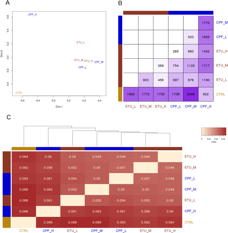
The genome-wide view of the PCCl3 transcriptome was obtained by high-throughput RNA-sequencing (RNAseq) data analysis. The differential gene expression was synthesized in only two relative values for each condition to draw a multi-dimensional scaling plot (Fig. 1A). A deep impact of both compounds on the transcriptome was observed. CPF had the stronger concentration-dependent effect as highlighted also in the related overview matrix (Fig. 1B), showing an increased number of deregulated genes in CPF treated samples vs control. ETU had a smaller concentration-dependent effect. Pairwise similarities between conditions were measured as JS distances across all genes22, reported in the heatmap and used to build a dendrogram between the conditions for CPF and ETU treatments (Fig. 1C). The JS distance between CPF-H and CPF-M (0.0625) or CPF-L (0.0609) was similar to the control (0.0662), underlining the non- monotonic concentration-response relationship. The reduced JS distance in co-exposed samples, MxM, LxL, indicated an alleviated impact on the transcriptome, also evidenced in Fig. 1B. Tested genes passing the fold change (FC) and corrected p-value filters (absolute log2FC ≥ 1, corrected p-value ≤ 0.05) were defined as Differentially Expressed Genes (DEGs) and represented in volcano plots (Supplementary Fig. 1).
Figure 1. Overview of gene expression analysis.
(A) Multi-dimensional scaling plot shows the two main components explaining the differences across the conditions. (B) Heatmap representing the number of significant differentially expressed genes (corrected p-value ≤ 0.05) for each contrast. (C) Jensen-Shannon distances between conditions were calculated to verify their similarities across all genes and represented by heatmap. Pairs with fairer colour are more similar and are represented nearer in the dendrogram.
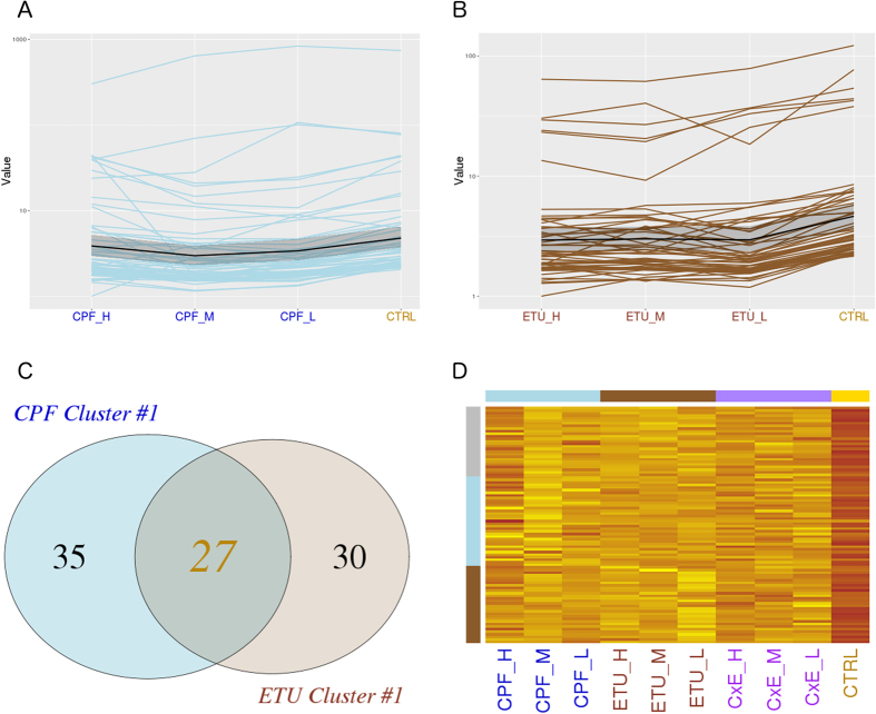
Gene cluster analysis was applied on DEGs retrieved at each dose, specifically for ETU and for CPF to study the dose-dependent effects of the two compounds. Partitioning of DEGs into clusters with similar expression profiles was achieved through K-means clustering analysis using JS distance as metric. Three different gene clusters were observed among the CPF-DEGs and three for ETU-DEGs (data not shown). Molecular signatures of toxicity were identified in the most significant cluster (cluster #1), including the higher number of genes. CPF-cluster #1 was composed of 62 transcripts (Fig. 2A and Supplementary Table 1) inhibited in CPF-L and/or CPF-M condition and not regulated at CPF-H. ETU-cluster #1 (Fig. 2B and Supplementary Table 2) consisted of 59 DEG, whose transcripts decreased at ETU-M and increased again at ETU-H. The two clusters were compared to selected common and specific genes through a Venn diagram (Fig. 2C). We defined a main signature of thyroid toxicity represented by 27 common DEGs and specific signatures composed of genes exclusively included in the CPF- or ETU- list. Their expression profiles were reported by heatmap for each dose of pesticides and their mixture (Fig. 2D). The heatmap showed the differences in the expression of the gene signatures and, strikingly, their deep changes in each condition of co-exposure.
Figure 2. CPF and ETU clusters #1 expression profiling.
DEGs were partitioned by k-means clustering analysis. The expression profiles of genes included in the most significant cluster is reported as line plot across the different doses for CPF (A), for ETU (B) and, finally, compared by Venn diagram (C). The expression profiles of selected common genes are represented by heatmap for CPF and ETU, including the different doses and combination treatments (D).
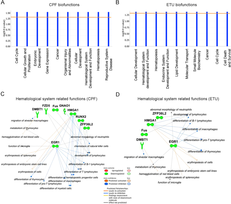
Insights into mechanisms of toxicity and unpredicted outcomes were obtained by Ingenuity Pathway Analysis (IPA), revealing the biological/toxicological functions over-represented in each gene cluster, in particular CPF- and ETU- cluster #1. The number of non-annotated genes in the rat genome assembly, used for read mapping (RGSC 5.0/rn5), was unfortunately too high to execute a functional analysis. Thus, we compared the sequences of non-annotated rat DEGs with human genes to label them with the corresponding human gene annotation having a high percentage of identity. We performed the IPA analyses on the new lists to identify the enriched biofunctions. The ten most significant biofunctions obtained from the CPF- cluster #1 and ETU- cluster #1 are reported in Fig. 3A and B, respectively. The low p-value was not surprising considering the small number of annotated genes. Unexpectedly, no damage of highly specific thyroid cell function was shown, although the impairment of thyroid cell growth was suggested by the biofunction “The Cell Cycle, Endocrine System Development and Function” (G2/M delay, CPF p-value = 2,86E-02 and ETU p-value = 4,24E-02, and S phase entry CPF p-value = 3,84E-02 ETU p-value = 4,84E-02). The same IPA analyses identified “Cellular Development, Hematological System Development and Function, Hematopoiesis” as a common deregulated biofunction (p-value = 2,86E-02 and p-value = 4,24E-02, respectively) for both compounds. The related gene networks are shown in Fig. 3C (CPF) and Fig. 3D (ETU).
Figure 3. CPF and ETU clusters #1 biofunctions.
Bio-functions identified by IPA analysis of CPF- cluster #1 (A) and ETU- cluster #1 (B). The left y-axis value is a significant score, −log10(BH corrected p-value), and the orange line evidences the threshold level (corrected p-value ≤ 0.05). Significant hematological system related functions are represented as molecular networks to evidence their relationships with deregulated genes included in CPF-cluster #1 (C) and ETU-cluster #1 (D).
Overall, in vitro transcriptomics distinguished between CPF- and ETU- treated samples, confirming a non-monotonic concentration-dependent effect (U-inverted shape) and the differences in co-exposure conditions. The signatures of thyroid toxicity common or specific for CPF and ETU were highlighted and both were modified by the co-exposure. The bioinformatics analysis suggested that both compounds could impair the growth of thyrocytes and predicted the hematopoietic dysfunctions. The bioinformatics predictions as well as the identified signatures were further assessed in vivo.
In vitro toxicogenomics points to mechanisms of thyreo-toxicity in vivo
To validate in vivo the in vitro toxicogenomics results, the mice were exposed from conception (GD 0) to CPF and ETU (10 mg/kg/day, 1 mg/kg/day and 0.1 mg/kg/day) and their combinations, as detailed in material and methods. Briefly, the dams were exposed to CPF and ETU, by feeding and watering respectively. We continued the exposure by the mother till the weaning and directly lifelong. ETU and CPF doses have been chosen to avoid systemic toxicity up to previously published reports10,11. For the mixture study we combined the lower dose of CPF with the lower dose of ETU (both 0, 1 mg/kg/day), known to not lead to thyroid phenotypic alterations to verify any additive effect, and the higher dose of CPF with the higher dose of ETU. The route and window of exposure have been selected in order to be relevant for humans. We have conducted a long-term exposure supposing that the damage of the thyroid due to foetal exposure could be compensated and that continued exposure would be necessary to reveal thyroid injuries in adulthood. As thyroid disorders are more common in women5, we analysed the thyroid molecular and phenotypic damages in females. General phenotypic analysis of the exposed females did not show any significant difference in survival or body weight (data not shown). Observed birth counts suggested reduced fertility of F1 generation, currently under investigation. Among the common deregulated genes, we assessed the expression of Egr1, Hmga1 and Zfp36l2 at post natal day (PND) 180 (Supplementary Table 3) and PND 360 (Table 2). The first two were reported as cell cycle regulators in thyroid and other cell types23,24, while the third was enriched in the thyroid primordium25. The exposure to CPF and ETU decreased their expression at PND 360, even if their inhibition was statistically significant only in some treatments (Table 2). The absence of a linear dose-response curve for the chemicals administrated was not surprising as both compounds are considered endocrine disruptors. A trend towards down-regulation was not observed at an earlier time point (Supplementary Table 3). We also tested the expression of CPF-DEGs (Runx2, Gnao1 and Fzd5) and ETU-DEGs (Ergic1, Zfp524 and Ifit3) at PND 360, respectively. The trend towards inhibition was confirmed in vivo but the compound specificity was hard to define. Indeed, the specific genes were inhibited with both compounds although the statistical significance was reached only in ETU samples for Ergic1 and Zfp524, identified as ETU specific, and Gnao1 among the CPF-specific genes.
Table 2. In vivo validation of thyroid toxicity signatures at PND360.
| Gene name | ETU mg/kg/day |
CPF mg/kg/day |
ETU + CPF mg/kg/day |
||||||
|---|---|---|---|---|---|---|---|---|---|
| 0.1 | 1 | 10 | 0.1 | 1 | 10 | 0.1 | 10 | ||
| 1 | Zfp36l2 | −1.51 | −1.99* | −2.17** | −1.14 | −1.83* | −1.49 | −1.78* | −3.14** |
| 2 | Hmga1 | 1.3 | −1.07 | −1.42* | −1.05 | −1.14 | −1.27 | −1.34 | −1.15 |
| 3 | Egr1 | 1.49 | −1.41 | −2.6** | −1.29 | −1.88* | −1.59 | −1.59 | −2.6* |
| 4 | Ergic1 | −1.2 | −1.3 | −1.47* | −1.12 | −1.16 | −1.19 | −1.23 | −1.59 |
| 5 | Zfp524 | −1.67 | 1.01 | −1.4* | 1.31 | 1.01 | 1.11 | 1.04 | −1.11 |
| 6 | Ifit3 | −1.61 | −1.17 | −2.68 | 1.04 | 1.05 | −1.12 | −2.14 | −1.01 |
| 7 | Fz5 | −2.55* | −2.8** | −4.6** | −1.2 | −3.04** | −3.24** | −2.01* | −2.27 |
| 8 | Gnao1 | −1.77 | −2.03 | −2.04 | −1.6 | −1.57 | −2.16* | −1.93 | −1.32 |
| 9 | Runx2 | 1.1 | 1 | −1.51 | 1.4 | −1.21 | −1.13 | −1.1 | −1.26 |
Transcript level of genes in common- (1–3), ETU- (4–6) and CPF- (7–9) signatures was validated by RT-qPCR. Data are reported as FC vs controls. In bold the ones reaching statistical significance.
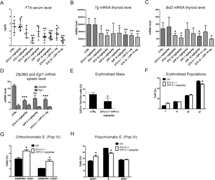
The in vivo phenotypic validation of thyroid effects was performed determining the free thyroxin (FT4) blood level in treated females. FT4 was reduced at PND 360 (Fig. 4A) but not at PND180 (Supplementary Fig. 2A). Furthermore, thyroglobulin (Tg) transcript was inhibited at high and medium doses of both pesticides at PND 360 (Fig. 4B) but not at earlier times (Supplementary Fig. 2B). A synergistic effect was observed when low-dose of both molecules was co-administered. Furthermore, we tested the thyroid expression level of the anti-apoptotic factor Bcl2 that plays a key role in thyrocyte survival26,27. Bcl2 transcript was inhibited by both compounds only at high doses and by their combination at PND 360 (Fig. 4C) but not at PND 180 (Supplementary Fig. 2C).
Figure 4. In vivo validation of the mechanisms of thyroid toxicity and hematopoietic dysfunction.
Thyroid toxicity was verified in females exposed to ETU or CPF (10, 1, 0.1 mg/kg/day) and their combination (10 mg/kg/day, 0.1 mg/kg/day) until PND 360. (A) FT4 serum level, each sign is a single mouse. Mean and standard deviation are reported. (B) Tg and (C) Bcl2 mRNA levels in the thyroid of the same females. (D) RT-qPCR analyses of Zfp36l2 and Egr1 transcripts in spleen. Data in panels B–D are reported as means ± SD of Gapdh-normalized mRNA levels. (E) Number of cells CD71 + TER119 + in bone marrow (F) Maturation pattern of erythroid precursors by FACS analysis using CD44 and TER119 as surface markers. The pro-erythroblasts (Pop I), basophilic erythroblasts (Pop II), polychromatic erythroblasts (Pop III) and orthochromatic erythroblasts (Pop IV) homogenous populations were gated. (G) Amount of Annexin V+-7AAD− cells (early apoptosis) and Annexin V+-7AAD+ (late apoptosis) in sorted Pop IV. (H) Cell cycle analysis of Pop III. Data are presented as means ± SD (n = 6). Haematologic dysfunctions reported in panels D-H were verified at CPF and ETU (0.1 mg/kg/day) and their mixture.
Overall the results show that the bioinformatics analysis of in vitro transcriptome can predict the in vivo cell response even if functional predictions are not fully confirmed by in vivo thyroid phenotyping. Thus, we have tried to validate the effects on thyrocytes growth/survival in vitro. We exposed the PCCl3 to different concentrations of CPF and ETU for 1 week to analyse their proliferation by MTT assay. Subsequently, we blocked their proliferation by thyroid stimulating hormone (TSH) deprivation for 3 days. Then, the cell growth was resumed by hormone addiction. The treatments with CPF and ETU resulted in the reduction in cell number, monitored by MTT, after 72 hrs of stimulation (Supplementary Fig. 3).
The presented data suggest that in vitro systems do not fully recapitulate the in vivo response because compensation mechanisms are active in vivo. Despite that, in vitro toxicogenomics predicts mechanism of thyroid toxicity confirmed in vivo if long exposures are conducted.
Toxicogenomics identifies unpredicted effects of the pesticides on hematopoiesis
The inhibition of Egr1, Hmga1 and Zfp36l2 transcripts was also related to hematological disease by IPA analysis, especially defects in erythropoiesis in CPF (Fig. 3A) and ETU (Fig. 3B).
Hematological dysfunctions have been reported in pesticide applicators28 and in treated mice29, although a mixture analysis has rarely been conducted. The synergistic effects of CPF and ETU on the hematological compartment were analysed in the sacrificed females exposed to 0.1 mg/kg/day of each compound administrated as single molecule or in combination. The hematologic parameters in the females within each treatment group were investigated and are reported in Table 3. Haemoglobin levels were significantly reduced in all exposed animals. A normocytic anaemia was present in ETU treated animals; whereas normocytic hypochromic anaemia was evident in CPF and co-exposed mice, also exhibiting a reduced reticulocyte count vs controls. All treated animals had a significant reduction in total white cell count vs controls, involving lymphocytes in all of them, monocytes in ETU and co-exposed mice and, finally, neutrophils only in co-treatment condition. A significant reduction in platelet count was present in all treated animals, reaching the lowest levels in co-treated mice. Overall, the exposure to ETU and CPF induced pancytopenia, with a more severe profile when molecules were co-administrated. It has been previously shown that the deletion of Zfp36l2 induced pancytopenia and impaired erythropoiesis30,31. Since we did not obtain enough sorted erythroid precursors for RT-PCR analysis, we validated its expression together with Egr1 transcript in the spleen that contained modest levels of erythropoiesis (data not shown). Zfp36l2 was significantly inhibited in the co-exposure conditions and in the single treatments by RT-qPCR (Fig. 4D). Although decreased, the inhibition of Egr1 transcript was not statistically significant (Fig. 4D).
Table 3. Hematological parameters and red cell indices at PND 360.
| Ctrl (n = 10) | ETU (0.1 mg/kg/day) (n = 4) | CPF (0.1 mg/kg/day) (n = 4) | ETU + CPF (0.1 mg/kg/day) (n = 10) | |
|---|---|---|---|---|
| Hct (%) | 47.2 ± 1.08 | 42.6 ± 2.2* | 41.0 ± 2.6* | 40.3 ± 1.6* |
| Hb (g/dl) | 15.6 ± 0.7 | 13.8 ± 1.2* | 13.6 ± 0.9* | 13.2 ± 0.4* |
| MCV (fl) | 53.4 ± 1.0 | 52.7 ± 1.1 | 52.3 ± 0.5 | 52.7 ± 0.7 |
| MCH (g/dl) | 16.7 ± 1.5 | 15.0 ± 0.7 | 14.8 ± 2.6* | 14.1 ± 0.5* |
| Retics (103 cells/μL) | 194 ± 48 | 135 ± 24 | 113 ± 15 | 73.9 ± 19* |
| WBC (cells/μL) | 1330 ± 324 | 450 ± 278* | 780 ± 451* | 862 ± 181* |
| N (cells/μL) | 378 ± 115 | 134 ± 69 | 273 ± 108 | 157 ± 27 |
| L (cells/μL) | 540 ± 279 | 240 ± 131 | 321 ± 158 | 433 ± 95 |
| M (cells/μL) | 16 ± 7.8 | 2.52 ± 0.9* | 14.8 ± 9.4 | 4.2 ± 0.1* |
| PLTs (103 cells/μL) | 847 ± 12 | 743 ± 5 | 731 ± 11 | 169 ± 9.0* |
Hct: hematocrit; Hb: hemoglobin; MCV: mean corpuscular volume; MCH: mean corpuscular hemoglobin; Retics: reticulocytes; MCVr: mean corpuscular volume reticulocytes; WBC: white blood cells; N: neutrophil; L: lymphocyte; M: monocyte; PLTs: platelets. Data are reported as means ± SD. In bold the ones reaching statistical significance.
The analyses of bone marrow evidenced a marked reduction of the erythroblast mass in co-exposed mice vs controls (Fig. 4E). The morphologic analysis of erythroid precursors revealed signs of dyserythropoiesis and erythrophagocytosis in co-treated mice (Supplementary Fig. 2D). The profile of erythroblast maturation showed an increased number of basophilic erythroblasts (Pop II) and a reduction in orthochromatic erythroblasts (Pop IV) (Fig. 4F), the latter displaying increased apoptosis (Fig. 4G). Thus, the co-exposure to CPF and ETU induced dyserythropoiesis and ineffective erythropoiesis characterized by a block in cell maturation and increased apoptosis in the late phase of erythropoiesis. The analyses of cell cycle for the different subpopulations revealed a general trend of cells to accumulate in G0/G1 phase with a reduction of cells in the S phase, reaching statistical significance only in polychromatic erythroblasts (Fig. 4H).
Collectively our results confirmed that in vitro toxicogenomics could predict cell type unrelated outcomes, their molecular mechanisms and markers in vivo.
Discussion
In a complex scenario involving cells physiologically adopting compensation processes, do the in vitro and in vivo toxicogenomics have the same predictive strength in revealing biomarkers or unpredicted outcomes of exposure valid in animals and, hopefully, in humans ? This answer is pivotal in transforming the Tox21c suggestion into a methodological approach in chemical testing.
Moving from our experience14,32,33, we planned an in vitro toxicogenomics experiment, in differentiated cells (immortalized thyrocytes), to identify gene signatures, mechanisms of toxicity or/and adverse outcomes potentially active in vivo of two pesticides, ETU and CPF. They were chosen for their known effects on thyroid physiology10,11, up to now not characterized at the level of thyroid gene expression.
As already reported, the first level of in vitro toxicogenomics data analysis, conducted in terms of “hits”, distinguished the compounds, their dose and mixtures. Some transcripts and mechanisms of toxicity common to CPF and ETU were identified and verified in vitro and in vivo. Thus, CPF could be molecularly classified as THDC as inhibiting Egr1, Hmga1 and Zfp36l2. These genes were not deregulated in the same cells exposed to BPA14. Therefore, the gene signature determined in vitro differentiated pesticides from BPA. The CPF and ETU signatures were not completely specific in vivo. In particular, some CPF DEGs were deregulated also in animals treated with ETU and vice versa. This happened because in the toxicogenomic analysis we applied a FC ≥ 2 as cut off excluding from the signature the genes deregulated less than 2 times, although statistically significant. Our data suggest that a better specificity could be reached through an all-encompassing approach in data analyses, using gene lists also including poorly regulated genes, that should be joined to the cross-platform integration of the omics data proposed for the classification of non-genotoxic compounds34. We considered a broad approach critical for organs subject to strong compensation processes, i.e. thyroid, because we expected that the full correspondence between doses and in vitro/in vivo effects could not be easy to obtain. Indeed in a previous study assessing the in vivo relevance of in vitro-obtained data for genotoxic carcinogens on hepatocytes, the correspondence between in vitro and in vivo results was obtained only for two out of three tested compounds35. Consequently, we conceived a multi-dose testing and a subsequent inclusive analysis of in vitro data as well as the in vivo evaluation of the effects of different doses and mixtures at different exposure times. Indeed, the in vitro results were confirmed in vivo only in some exposure conditions at longer exposure (PND 360), strengthening the need to overcome compensation processes not developed in vitro. We suggest that both aspects, multiple doses and long exposure windows, should be considered in planning experiments to investigate the causal link between high-throughput data produced in vitro and effects in vivo, not easily established till now. Notably, in vitro toxicogenomics can reduce the time required for a first evaluation of different classes of compounds, including THDCs.
We were surprised that no thyroid specific gene/pathway was revealed in vitro although we found reduced levels of FT4 in blood and of the thyroid specific Tg transcript. We noticed that IPA predicted several shared biofunctions, cell cycle, etc., as mechanism of toxicity for endocrine system, identified by Egr1 and Hmga1. These genes are known players of cellular proliferation in thyrocytes or other cell types23,24. We showed that both pesticides increased the apoptosis in vivo (inhibition of Bcl2 transcript) that could be responsible for the decrease in circulating FT4. These results are in agreement with previous studies showing a higher number of pyknotic nuclei and dead cells in the thyroid follicular epithelium of rodents exposed to ETU11 and CPF10 associated to low levels of serum thyroid hormones. Furthermore, our data provide candidate molecular mechanisms (deregulated expression of Bcl2, Zfp36l2, Egr1 and Hmga1) for the pesticides-induced hypothyroidism. Despite the fact that the weight of evidence (WoE) evaluation for chlorpyrifos evidenced no potential interaction with thyroid pathways at doses below the ones inhibiting the cholinesterase36, our findings are in agreement with epidemiological studies. Indeed, impairment of thyroid activity has been described among farmers using mancozeb (of which the ETU is the metabolite)37, among orgonophosphate pesticide formulators (CPF)38, and among men who were partners in subfertile couples and having high urine levels of 3,5,6-trichloro-2-pyridinol (TCPY), a metabolite of chlorpyrifos39. Our transcriptomic IPA analysis drove towards the characterization of the response to proliferation stimuli of the pesticides-treated thyrocytes in vitro, suggesting the molecular involvement of Egr1 and Hmga1 in the observed phenotype. This alteration of cell proliferation was revealed clearly after deprivation from TSH, mimicking an adverse condition that could be modelled in vivo by the co-exposure conditions. These data suggest that in vivo phenotypic validation of in vitro molecular data should be conducted considering that, in a real scenario, different insults can synergize lowering the adaptation response and producing, finally, phenotypic damages.
Our work evidenced the power of in vitro toxicogenomics in predicting pesticide effects not related to thyrocyte biology such as damages in erythropoiesis. Indeed the bioinformatic prediction obtained on thyroid cells resulted to be relevant when assessing the erythropoietic function in vivo. In our experimental settings the exposure to CPF or ETU and to their mixture reduced the number of mature white and red blood cells. Specifically, the reduction in circulating erythrocytes was due to a block in cell maturation and increased apoptosis in the late phase of erythropoiesis. Although in our experiment we only analysed the erythroblast populations, a recent publication demonstrated defective maturation of different marrow cell lineages due to a block in cell proliferation and increased apoptosis of hematopoietic precursor cells following exposure to a mixture of pesticides (including chlorpyrifos). The pancytopenia observed in the exposed mice was linked to defects of the bone marrow stromal cells29. In addition the exposure of hematopoietic stem cells from human cord blood to mancozeb reduced the clonogenic potential of erythroid and granulocyte-macrophage progenitors40. Here, we confirmed these effects in a realistic exposure scenario and we propose a molecular mechanism behind the observed phenotype consisting in the inhibition of Egr1, Hmga1 and Zfp36l2 transcripts. These genes are evidently part of complex and different gene networks regulating cell cycle/apoptosis in the thyroid as well as in the hematopoietic system.
In conclusion, the in vitro transcriptomic can support the identification of gene signatures, toxicity mechanisms and adverse effects also active in vivo. Although lacking in specificity, it shows a good predictive strength for mechanisms involving broad cell functions (i.e. cell cycle regulation, apoptosis) also when compensation processes are possible. Furthermore, the in vitro prediction of unexpected outcomes could allow a better planning for in vivo experiments, matching the 3 R rules.
Overall the presented data highlight the strength and limits of the Tox21c vision pointing to experimental approaches that can be undertaken to improve both in vitro toxicogenomics testing and in vivo phenotypic validation in order to establish cause-effect links. This is pivotal to transform the Tox21c suggestion into a real strategy in chemical testing.
Methods
Cell culture and treatment
PCCl3 were maintained in Coon’s modified F12 medium (EuroClone) supplemented with 5% newborn bovine serum (HyClone Laboratories) and six hormones16. Cells were exposed for 7 days to CPF (6 × 10⁻⁷ M, 6 × 10⁻⁸ M, 6 × 10⁻⁹ M, Greyhound Chromatography F2057) and ETU (6 × 10⁻⁸ M, 6 × 10⁻⁹ M, 6 × 10⁻¹⁰ M, 2-Imidazolidinethione, Sigma-Aldrich I504) (Table 1) and their combinations. Cell viability was analysed by MTT assay13. Briefly, 1 × 10³ cells were plated in 96-well plates and treated for 7 days. After 3-day long TSH deprivation, TSH was added and proliferation assessed at 24, 48 and 72 hrs.
Animals and treatments
Animal experiments were performed in accordance with the European Council Directive 86/609/EEC following the rules of the D.Lvo 116/92 (ID number 25–10) and procedures were approved by the Ethical committee named CESA (Committee for the Ethics of the Experimentations on Animals) of the Biogem Institute of Genetics Research “Gaetano Salvatore” (IRGS). Mice were kept under standard facility conditions and received water and standard diet (4RF21, Mucedola) “ad libitum”. CD1 dams (outbred strain, 8 mice/treatment group) were exposed, 7 days before the mating, to pesticides dosed at 10 mg/kg/day, 1 mg/kg/day, 0.1 mg/kg/day, and the combinations of higher and lower doses, administrating ETU by drinking water (59, 5.9 and 0.59 mg/L), and CPF by food at 44, 4.4 and 0.44 mg/kg (Mucedola) till the weaning. Therefore, the offspring were exposed through the mothers from gestational day 0 (GD0) till the weaning. Then, the offspring (10 females and males) were directly exposed. Animals were sacrificed at 6 months, 4 mice/group, and at 12 months, 6 mice/group, for blood and organs collection, by carbon dioxide inhalation.
RNA extraction, sequencing and RT-qPCR
RNA was prepared as already reported13. For RNA-seq samples were treated with DNasi (NEB), purified with RNasy Mini Kit (Qiagen) and controlled with Agilent 2100 bioanalyzer (Agilent Technologies). RNAs, 3 μg/each obtained from three biological replicates, were pooled to minimize sample-to-sample variations. 1 μg of each RNA pool was used for library preparation with TruSeq Stranded total RNA Sample Prep Kit (Illumina Inc.) according to the manufacturer’s instructions. Libraries were sequenced in triplicates (paired-end, 2 × 100 cycles) at a concentration of 8 pmol/L per lane on HiSeq2500 platform (Illumina Inc.).
The generated raw sequence files (.fastq files) underwent quality control analysis using FastQC (http://www.bioinformatics.babraham.ac.uk/projects/fastqc/) and will be available at the ArrayExpress database (http://www.ebi.ac.uk/arrayexpress.).
RT-qPCR and primer design were conducted as already described13 using QuantiTect Reverse Transcription Kit (QIAGEN), Power SYBR Green Master Mix (Applied Biosystems with Applied Biosystem 7900 Real-Time PCR System) and NCBI Primer Blast, respectively. See Supplementary Table S1 for primer sequences. The expression values were normalized on the relative expression of Gapdh.
RNA sequencing data and bioinformatics analyses
For RNA seq data analysis about 3 × 10⁷ high quality sequences reads pairs were produced for each sample. The rat UCSC rn5 genome was used as reference for read mapping, by TopHat aligner, further processed using Cufflinks package41. Normalized counts were represented by a box plot across the conditions to assess the quality of processed data (see Fig. S1). Selected reads were assembled in transcripts, compared among the samples and the unique final transcriptome generated. Differential expression was tested for the resulting 23762 genes comparing each pesticide treatment vs control. Significantly deregulated genes were selected by corrected p-value (FDR) ≤ 0.05 and absolute FC ≥ 2. Genes significantly deregulated at least in one dose were partitioned in three different sets through K-means clustering analysis using Jensen-Shannon (JS) distance as metric22.
Assembled rat genes without annotation were compared with the human genome to find their putative orthologous, using UCSC xenoRefGene table. The human gene with highest base identity was proposed to annotate the corresponding rat gene.
Functional enrichment analysis was performed using IPA (http://www.ingenuity.com). Over-represented biological and toxicological functions were found by Fisher exact test. Significant categories were selected by B–H corrected p-value ≤ 0.05.
FT4 determination, blood and bone marrow analysis
The serum FT4 level was determined with the FT4 ELISA kit (Diametra) following the manufacturer instructions.
Hematological parameters and red cell indices were evaluated with ADVIA 2120 Hematology System (Siemens Healthcare GmbH). Hematocrit and hemoglobin were manually determined42. Flow cytometric analysis of erythroid precursors from bone marrow was carried out using the CD44-TER-119 using the FACSCanto IITM flow cytometer (Becton Dickinson). Population II, III and IV were sorted from bone marrow, as previously reported43. Morphological analysis of sorted erythroblasts was performed on cytospin preparations stained with May Grunwald-Giemsa. Apoptosis in orthochromatic erythroblasts was carried out on CD44-Ter-119 gated cells (Annexin-V PE Apoptosis detection kit, eBioscience,). Cell cycle analysis was carried out by DAPI staining and data analyzed with FlowJo (Tree Star, Ashland) using Watson model.
Statistical and bioinformatics analyses
Statistical analyses were performed using Student’s t-test. Probability p-values below 0.05 were considered significant. *, **, ***Indicate p-value < 0.05, <0.01 and <0.001, respectively. Unless otherwise indicated, at least three independent experiments were considered for in vitro data and four animals for in vivo experiments. The results are expressed as the mean ± standard deviation.
Additional Information
How to cite this article: Porreca, I. et al. Pesticide toxicogenomics across scales: in vitro transcriptome predicts mechanisms and outcomes of exposure in vivo. Sci. Rep. 6, 38131; doi: 10.1038/srep38131 (2016).
Publisher's note: Springer Nature remains neutral with regard to jurisdictional claims in published maps and institutional affiliations.
Supplementary Material
Acknowledgments
This work was supported by: the Italian Workers’ Compensation Authority (grant. n. 12010), the Italian Ministry of Health (grant n. RF 2010-2309484), Sensor Regione Campania (grant n. 23), PRIN (201228PNX83), FUR_UNIVR and AIRC (grant n. IG-17426). We thank IRGS (Biogem) for support and the mouse facility for the technical support. We wish to thank R. De Felice for linguistic editing of the manuscript.
Footnotes
Author Contributions M.M., M.D.F. and C.A. developed the rationale of the study; M.M., F.D.A., I.P., L.D.F. and C.A. wrote the manuscript, which was commented on by all authors; I.P. performed the majority of the cell culture and mouse experiments with additional experimental contributions of M.S. and F.R.; M.R., R.T. and A.W. performed the RNA seq; F.D.A. and M.C. performed the RNAseq data and bioinformatic analysis; L.D.F., A.M., A.Z. and A.I. performed the hematopoiesis experiments; C.A. supervised all the experiments.
References
- Ellinger-Ziegelbauer H., Aubrecht J., Kleinjans J. C. & Ahr H. J. Application of toxicogenomics to study mechanisms of genotoxicity and carcinogenicity. Toxicol Lett 186, 36–44 (2009). [DOI] [PubMed] [Google Scholar]
- Research C. N. Toxicity Testing in the 21st Century: A Vision and a Strategy. National Academy Press (2007). [Google Scholar]
- Marx-Stoelting P. et al. Application of omics data in regulatory toxicology: report of an international BfR expert workshop. Arch Toxicol 89, 2177–2184 (2015). [DOI] [PubMed] [Google Scholar]
- Rouquie D. et al. Contribution of new technologies to characterization and prediction of adverse effects. Crit Rev Toxicol 45, 172–183 (2015). [DOI] [PubMed] [Google Scholar]
- Vanderpump M. P. The epidemiology of thyroid disease. Br Med Bull 99, 39–51 (2011). [DOI] [PubMed] [Google Scholar]
- Gutleb A. C., Cambier S. & Serchi T. Impact of Endocrine Disruptors on the Thyroid Hormone System. Horm Res Paediatr (2016). [DOI] [PubMed] [Google Scholar]
- Zoeller T. R. Environmental chemicals targeting thyroid. Hormones (Athens) 9, 28–40 (2010). [DOI] [PubMed] [Google Scholar]
- Goldner W. S. et al. Hypothyroidism and pesticide use among male private pesticide applicators in the agricultural health study. J Occup Environ Med 55, 1171–1178 (2013). [DOI] [PMC free article] [PubMed] [Google Scholar]
- Toft G., Flyvbjerg A. & Bonde J. P. Thyroid function in Danish greenhouse workers. Environ Health 5, 32 (2006). [DOI] [PMC free article] [PubMed] [Google Scholar]
- De Angelis S. et al. Developmental exposure to chlorpyrifos induces alterations in thyroid and thyroid hormone levels without other toxicity signs in CD-1 mice. Toxicol Sci 108, 311–319 (2009). [DOI] [PubMed] [Google Scholar]
- Maranghi F. et al. Reproductive toxicity and thyroid effects in Sprague Dawley rats exposed to low doses of ethylenethiourea. Food Chem Toxicol 59, 261–271 (2013). [DOI] [PubMed] [Google Scholar]
- Crofton K. M. Thyroid disrupting chemicals: mechanisms and mixtures. Int J Androl 31, 209–223 (2008). [DOI] [PubMed] [Google Scholar]
- Gentilcore D. et al. Bisphenol A interferes with thyroid specific gene expression. Toxicology 304, 21–31 (2013). [DOI] [PubMed] [Google Scholar]
- Porreca I. et al. “Stockpile” of Slight Transcriptomic Changes Determines the Indirect Genotoxicity of Low-Dose BPA in Thyroid Cells. PLoS One 11, e0151618 (2016). [DOI] [PMC free article] [PubMed] [Google Scholar]
- Axelstad M. et al. Exposure to the widely used fungicide mancozeb causes thyroid hormone disruption in rat dams but no behavioral effects in the offspring. Toxicol Sci 120, 439–446 (2011). [DOI] [PubMed] [Google Scholar]
- Kimura T., Van Keymeulen A., Golstein J., Fusco A., Dumont J. E. & Roger P. P. Regulation of thyroid cell proliferation by TSH and other factors: a critical evaluation of in vitro models. Endocr Rev 22, 631–656 (2001). [DOI] [PubMed] [Google Scholar]
- Jomaa B. et al. In vitro pituitary and thyroid cell proliferation assays and their relevance as alternatives to animal testing. ALTEX 30, 293–307 (2013). [DOI] [PubMed] [Google Scholar]
- Howell Iii G. E., Mulligan C., Young D. & Kondakala S. Exposure to chlorpyrifos increases neutral lipid accumulation with accompanying increased de novo lipogenesis and decreased triglyceride secretion in McArdle-RH7777 hepatoma cells. Toxicol In Vitro 32, 181–189 (2016). [DOI] [PubMed] [Google Scholar]
- CDC. Fourth National Report on Human Exposure to Environmental Chemicals (ed^(eds) (2009). [PubMed]
- CDC. Fourth National Report on Human Exposure to Environmental Chemicals (ed^(eds) (2015). [PubMed]
- Huen K. et al. Organophosphate pesticide levels in blood and urine of women and newborns living in an agricultural community. Environ Res 117, 8–16 (2012). [DOI] [PMC free article] [PubMed] [Google Scholar]
- Reynolds A. P., Richards G., de la Iglesia B. & Rayward-Smith V. J. Clustering Rules: A Comparison of Partitioning and Hierarchical Clustering Algorithms. Journal of Mathematical Modelling and Algorithms 5, 475–504 (2006). [Google Scholar]
- Pritchard M. T., Malinak R. N. & Nagy L. E. Early growth response (EGR)-1 is required for timely cell-cycle entry and progression in hepatocytes after acute carbon tetrachloride exposure in mice. Am J Physiol Gastrointest Liver Physiol 300, G1124–1131 (2011). [DOI] [PMC free article] [PubMed] [Google Scholar]
- Zhong J. et al. The association between S100A13 and HMGA1 in the modulation of thyroid cancer proliferation and invasion. J Transl Med 14, 80 (2016). [DOI] [PMC free article] [PubMed] [Google Scholar]
- Fagman H. et al. Gene expression profiling at early organogenesis reveals both common and diverse mechanisms in foregut patterning. Dev Biol 359, 163–175 (2011). [DOI] [PMC free article] [PubMed] [Google Scholar]
- Marotta P. et al. The paired box transcription factor Pax8 is essential for function and survival of adult thyroid cells. Mol Cell Endocrinol 396, 26–36 (2014). [DOI] [PubMed] [Google Scholar]
- Porreca I., De Felice E., Fagman H., Di Lauro R. & Sordino P. Zebrafish bcl2l is a survival factor in thyroid development. Dev Biol 366, 142–152 (2012). [DOI] [PubMed] [Google Scholar]
- Fareed M., Pathak M. K., Bihari V., Kamal R., Srivastava A. K. & Kesavachandran C. N. Adverse respiratory health and hematological alterations among agricultural workers occupationally exposed to organophosphate pesticides: a cross-sectional study in North India. PLoS One 8, e69755 (2013). [DOI] [PMC free article] [PubMed] [Google Scholar]
- Chatterjee S. et al. Pesticide induced alterations in marrow physiology and depletion of stem and stromal progenitor population: an experimental model to study the toxic effects of pesticide. Environ Toxicol 29, 84–97 (2014). [DOI] [PubMed] [Google Scholar]
- Stumpo D. J. et al. Targeted disruption of Zfp36l2, encoding a CCCH tandem zinc finger RNA-binding protein, results in defective hematopoiesis. Blood 114, 2401–2410 (2009). [DOI] [PMC free article] [PubMed] [Google Scholar]
- Zhang L. et al. ZFP36L2 is required for self-renewal of early burst-forming unit erythroid progenitors. Nature 499, 92–96 (2013). [DOI] [PMC free article] [PubMed] [Google Scholar]
- Carchia E. et al. Evaluation of low doses BPA-induced perturbation of glycemia by toxicogenomics points to a primary role of pancreatic islets and to the mechanism of toxicity. Cell Death Dis 6, e1959 (2015). [DOI] [PMC free article] [PubMed] [Google Scholar]
- Porreca I. et al. Cross-species toxicogenomic analyses and phenotypic anchoring in response to groundwater low-level pollution. BMC Genomics 15, 1067 (2014). [DOI] [PMC free article] [PubMed] [Google Scholar]
- Romer M. et al. Cross-platform toxicogenomics for the prediction of non-genotoxic hepatocarcinogenesis in rat. PLoS One 9, e97640 (2014). [DOI] [PMC free article] [PubMed] [Google Scholar]
- Doktorova T. Y. et al. Comparison of genotoxicant-modified transcriptomic responses in conventional and epigenetically stabilized primary rat hepatocytes with in vivo rat liver data. Arch Toxicol 86, 1703–1715 (2012). [DOI] [PubMed] [Google Scholar]
- Juberg D. R. et al. Chlorpyrifos: weight of evidence evaluation of potential interaction with the estrogen, androgen, or thyroid pathways. Regul Toxicol Pharmacol 66, 249–263 (2013). [DOI] [PubMed] [Google Scholar]
- Goldner W. S., Sandler D. P., Yu F., Hoppin J. A., Kamel F. & Levan T. D. Pesticide use and thyroid disease among women in the Agricultural Health Study. Am J Epidemiol 171, 455–464 (2010). [DOI] [PMC free article] [PubMed] [Google Scholar]
- Zaidi S. S., Bhatnagar V. K., Gandhi S. J., Shah M. P., Kulkarni P. K. & Saiyed H. N. Assessment of thyroid function in pesticide formulators. Hum Exp Toxicol 19, 497–501 (2000). [DOI] [PubMed] [Google Scholar]
- Meeker J. D., Barr D. B. & Hauser R. Thyroid hormones in relation to urinary metabolites of non-persistent insecticides in men of reproductive age. Reprod Toxicol 22, 437–442 (2006). [DOI] [PubMed] [Google Scholar]
- Mandarapu R. & Prakhya B. M. In vitro myelotoxic effects of cypermethrin and mancozeb on human hematopoietic progenitor cells. J Immunotoxicol 12, 48–55 (2015). [DOI] [PubMed] [Google Scholar]
- Trapnell C. et al. Differential gene and transcript expression analysis of RNA-seq experiments with TopHat and Cufflinks. Nat Protoc 7, 562–578 (2012). [DOI] [PMC free article] [PubMed] [Google Scholar]
- Franco S. S. et al. Resveratrol accelerates erythroid maturation by activation of FoxO3 and ameliorates anemia in beta-thalassemic mice. Haematologica 99, 267–275 (2014). [DOI] [PMC free article] [PubMed] [Google Scholar]
- Matte A. et al. The Interplay Between Peroxiredoxin-2 and Nuclear Factor-Erythroid 2 Is Important in Limiting Oxidative Mediated Dysfunction in beta-Thalassemic Erythropoiesis. Antioxid Redox Signal 23, 1284–1297 (2015). [DOI] [PMC free article] [PubMed] [Google Scholar]
Associated Data
This section collects any data citations, data availability statements, or supplementary materials included in this article.


