Abstract
Mutations in the X-linked gene methyl-CpG-binding protein 2 (MECP2) are the principal cause of Rett syndrome, a progressive neurodevelopmental disorder afflicting 1 in 10,000 to 15,000 females. Studies using hemizygous Mecp2 mouse models have revealed disruptions to some aspects of their lipid metabolism including a partial suppression of cholesterol synthesis in the brains of mature Mecp2 mutants. The present studies investigated whether this suppression is evident from early neonatal life, or becomes manifest at a later stage of development. We measured the rate of cholesterol synthesis, in vivo, in the brains of male Mecp2−/y and their Mecp2+/y littermates at 7, 14, 21, 28, 42 and 56 days of age. Brain weight was consistently lower in the Mecp2−/y mice than in their Mecp2+/y controls except at 7 days of age. In the 7- and 14-day-old mice there was no genotypic difference in the rate of brain cholesterol synthesis but, from 21 days and later, it was always marginally lower in the Mecp2−/y mice than in age-matched Mecp2+/y littermates. At no age was a genotypic difference detected in either the rate of fatty acid synthesis or cholesterol concentration in the brain. Cholesterol synthesis rates in the liver and lungs of 56-day-old Mecp2−/y mice were normal. The onset of lower rates of brain cholesterol synthesis at about the time closure of the blood brain barrier purportedly occurs might signify a disruption to mechanism(s) that dictate intracellular levels of cholesterol metabolites including oxysterols known to exert a regulatory influence on the cholesterol biosynthetic pathway.
Keywords: Brain weight, Desmosterol, Fatty acid synthesis, Ontogeny, Rett syndrome
1. Introduction
The bulk of cholesterol in the central nervous system (CNS) is present in myelin, having been synthesized locally in the earlier stages of development (Jurevics and Morell, 1995; Snipes and Suter, 1997). Disruption of the cholesterol biosynthetic pathway in the brain during development is the cause of several rare diseases (Porter and Herman, 2011). These include the Smith-Lemli-Optiz syndrome which results from a block in the pathway at the step in which 7-dehydrocholesterol is converted to cholesterol (Tint et al., 1994). In another part of the biosynthetic pathway, mutations in the gene SC4mol, which encodes a sterol-C4 methyl oxidase-like (also known as methylsterol monooxygenase), result in multiple disorders including microcephaly and delayed development (He et al., 2011). Aberrations in other pathways involved in regulating cholesterol homeostasis in the CNS can be as devastating as those that disrupt the biosynthetic pathway. Such is the case in Niemann-Pick type C disease where mutations in the lysosomal transporters NPC1 and NPC2 prevent the movement of unesterified cholesterol from the late endosomal/lysosomal compartment into the cytosol in every cell (Vanier, 2010). This results in a pronounced increase in the tissue content of unesterified cholesterol and other classes of lipids in all organs, leading to debilitation and death, most frequently from neurodegeneration but also from pulmonary dysfunction or liver failure.
In more common neurodegenerative disorders, especially Alzheimer’s disease, there is a growing body of evidence that various aspects of cholesterol homeostasis are awry (Bryleva et al., 2010; Orth and Bellosta, 2012; Vance, 2012; Martin et al., 2014). This is now also the case with Rett syndrome, a pervasive neurodevelopmental disorder caused in most cases by loss-of-function mutations in the X-linked gene, methyl-CpG-binding protein 2 (MECP2) (Lombardi et al., 2015; Lyst and Bird, 2015). Evidence for a disruption of some aspects of cholesterol metabolism in Mecp2 deficiency, in particular the biosynthetic pathway, has come mainly from studies in two mouse models for this disease. Specifically, these are widely referred to as the Bird (Guy et al., 2001) and Jaenisch (Chen et al., 2001) models, in recognition of the investigators whose laboratories developed them. In 2008, a gene expression analysis by microarray using whole brains from Mecp2 mutant mice of the Bird strain at 6–10 weeks of age revealed significant reductions in the mRNA expression levels for two of the genes that encode proteins involved in cholesterol biosynthesis, squalene monooxygenase (previously epoxidase) (Sqle), and sterol-C4-methyl-oxidase-like (SC4mol) (Urdinguio et al., 2008). At about the same time, another lab similarly reported a lower mRNA level in the cerebellum for Cyp46a1, which encodes the enzyme that converts cholesterol to 24(S)-hydroxycholesterol (24S-OHC) exclusively in the brain (Ben-Shachar et al., 2009). However, as noted in this publication, the loss of Mecp2 function results in reduced expression levels of hundreds of genes in the brain (Ben-Shachar et al., 2009). Thus, the physiological significance of the reductions in mRNA levels for two enzymes in the cholesterol biosynthetic pathway was not clear, especially as another study at that time involving a meticulous analysis of brain lipid composition in old Mecp2 mutant mice showed that the concentration of cholesterol across multiple regions of their CNS was the same as in their Mecp2+/y controls (Seyfried et al., 2009). More compelling evidence for a disruption of cholesterol metabolism in Rett syndrome came from detailed studies by the Justice laboratory using both the Jaeneisch and Bird mouse models, together with an array of techniques including a mutagenesis suppressor screen (Buchovecky et al., 2013). These experiments showed that cholesterol biosynthesis in Mecp2 mutant mice was disrupted at the squalene monoxygenase step, and more importantly, that overcoming this defect alleviated many symptoms and increased longevity. One particular experiment in these studies involving the measurement in vivo of whole brain cholesterol synthesis showed that it was suppressed about 20% in the Mecp2−/y mice at 56 days of age.
The primary objective of the present studies was to establish whether the lower rate of brain cholesterol synthesis in Mecp2 mutant mice is congenital, and if not, at what stage of development this becomes evident.
2. Results
2.1 Mecp2−/y mice exhibited prototypical shortened lifespan and reduced brain weight
As shown in Fig. 1A, the lifespan of the Mecp2−/y mice ranged from 53 to 82 days (n=12), with the median survival time being 59 days. This agrees closely with the findings of Guy and her colleagues (Guy et al., 2001), and thereby confirms that Mecp2−/y mice of the Bird strain have a slightly shorter lifespan than those of the Jaenisch line (Chang et al., 2006). The data presented in the lower panels of Fig. 1 were generated from the metabolic studies. At most age points, the body weights of the Mecp2−/y mice were less than those of their Mecp2+/y controls (Fig. 1B). As illustrated in Fig. 1C, from the second week after birth, the weight of the brain in the Mecp2−/y mice was consistently lower than in their Mecp2+/y controls. This is in agreement with the findings other investigators for the both Bird and Jaenisch models (Chen et al., 2001; Guy et al., 2001; Belichenko et al., 2008; Nguyen et al., 2012) and also replicates the reduced brain mass described in Rett syndrome subjects over a wide age range (Armstrong, 2005). There was a steady age-related decline in the brain-to-body weight ratio which, irrespective of genotype, fell from about 5.4% in the 7-day old mice to 1.9% in their 56-day old counterparts (data not shown).
Fig. 1.
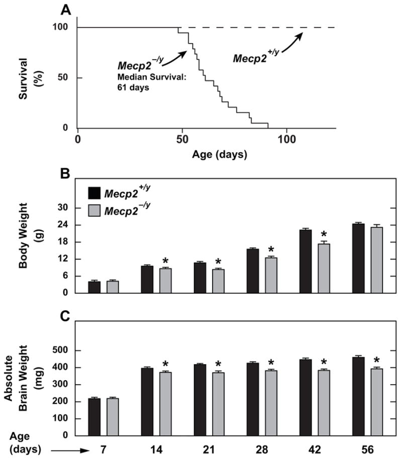
Longevity of Mecp2−/y mice, and body and whole brain weights in Mecp2−/y mice and their Mecp2+/y controls from 7 to 56 days of age. A. The lifespan data are for 19 Mecp2−/y mice and 15 Mecp2+/y controls. B and C. Values are the mean ± SEM of data from a minimum of 6 mice per group except at 14d Mecp2+/y and 21d Mecp2−/y where n=4 in both cases, and 42d Mecp2+/y where n=5. *Significantly different from the value for the age-matched Mecp2+/y controls (p < 0.05).
2.2 Suppression of brain cholesterol synthesis in Mecp2−/y mice was not evident until the third week after birth
The data presented in Fig. 2 reveal a number of key findings regarding sterol synthesis and lipogenesis in the developing brain of Mecp2−/y mice and their Mecp2+/y controls. The most striking of these is that suppression of brain cholesterol synthesis in the mutants did not become evident until the third week after birth (Fig. 2A). No genotypic difference in the rate of cholesterol synthesis was discernible at 7 or 14 days postpartum, but clearly by 21 days, the rate was lower in the mutants, and this remained the case at 28, 42 and 56 days of age. In mice of both genotypes, the rate of cholesterol synthesis declined markedly after 21 days, in keeping with what has been well documented for the developing brain in several species (Jurevics and Morell, 1995; Quan et al., 2003). It is particularly noteworthy that, against this backdrop of a dramatic age-related decline in synthesis, the lower rate of sterol synthesis in the mutant vs. wild-type mice beyond 14 days was still clearly discernible. It should be also noted here that the lower rate of sterol synthesis seen in the mutants at 56 days is fully consistent with what Mecp2−/y males of the Jaenisch model, on a mixed strain background, exhibited at this age (Buchovecky et al., 2013).
Fig. 2.
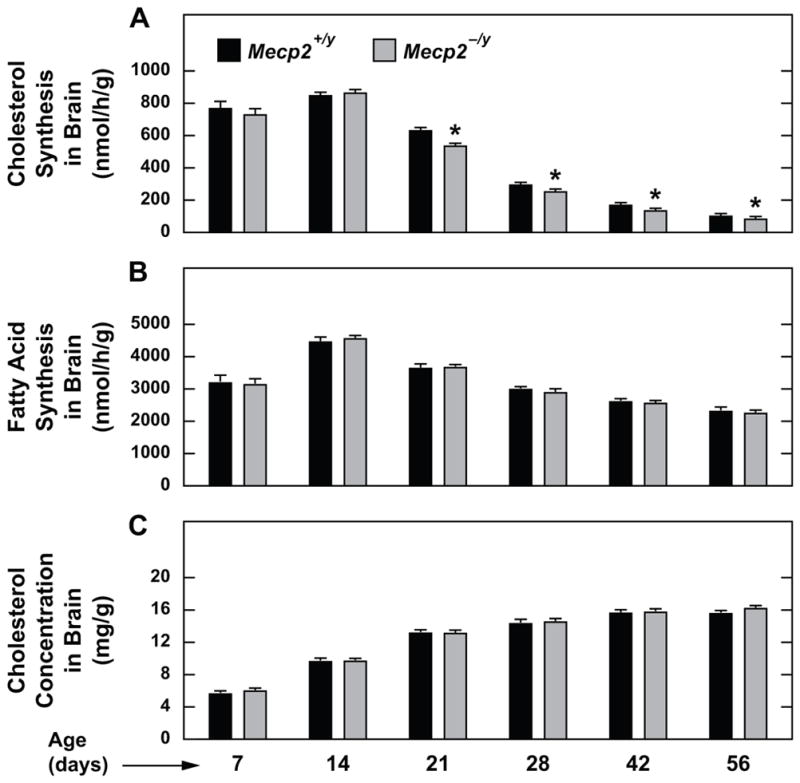
Rates of cholesterol and fatty acid synthesis, and cholesterol concentrations, in the whole brains of Mecp2−/y mice and their Mecp2+/y controls from 7 to 56 days of age. Cholesterol and fatty acid synthesis rates were measured in vivo using [3H]water as described in Experimental procedures. Extracts of the same brains were used to determine the total cholesterol concentration by gas chromatography. Values are the mean ± SEM of data from a minimum of 6 mice per group except at 14d Mecp2+/y, 21d Mecp2−/y, and 42d Mecp2+/y where n=4 in each case. *Significantly different from the value for the age-matched Mecp2+/y controls (p < 0.05).
The next important finding was that, at no age, did a genotypic difference in the rate of fatty acid synthesis in these same brains become manifest (Fig. 2B). Furthermore, when compared to the data in Fig. 2A, it is seen first, that the rate of fatty acid synthesis at any age was much higher than the rate of cholesterol synthesis, and second, that the age-related decline in lipogenesis was much more gradual than was the case for sterol synthesis. This dissociation partly reflects the extraordinarily high demand for newly generated cholesterol for use in myelin formation in the early stages of development (Dietschy and Turley, 2004). Once this process begins to reach completion, the requirement for locally synthesized cholesterol falls dramatically (Jurevics and Morell, 1995; Quan et al., 2003). Despite the lower rate of cholesterol synthesis in the brains of Mecp2−/y mice after 14 days, the brain cholesterol concentration did not show a genotypic difference at any age (Fig. 2C).
Another observation from the studies described in Fig. 2 concerns the content of desmosterol, an immediate precursor of cholesterol in its biosynthetic pathway, in the brains of mice at 7 and 14 days of age. An important branch point in this pathway occurs at the lanosterol step. Ultimately, lanosterol can be converted to cholesterol via desmosterol in the Bloch pathway, or via 7-dehydrocholesterol in the Kandutsch-Russell (Sharpe and Brown, 2013). It was shown decades ago that in early development, the content of desmosterol in the CNS is quite pronounced but then rapidly dissipates (Paoletti et al., 1965). In the mouse brain at 7 days after birth, desmosterol accounts for about ~20–25% of the total sterol (Paoletti et al., 1965; Quan et al., 2003). At 14 days this figure is only ~4%, and by adulthood desmosterol accounts for less than ~0.2%. As shown in Fig. 3, there was no genotypic difference in the brain desmosterol concentration at 7 or 14 days, just as there was no difference in the rate of brain cholesterol synthesis at these two ages (Fig. 2A). Our conventional gas chromatographic method was not sufficiently sensitive to determine desmosterol levels beyond 14 days of age. This requires the combined use of gas chromatography and mass spectrometry. An earlier publication utilizing this technique in Mecp2−/y mice and their wildtype controls at 56 and 70 days showed significantly lower brain desmosterol concentrations in the mutants. That finding was consistent with the lower rates of brain cholesterol synthesis in older Mecp2 mutant mice (Fig. 2A) (Buchovecky et al., 2013). In the present studies, the diminished concentration of desmosterol in the brains of the 14- vs 7-day-old mice of both genotypes (Fig. 3) in the face of comparable high rates of cholesterol synthesis at these two age points (Fig. 2A), may reflect either an increased rate of conversion of desmosterol to cholesterol at the later age, or/and an increased rate of cholesterol production via the Kandutsch-Russell pathway in the 14- vs 7-day-old mice. The latter scenario appears to exist in mice with genetically altered levels of Cyp27A1 expression (Ali et al., 2013).
Fig. 3.
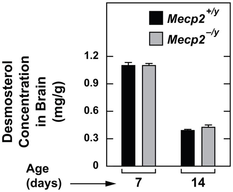
Desmosterol concentrations in the whole brains of Mecp2−/y mice and their Mecp2+/y controls at 7 and 14 days of age, respectively. Values are the mean ± SEM of data from 6 mice of each genotype at 7 days, and 4 Mecp2+/y and 7 Mecp2−/y mice at 14 days.
The earlier studies in 56-day-old Jaenisch mice showed that, unlike the brain, other organs including the liver did not manifest a lower rate of cholesterol synthesis (Buchovecky et al., 2013). Therefore, in the current study with Bird strain mice, sterol and fatty acid synthesis were measured in the liver and lungs of 56-day-old mutants and wildtypes. The lungs were of particular interest because pulmonary dysfunction is a hallmark feature of Rett syndrome, and also the expression level of Mecp2 protein in lung tissue is very high, as it is in the mature CNS (Shahbazian et al., 2002). The data for the liver and lungs are presented in Fig. 4, along with those for the brain in these same mice. The brain data are from Fig. 1 and 2, and in the case of cholesterol synthesis, are presented on an adjusted scale so as to accent the lower rate of sterol synthesis in the mutants (Fig. 4D). In the case of the liver (Fig. 4E) and lungs (Fig. 4F), cholesterol synthesis rates did not show a genotypic difference. The fatty acid synthesis rates in the brain (Fig. 4G) and lungs (Fig. 4I) also did not vary with genotype, but in the liver, the rate was clearly elevated in the mutants (Fig. 4H). In 56-day-old mice, irrespective of their Mecp2 genotype, the total cholesterol concentration in the liver and lungs averaged 2.1 and 4.7 mg/g, respectively (data not shown). Plasma total cholesterol levels were determined in 43-day-old mice. The concentrations in the Mecp2−/y mice (98.9 ± 0.8 mg/dl, n=9) were not significantly different than in their Mecp2+/y controls (89.2 ± 11.2 mg/dl, n=8). A marginal increase in plasma cholesterol concentrations was reported for 49-day-old Mecp2−/y mice (Jaenisch allele) on a mixed strain background (129/SvEv:C57BL/6:BALB/c) (Park et al., 2014). In 70-day-old Mecp2−/y mice (Bird allele) on a 129S6/SvEv background, plasma total cholesterols were about 50% higher than in their wildtype controls (Buchovecky et al., 2013).
Fig. 4.
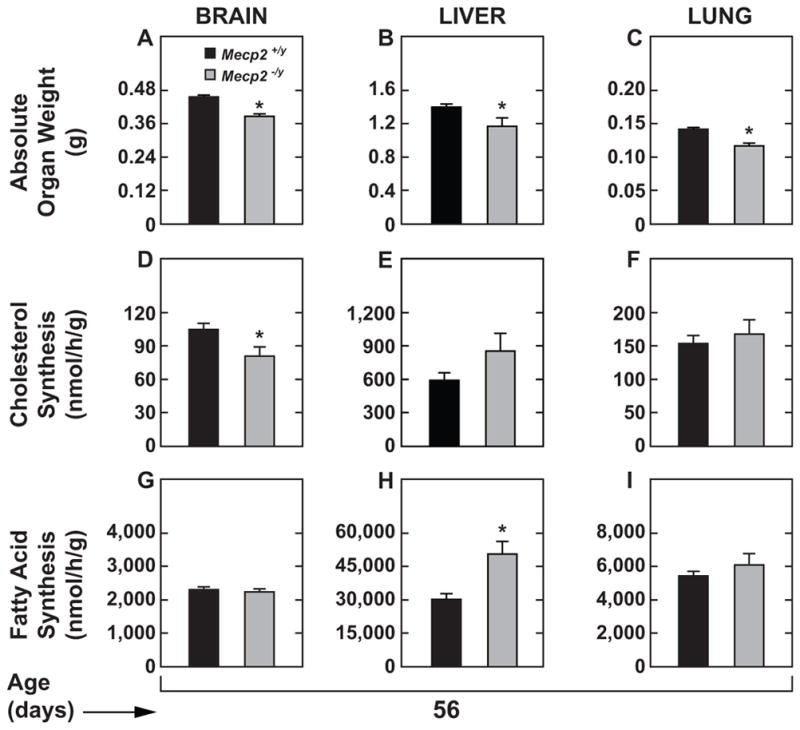
Brain, liver and lung weights, and rates of cholesterol and fatty acid synthesis in these organs in Mecp2−/y mice and their Mecp2+/y controls at 56 days of age. These data are from the same sets of 56-day-old mice described in Figs. 1B and 1C, and 2. Values are the mean ± SEM of data from 10 Mecp2+/y controls and 7 Mecp2−/y mice. *Significantly different from the value for their Mecp2+/y controls (p < 0.05).
2.3 Mecp2−/y mice showed age-related changes in the brain mRNA expression levels for several enzymes in the cholesterol biosynthetic pathway but not for either Apoe or Cyp46a1
At 14 days of age, the brain cholesterol synthesis rate (nmol/h/g) in the Mecp2−/y mice (862 ± 19) was not different from that in their Mecp2+/y littermates (846 ± 18) (Fig. 2A). However, at 42 days, the rate in the Mecp2−/y mice (133 ± 5) was 21% lower than in their Mecp2+/y littermates (168 ± 2). A comparable genotypic difference was also found at 56 days (81 ± 8 in the Mecp2−/y mice vs. 105 ± 5 in the Mecp2+/y controls). In separate groups of mice at these same three age points, the brain mRNA expression levels of five genes involved in regulating cholesterol homeostasis in the CNS, including three within the biosynthetic pathway, were determined. As shown in Fig. 5A, the absence of Mecp2 in the mutants at each age was confirmed. For Hmgcr (Fig. 5B), there were clearly lower levels of mRNA expression in the 43- and 56-day-old mice, but not in their 14-day-old counterparts. The mRNA level for Sqle (Fig. 5C) was lower in the mutants only at 43 days. For SC4mol (Fig. 5D), the mRNA level was lower in the Mecp2−/y mice at 43 and 56 days, but this achieved statistical significance only at the latter age. While the focus was on genes regulating cholesterol synthesis and degradation, the mRNA expression level of apolipoprotein E was also included because of the major role that it plays in the maintenance of cholesterol homeostasis in the CNS (Vitali et al., 2014; Zhang and Liu, 2015). The mRNA expression level for both Apoe (Fig. 5E), and Cyp46a1 (Fig. 5F), showed no genotypic difference at any of the three ages.
Fig. 5.
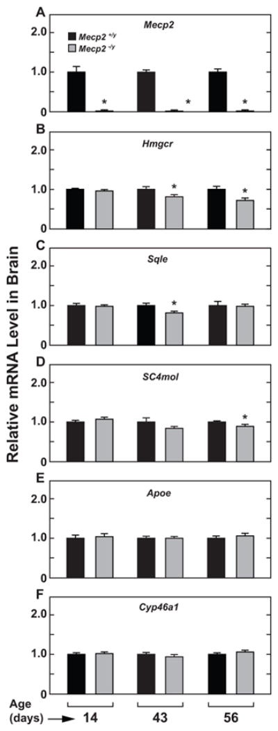
Relative levels of expression of mRNA for several genes involved in cholesterol metabolism in the brains of Mecp2−/y mice and their Mecp2+/y controls at 14, 43 and 56 days of age, respectively. These analyses were carried out using whole brains as described in Experimental procedures. The primer sequences for each of these genes are given in Table 1. Mecp2, methyl CpG-binding protein 2; Hmgcr, 3-hydroxy-3-methylglutaryl-CoA reductase; SC4mol, sterol-C4-methyl-oxidase-like (also known as methylsterol monooxygenase with the abbreviation msmol); Sqle, squalene monooxygenase; Apoe, apolipoprotein E; Cyp46a1, cholesterol 24-hydroxylase. Values are the mean ± SEM of data from 3 Mecp2+/y and 5 Mecp2−/y mice at 14 days, 4 Mecp2+/y and 5 Mecp2−/y mice at 43 days, and 6 mice of each genotype at 56 days. *Significantly different from the value for the age-matched Mecp2+/y controls (p < 0.05).
3. Discussion
Together, the present data confirm and extend earlier published findings that specific aspects of cholesterol metabolism in the CNS, particularly the biosynthetic pathway, are disrupted in mouse models with loss-of-function mutations in Mecp2 (Buchovecky et al., 2013). A similar conclusion was reached in studies with primary cultured fibroblasts from Rett syndrome patients (Segatto et al., 2014). In evaluating the pathophysiological significance of the findings presented here, a number of points pertaining to technical aspects of the studies warrant comment. The first relates to the challenge of being able to detect genotypic differences in absolute rates of whole brain cholesterol synthesis against a background of pronounced ontogenic changes in this parameter. In the case of the mouse, the changes in cholesterol synthesis rates in the CNS from early neonatal life to adulthood seen here (Fig. 2A), and in earlier studies (Quan et al., 2003), far exceed the magnitude of the difference in brain cholesterol synthesis rates between Mecp2−/y and Mecp2+/y mice. The use of [3H]water to measure the absolute cholesterol biosynthesis rate in the whole brain in vivo provided the best strategy for answering the key question of whether Mecp2−/y mice have lower rates of brain sterol synthesis from birth, or manifest this feature at a later stage of development. A limitation of this approach is that it did not yield information about whether the lower rate of brain synthesis detected in the mutants after 14 days of age was confined to specific regions or cell types in the CNS. We have previously utilized this method to quantitate broad regional differences in brain cholesterol synthesis in other mouse models (Xie et al., 2000; Xie et al., 2003), but when the focus is on specific subcortical structures that each represent only a small proportion of whole brain mass, exceptionally large quantities of [3H]water are needed to obtain measurable rates of cholesterol synthesis. The deuterated water technique could be an alternative for such measurements (Saeed et al., 2014b). It would probably be even more challenging to investigate these genotypic differences in brain cholesterol synthesis at the level of individual cell types partly because of prevailing uncertainty about how the quantitative importance of specific cells such as neurons as sites for cholesterol synthesis may shift with age (Pfrieger, 2003; Björkhem and Meaney, 2004; Orth and Bellosta, 2012). Now that a Mecp2-knockout rat model has been described (Wu et al., 2016), the larger brain mass in this species might be an advantage for studying genotypic differences in the rate of cholesterol synthesis in different regions of the CNS.
Another advantage of the technique used here was that it allowed measurement of the rate of fatty acid synthesis in the same brains where cholesterol synthesis was determined. The biosynthetic pathways for cholesterol and fatty acids share a common initiating step, and in some instances are coordinately regulated, at least in the liver (Gibbons, 2003). The finding that at no stage of development was a genotypic difference in brain fatty acid synthesis detected points to a selective, albeit modest, downregulation of sterol biosynthesis, starting in the third postnatal week. Up to that point, not only were the rates of cholesterol synthesis comparable in the mutants and matching wildtypes, but also there was no discernible genotypic difference in the relative mRNA expression levels in brain of three key enzymes in the cholesterol biosynthetic pathway. By 43-days of age, there was however a clear trend towards lower levels of mRNA for these enzymes (Fig. 5). Together then, these data strongly indicate that beginning in the third postnatal week there is an adaptive suppression of brain sterol synthesis in the Mecp2−/y mice that persists through all subsequent stages of development. There was not a detectable impact of the lower sterol synthesis rate on the cholesterol concentration across the brain as a whole. This suggests there was no disruption to the supply of cholesterol for myelin formation which underscores the fact that Rett syndrome is a neurodevelopmental, not a neurodegenerative disorder. One further point to be made regarding the advantage of measuring brain cholesterol synthesis in vivo using [3H]water is that synthesis rates in other organs can be determined. Although the technique has yet to be applied to obtain rates of cholesterol synthesis in all the major organs in Mecp2−/y mice and their controls at different ages, data presented here (Fig. 3), and earlier (Buchovecky et al., 2013), show that the fall in brain cholesterol synthesis in the mutants is not seen in organs like the liver and lung. Taken together then, all these findings point to a selective suppression in cholesterol synthesis in the developing brain starting at about the time that closure of the blood brain barrier is purported to occur in the mouse (Vorbrodt et al., 2001).
The signal responsible for causing the cholesterol synthesis rate in the Mecp2−/y mice to fall is unknown but it could conceivably be related to a change in the rate of transformation of cholesterol to 24(S)-hydroxycholesterol (24S-OHC) and its subsequent transport out of the brain for eventual conversion to bile acids in the liver. This catabolic pathway, which is facilitated by cholesterol 24-hydroxylase (CYP46A1), is the major route for eliminating cholesterol from the CNS, and is therefore a key player in the maintenance of cholesterol homeostasis in the brain (Lütjohann et al., 1996; Russell et al., 2009). There is compelling evidence that the rate of cholesterol synthesis in the CNS shifts in accordance with loss- or gain-of-function changes in Cyp46a1 expression. In the Cyp46a1 knockout mouse, the marked reduction in the production of 24S-OHC and its elimination from the brain is offset by a substantial fall in cholesterol synthesis in all the major regions (Xie et al., 2003). This fine balance between synthesis and degradation prevents the accumulation of excess unesterified cholesterol, while at the same time ensuring that the supply of newly synthesized cholesterol for myelin formation is not disrupted (Jurevics and Morell, 1995; Snipes and Suter, 1997). In the converse situation where 24S-OHC production and elimination from the brain increases, as occurs when the blood brain barrier becomes more permeable because of defects induced by genetic manipulation, there is a compensatory increase in cholesterol synthesis within the brain (Saeed et al., 2014b). Such data raise the question of what is known about Cyp46a1 expression levels and tissue concentrations of 24S-OHC in the brains of Mecp2 mutant mice. One study using microarray analysis of cerebella from 6-week-old Mecp2 null mice, as well as mice with an increased expression of human MECP2, showed a 28% fall versus a 21% increase in the relative mRNA level for Cyp46a1 in the knockouts and transgenic mice, respectively (Ben-Shachar et al., 2009). However, the physiological significance of these findings was unclear given that the expression levels of hundreds of other genes changed in both the knockout and transgenic mice. In a subsequent study using qRT-PCR, the Justice laboratory (Buchovecky et al., 2013) showed that in the subcortical regions (containing the corpus callosum, striatum, thalamus, hypothalamus and hippocampus) of brains from 56-day-old mice, the expression level of mRNA for Cyp46a1 was about 40% lower in the Mecp2−/y mice than in their Mecp2+/y controls but in 28-day-old mice the converse was found. In the present studies, no genotypic difference in brain Cyp46a1 mRNA levels was detected over the age span of 14 to 56 days.
Taken together, these various sets of data suggest that a more extensive study of brain oxysterol metabolism in Mecp2 mutant mice and their wildtype controls is now warranted. This should be conducted not only at 56 days of age but also at 14 days or less before closure of the blood brain barrier. The absolute levels of not only 24S-OHC but other oxysterols such as 27-hydroxycholesterol should be determined along with the plasma and liver levels of these and related oxysterols. Inclusion of 27-hydroxycholesterol is warranted based on studies in other models suggesting that this oxysterol exerts regulatory effects on brain cholesterol homeostasis (Ali et al., 2013; Zhang et al., 2015). Be that as it may, these new data may help to explain why the brain cholesterol concentration in the mutant mice is unchanged from that seen in their matching wildtypes at all ages even though their rate of cholesterol synthesis falls below normal beginning in the third week after birth. Given that the effect of Mecp2 dysfunction on brain cholesterol synthesis rates is modest, it could be that this is also the case with any genotypic differences in oxysterol concentrations that are detected.
The final point of emphasis centers on the incongruity between the present finding of lower rates of brain cholesterol synthesis in the Mecp2 mutant mice and earlier data detailing the favorable response of this model to the subcutaneous administration of fluvastatin or lovastatin, starting at the beginning of the fifth or sixth week after birth (Buchovecky et al., 2013). Such treatment resulted in multiple beneficial effects including an alleviation of motor symptoms and increased longevity. Based on these findings, a small trial evaluating lovastatin treatment in Rett syndrome patients is now being carried out (ClinicalTrials.gov Identifier:NCT02563860). As statins were designed to act primarily by inhibiting cholesterol synthesis, their beneficial effects in the Mecp2-deficient mouse presumably reflect other mechanisms of action given that brain cholesterol synthesis is already moderately suppressed in these mice (Orth and Bellosta, 2012; Martin et al., 2014). In their commentary on the findings of Buchovecky et al., Nagy and Ackerman provide an insightful discussion of the possible explanations for the therapeutic benefit of statins in Mecp2−/y mice, including actions at the level of membrane fluidity and function (Nagy and Ackerman, 2013). If it were ultimately demonstrated that there is a disruption to the mechanism(s) that dictate the steady-state levels of either 24S-OHC or 27-hydroxycholesterol in the brain in Mecp2 deficiency, then an important next step would be to determine if this could be reversed by statin treatment using the protocols developed by the Justice laboratory.
4. Experimental procedures
4.1 Animals
Six pairs of female Mecp2+/− mice (Mecp2tm.1Bird) and matching Mecp2+/y males, all on C57BL/6 background, in the age range of 3 to 4 weeks were purchased from The Jackson Laboratory (Bar Harbor, ME). These were used to generate the Mecp2−/y or Mecp2+/y males for the metabolic studies as well as new replacement breeding stock. All new-born litters were, within 24 hours, transferred to BALB/c surrogate dams which were maintained on a low-cholesterol pelleted rodent chow diet with a fat content of ~6% (Teklad diet 7002) (Harlan Laboratories, Inc., Indianapolis, IN). The pups were weaned at 22 days unless studied before that age. Genotyping was performed by Transnetyx Inc. (Cordova, TN) using a small notch of ear tissue taken either on the day of study (for mice studied at 7, 14, 21 days), or at weaning (for mice to be studied at 28, days or later). Pups to be studied at these latter ages were weaned on to a low-cholesterol, cereal-based, chow diet with a fat content of ~4% (Teklad diet 7001). The mice were group housed in plastic colony cages containing wood shavings, and had continual access to drinking water. They were kept in a light-cycled room with 12 hours of light (1200–2400h) and darkness (0–1200h). The cholesterol synthesis experiments were consistently carried out in about the last 2 hours of the dark phase even though there is no diurnal variation in the rate of cholesterol synthesis in the developing brain (Jurevics et al., 2000). All mice were studied in the fed state. During the running of the ontogenic studies, some litters were born on dates that did not synchronize with the scheduling of experiments at 7 days of age or later. The male pups from such litters were used for lifespan measurements. All experimental protocols were approved by the Institutional Animal Care and Use Committee of the University of Texas Southwestern Medical Center.
4.2 Cholesterol and fatty acid synthesis
The technique employed for these measurements has been utilized in several laboratories for measuring, in vivo, the rate of cholesterol synthesis in either the whole brain, or specific regions of the CNS, in an array of genetically altered mouse models (Xie et al., 2003; Halford and Russell, 2009; Bryleva et al., 2010; Liu et al., 2010; Aqul et al., 2011; Suzuki et al., 2013). In the current studies, whole brain cholesterol and fatty acid synthesis were measured at 7, 14, 21, 28, 42 and 56 days of age. Tritiated water, custom generated with an activity of 5 Ci/ml by PerkinElmer New England Nuclear Radiochemicals, (Waltham, MA) was diluted in sterile saline to an activity of approx 200 mCi/ml. The mice were weighed, given an intraperitoneal injection of [3H]water (approx 2 mCi/g body weight), and then kept in a warm environment under a well ventilated fume hood for the next 60 minutes. They were then anesthetized and exsanguinated from the inferior vena cava into a heparinzed syringe. The brain was carefully excised, weighed, and placed in 5 ml of ethanolic KOH. In the case of the experiment with the 56-day-old mice, the liver and lungs were also removed, rinsed in saline, blotted and weighed. Duplicate aliquots of liver, and both lungs combined, were added to 5 ml of ethanolic KOH. From each mouse, a 100 ul aliquot of plasma was taken through a series of dilutions for the measurement of the plasma water specific activity. The labeled sterols within the brain, liver and lung aliquots were extracted and quantitated as detailed earlier (Jeske and Dietschy, 1980). The rate of cholesterol synthesis was expressed as nmol of water incorporated into sterol per hour per gram wet weight of tissue (nmol/h/g). It should be emphasized here that digitonin precipitates not only cholesterol but also several of the intermediates in the biosynthetic pathway including desmosterol, lathosterol and lanosterol (Dietschy and Siperstein, 1967). These three non-cholesterol sterols have been detected in the brains of mice over a wide age range (Lütjohann et al., 2002). The tritiated fatty acid content of the brain, liver and lung extracts was determined as described (Repa et al., 2000). The rate of fatty acid synthesis was expressed in the same units as for sterol synthesis.
4.3 Brain cholesterol concentrations
A portion of the same brain extract used for isolation of labeled sterols was taken for the measurement of the total cholesterol concentration (as mg/g wet weight of tissue) by gas chromatography using stigmastanol as an internal standard (Schwarz et al., 1998). The fraction of tissue cholesterol present in the esterified form could not be determined because the digestion of the brain tissue in alcoholic KOH hydrolyzed any cholesteryl esters present. Another point regarding the brain cholesterol concentration measurements is that the values for the 7-day-old animals in particular, and to some extent for the 14-day-old mice, include desmosterol, an immediate precursor of cholesterol (Paoletti et al., 1965; Sharpe and Brown, 2013). The gas chromatographic method used for cholesterol quantitation was sensitive enough to accurately determine the concentration of desmosterol (only at 7 and 14 days after birth), but not of other cholesterol precursors such as lanosterol, the detection of which at any age requires methods employing gas chromatography-mass spectrometry (Lütjohann et al., 2002).
4.4 Liver, lung and plasma cholesterol concentrations
The same method used for measuring brain total cholesterol concentrations was applied to such measurements in the livers and lungs from groups of Mecp2+/y and Mecp2−/y mice at 56 days of age, and in the plasma in 44-day old mice of both genotypes.
4.5 Relative mRNA expression analysis
Whole brains from 14-, 43- and 56-day-old mice were quickly frozen in liquid nitrogen. mRNA levels were measured using a Bio-Rad CFX384 Real-Time PCR detection system. The primer sequences used to measure mRNA levels for each gene are given in Table 1. This list of genes does not include Cyp27A1 which encodes the production of 27-hydroxycholesterol, a putative regulator of cholesterol synthesis in the brain (Ali et al., 2013). This oxysterol is generated at multiple sites in the body and readily transverses the blood-brain barrier (Saeed et al., 2014a). All analyses were determined by the comparative cycle number at threshold method with cyclophilin as the internal control (Valasek and Repa, 2005). The mRNA levels were normalized to cyclophilin and values for each mouse were then expressed relative to that obtained for their matching Mecp2+/y controls, which, in each case, were arbitrarily set to 1.0.
Table 1.
Primer sequences for brain mRNA expression analysis
| Gene | Accession # | Forward Primer (5′-3)′ | Reverse Primer (5′-3′) |
|---|---|---|---|
| Mecp2 | NM_010788 | ATGGTAGCTGGGATGTTAGGG | TGAGCTTTCTGATGTTTCTGCTT |
| Hmgcr | NM_008255 | AGCTTGCCCGAATTGTATGTG | TCTGTTGTGAACCATGTGACTTC |
| SC4mol | NM_025436 | AAACAAAAGTGTTGGCGTGTTC | AAGCATTCTTAAAGGGCTCCTG |
| Sqle | NM_009270 | ATAAGAAATGCGGGGATGTCAC | ATATCCGAGAAGGCAGCGAAC |
| Apoe | NM_009696 | GCAGGCGGAGATCTTCCA | CCACTGGCGATGCATGTC |
| Cyp46a1 | NM_010010 | GCGCGCTTCAGACTGTGTT | GCGCCCATAGTCACATTCAG |
4.6 Analysis of data
All values are presented as the mean ± SEM of data from the specified number of animals. Graphpad Prism 6.02 software (GraphPad, San Diego, CA) was used to perform all statistical analyses. Differences between means were tested for statistical significance (p < 0.05) using an unpaired Student’s t-test to compare the values for the Mecp2−/y mice and their age-matched Mecp2+/y controls. The data from the lifespan study were presented as Kaplan-Meier survival curves.
Highlights.
Adult Mecp2−/Y mice have reduced rates of whole brain cholesterol synthesis.
Lower brain sterol synthesis rates in −/Y mice start in the third week after birth.
Brain fatty acid synthesis and cholesterol content remain normal in Mecp2−/Y mice.
Cholesterol synthesis rates in liver and lung are unchanged in Mecp2−/Y mice.
Acknowledgments
This research was supported by the Rett Syndrome Research Trust. During the time these studies were performed, three of the authors (AML, KSP, and SDT) received salary support from US Public Health Service Grant RO1HL009610. SDT also received partial salary support from the Department of Internal Medicine. We thank Drs. Monica Justice and Christie Buchovecky for insightful conversations about brain sterol metabolism in Mecp2 deficiency, and also for helpful advice regarding the generation and rearing of Mecp2 mutant mice. The assistance of Monti Schneiderman with the maintenance of the mouse colony is gratefully acknowledged.
Abbreviations
- 24S-OHC
24(S)-hydroxycholesterol
- Apoe
apolipoprotein E
- CNS
central nervous system
- Cyp27a1
cholesterol 27-hydroxylase
- Cyp46a1
cholesterol 24-hydroxylase
- Hmgcr
3-hydroxy-3-methylglutaryl-CoA reductase
- Mecp2
methyl CpG-binding protein 2
- SC4mol
sterol-C4-methyl-oxidase-like
- Sqle
squalene monooxygenase
Footnotes
Author contributions
AML: designed experiments, generated mice for all experiments, performed sample analyses as well as statistical calculations on raw data, prepared figures and wrote text for manuscript. J-CC: contributed to study design and performed all mRNA analyses and calculations, as well as writing of sections of text for manuscript. KSP: performed sample analyses and data calculations as well as literature searches. SDT: designed and carried out experiments, analyzed data, and wrote the bulk of the manuscript.
Publisher's Disclaimer: This is a PDF file of an unedited manuscript that has been accepted for publication. As a service to our customers we are providing this early version of the manuscript. The manuscript will undergo copyediting, typesetting, and review of the resulting proof before it is published in its final citable form. Please note that during the production process errors may be discovered which could affect the content, and all legal disclaimers that apply to the journal pertain.
Contributor Information
Adam M. Lopez, Email: adam.lopez@utsouthwestern.edu.
Jen-Chieh Chuang, Email: Jen-Chieh.Chuang@gilead.com.
Kenneth S. Posey, Email: kennethsposey@gmail.com.
Stephen D. Turley, Email: stephen.turley@utsouthwestern.edu.
References
- Ali Z, Heverin M, Olin M, Acimovic J, Lovgren-Sandblom A, Shafaati M, Bavner A, Meiner V, Leitersdorf E, Bjorkhem I. On the regulatory role of side-chain hydroxylated oxysterols in the brain. Lessons from CYP27A1 transgenic and Cyp27a1(−/−) mice. J Lipid Res. 2013;54:1033–1043. doi: 10.1194/jlr.M034124. [DOI] [PMC free article] [PubMed] [Google Scholar]
- Aqul A, Liu B, Ramirez CM, Pieper AA, Estill SJ, Burns DK, Liu B, Repa JJ, Turley SD, Dietschy JM. Unesterified cholesterol accumulation in late endosomes/lysosomes causes neurodegeneration and is prevented by driving cholesterol export from this compartment. J Neurosci. 2011;31:9404–9413. doi: 10.1523/JNEUROSCI.1317-11.2011. [DOI] [PMC free article] [PubMed] [Google Scholar]
- Armstrong DD. Neuropathology of Rett syndrome. J Child Neurol. 2005;20:747–753. doi: 10.1177/08830738050200090901. [DOI] [PubMed] [Google Scholar]
- Belichenko NP, Belichenko PV, Li HH, Mobley WC, Francke U. Comparative study of brain morphology in Mecp2 mutant mouse models of Rett syndrome. J Comp Neurol. 2008;508:184–195. doi: 10.1002/cne.21673. [DOI] [PubMed] [Google Scholar]
- Ben-Shachar S, Chahrour M, Thaller C, Shaw CA, Zoghbi HY. Mouse models of MeCP2 disorders share gene expression changes in the cerebellum and hypothalamus. Hum Mol Genet. 2009;18:2431–2442. doi: 10.1093/hmg/ddp181. [DOI] [PMC free article] [PubMed] [Google Scholar]
- Björkhem I, Meaney S. Brain cholesterol: long secret life behind a barrier. Arterioscler Thromb Vasc Biol. 2004;24:806–815. doi: 10.1161/01.ATV.0000120374.59826.1b. [DOI] [PubMed] [Google Scholar]
- Bryleva EY, Rogers MA, Chang CC, Buen F, Harris BT, Rousselet E, Seidah NG, Oddo S, LaFerla FM, Spencer TA, Hickey WF, Chang TY. ACAT1 gene ablation increases 24(S)-hydroxycholesterol content in the brain and ameliorates amyloid pathology in mice with AD. Proc Natl Acad Sci U S A. 2010;107:3081–3086. doi: 10.1073/pnas.0913828107. [DOI] [PMC free article] [PubMed] [Google Scholar]
- Buchovecky CM, Turley SD, Brown HM, Kyle SM, McDonald JG, Liu B, Pieper AA, Huang W, Katz DM, Russell DW, Shendure J, Justice MJ. A suppressor screen in Mecp2 mutant mice implicates cholesterol metabolism in Rett syndrome. Nat Genet. 2013;45:1013–1020. doi: 10.1038/ng.2714. [DOI] [PMC free article] [PubMed] [Google Scholar]
- Chang Q, Khare G, Dani V, Nelson S, Jaenisch R. The disease progression of Mecp2 mutant mice is affected by the level of BDNF expression. Neuron. 2006;49:341–348. doi: 10.1016/j.neuron.2005.12.027. [DOI] [PubMed] [Google Scholar]
- Chen RZ, Akbarian S, Tudor M, Jaenisch R. Deficiency of methyl-CpG binding protein-2 in CNS neurons results in a Rett-like phenotype in mice. Nat Genet. 2001;27:327–331. doi: 10.1038/85906. [DOI] [PubMed] [Google Scholar]
- Dietschy JM, Siperstein MD. Effect of cholesterol feeding and fasting on sterol synthesis in seventeen tissues of the rat. J Lipid Res. 1967;8:97–104. [PubMed] [Google Scholar]
- Dietschy JM, Turley SD. Cholesterol metabolism in the central nervous system during early development and in the mature animal. J Lipid Res. 2004;45:1375–1397. doi: 10.1194/jlr.R400004-JLR200. [DOI] [PubMed] [Google Scholar]
- Gibbons GF. Regulation of fatty acid and cholesterol synthesis: co-operation or competition? Prog Lipid Res. 2003;42:479–497. doi: 10.1016/s0163-7827(03)00034-1. [DOI] [PubMed] [Google Scholar]
- Guy J, Hendrich B, Holmes M, Martin JE, Bird A. A mouse Mecp2-null mutation causes neurological symptoms that mimic Rett syndrome. Nat Genet. 2001;27:322–326. doi: 10.1038/85899. [DOI] [PubMed] [Google Scholar]
- Halford RW, Russell DW. Reduction of cholesterol synthesis in the mouse brain does not affect amyloid formation in Alzheimer’s disease, but does extend lifespan. Proc Natl Acad Sci U S A. 2009;106:3502–3506. doi: 10.1073/pnas.0813349106. [DOI] [PMC free article] [PubMed] [Google Scholar]
- He M, Kratz LE, Michel JJ, Vallejo AN, Ferris L, Kelley RI, Hoover JJ, Jukic D, Gibson KM, Wolfe LA, Ramachandran D, Zwick ME, Vockley J. Mutations in the human SC4MOL gene encoding a methyl sterol oxidase cause psoriasiform dermatitis, microcephaly, and developmental delay. J Clin Invest. 2011;121:976–984. doi: 10.1172/JCI42650. [DOI] [PMC free article] [PubMed] [Google Scholar]
- Jeske DJ, Dietschy JM. Regulation of rates of cholesterol synthesis in vivo in the liver and carcass of the rat measured using [3H]water. J Lipid Res. 1980;21:364–376. [PubMed] [Google Scholar]
- Jurevics H, Morell P. Cholesterol for synthesis of myelin is made locally, not imported into brain. J Neurochem. 1995;64:895–901. doi: 10.1046/j.1471-4159.1995.64020895.x. [DOI] [PubMed] [Google Scholar]
- Jurevics H, Hostettler J, Barrett C, Morell P, Toews AD. Diurnal and dietary-induced changes in cholesterol synthesis correlate with levels of mRNA for HMG-CoA reductase. J Lipid Res. 2000;41:1048–1054. [PubMed] [Google Scholar]
- Liu B, Ramirez CM, Miller AM, Repa JJ, Turley SD, Dietschy JM. Cyclodextrin overcomes the transport defect in nearly every organ of NPC1 mice leading to excretion of sequestered cholesterol as bile acid. J Lipid Res. 2010;51:933–944. doi: 10.1194/jlr.M000257. [DOI] [PMC free article] [PubMed] [Google Scholar]
- Lombardi LM, Baker SA, Zoghbi HY. MECP2 disorders: from the clinic to mice and back. J Clin Invest. 2015;125:2914–2923. doi: 10.1172/JCI78167. [DOI] [PMC free article] [PubMed] [Google Scholar]
- Lütjohann D, Breuer O, Ahlborg G, Nennesmo I, Sidén Å, Diczfalusy U, Björkhem I. Cholesterol homeostasis in human brain: evidence for an age-dependent flux of 24S-hydroxycholesterol from the brain into the circulation. Proc Natl Acad Sci USA. 1996;93:9799–9804. doi: 10.1073/pnas.93.18.9799. [DOI] [PMC free article] [PubMed] [Google Scholar]
- Lütjohann D, Brzezinka A, Barth E, Abramowski D, Staufenbiel M, von Bergmann K, Beyreuther K, Multhaup G, Bayer TA. Profile of cholesterol-related sterols in aged amyloid precursor protein transgenic mouse brain. J Lipid Res. 2002;43:1078–1085. doi: 10.1194/jlr.m200071-jlr200. [DOI] [PubMed] [Google Scholar]
- Lyst MJ, Bird A. Rett syndrome: a complex disorder with simple roots. Nat Rev Genet. 2015;16:261–275. doi: 10.1038/nrg3897. [DOI] [PubMed] [Google Scholar]
- Martin MG, Pfrieger F, Dotti CG. Cholesterol in brain disease: sometimes determinant and frequently implicated. EMBO Rep. 2014;15:1036–1052. doi: 10.15252/embr.201439225. [DOI] [PMC free article] [PubMed] [Google Scholar]
- Nagy G, Ackerman SL. Cholesterol metabolism and Rett syndrome pathogenesis. Nat Genet. 2013;45:965–967. doi: 10.1038/ng.2738. [DOI] [PubMed] [Google Scholar]
- Nguyen MV, Du F, Felice CA, Shan X, Nigam A, Mandel G, Robinson JK, Ballas N. MeCP2 is critical for maintaining mature neuronal networks and global brain anatomy during late stages of postnatal brain development and in the mature adult brain. J Neurosci. 2012;32:10021–10034. doi: 10.1523/JNEUROSCI.1316-12.2012. [DOI] [PMC free article] [PubMed] [Google Scholar]
- Orth M, Bellosta S. Cholesterol: its regulation and role in central nervous system disorders. Cholesterol. 2012;2012:292598. doi: 10.1155/2012/292598. [DOI] [PMC free article] [PubMed] [Google Scholar]
- Paoletti R, Fumagalli R, Grossi E, Paoletti P. Studies on brain sterols in normal and pathological conditions. J Am Oil Chem Soc. 1965;42:400–404. doi: 10.1007/BF02635575. [DOI] [PubMed] [Google Scholar]
- Park MJ, Aja S, Li Q, Degano AL, Penati J, Zhuo J, Roe CR, Ronnett GV. Anaplerotic triheptanoin diet enhances mitochondrial substrate use to remodel the metabolome and improve lifespan, motor function, and sociability in MeCP2-null mice. PLoS ONE. 2014;9:e109527. doi: 10.1371/journal.pone.0109527. [DOI] [PMC free article] [PubMed] [Google Scholar]
- Pfrieger FW. Outsourcing in the brain: do neurons depend on cholesterol delivery by astrocytes? BioEssays. 2003;25:72–78. doi: 10.1002/bies.10195. [DOI] [PubMed] [Google Scholar]
- Porter FD, Herman GE. Malformation syndromes caused by disorders of cholesterol synthesis. J Lipid Res. 2011;52:6–34. doi: 10.1194/jlr.R009548. [DOI] [PMC free article] [PubMed] [Google Scholar]
- Quan G, Xie C, Dietschy JM, Turley SD. Ontogenesis and regulation of cholesterol metabolism in the central nervous system of the mouse. Brain Res Dev. 2003;146:87–98. doi: 10.1016/j.devbrainres.2003.09.015. [DOI] [PubMed] [Google Scholar]
- Repa JJ, Lund EG, Horton JD, Leitersdorf E, Russell DW, Dietschy JM, Turley SD. Disruption of the sterol 27-hydroxylase gene in mice results in hepatomegaly and hypertriglyceridemia. Reversal by cholic acid feeding. J Biol Chem. 2000;275:39685–39692. doi: 10.1074/jbc.M007653200. [DOI] [PubMed] [Google Scholar]
- Russell DW, Halford RW, Ramirez DMO, Shah R, Kotti T. Cholesterol 24-Hydroxylase: An Enzyme of Cholesterol Turnover in the Brain. Annu Rev Biochem. 2009;78:1017–1040. doi: 10.1146/annurev.biochem.78.072407.103859. [DOI] [PMC free article] [PubMed] [Google Scholar]
- Saeed A, Floris F, Andersson U, Pikuleva I, Lovgren-Sandblom A, Bjerke M, Paucar M, Wallin A, Svenningsson P, Bjorkhem I. 7α-hydroxy-3-oxo-4-cholestenoic acid in cerebrospinal fluid reflects the integrity of the blood-brain barrier. J Lipid Res. 2014a;55:313–318. doi: 10.1194/jlr.P044982. [DOI] [PMC free article] [PubMed] [Google Scholar]
- Saeed AA, Genove G, Li T, Lutjohann D, Olin M, Mast N, Pikuleva IA, Crick P, Wang Y, Griffiths W, Betsholtz C, Bjorkhem I. Effects of a disrupted blood-brain barrier on cholesterol homeostasis in the brain. J Biol Chem. 2014b;289:23712–23722. doi: 10.1074/jbc.M114.556159. [DOI] [PMC free article] [PubMed] [Google Scholar]
- Schwarz M, Russell DW, Dietschy JM, Turley SD. Marked reduction in bile acid synthesis in cholesterol 7α-hydroxylase-deficient mice does not lead to diminished tissue cholesterol turnover or to hypercholesterolemia. J Lipid Res. 1998;39:1833–1843. [PubMed] [Google Scholar]
- Segatto M, Trapani L, Di Tunno I, Sticozzi C, Valacchi G, Hayek J, Pallottini V. Cholesterol metabolism is altered in Rett syndrome: a study on plasma and primary cultured fibroblasts derived from patients. PLoS ONE. 2014;9:e104834. doi: 10.1371/journal.pone.0104834. [DOI] [PMC free article] [PubMed] [Google Scholar]
- Seyfried TN, Heinecke KA, Mantis JG, Denny CA. Brain lipid analysis in mice with Rett syndrome. Neurochem Res. 2009;34:1057–1065. doi: 10.1007/s11064-008-9874-7. [DOI] [PMC free article] [PubMed] [Google Scholar]
- Shahbazian MD, Antalffy B, Armstrong DL, Zoghbi HY. Insight into Rett syndrome: MeCP2 levels display tissue- and cell-specific differences and correlate with neuronal maturation. Hum Mol Genet. 2002;11:115–124. doi: 10.1093/hmg/11.2.115. [DOI] [PubMed] [Google Scholar]
- Sharpe LJ, Brown AJ. Controlling cholesterol synthesis beyond 3-hydroxy-3-methylglutaryl-CoA Reductase (HMGCR) J Biol Chem. 2013;288:18707–18715. doi: 10.1074/jbc.R113.479808. [DOI] [PMC free article] [PubMed] [Google Scholar]
- Snipes GJ, Suter U. Subcellular Biochemistry. Plenum Press; New York: 1997. Cholesterol and Myelin; pp. 173–204. [DOI] [PubMed] [Google Scholar]
- Suzuki R, Ferris HA, Chee MJ, Maratos-Flier E, Kahn CR. Reduction of the cholesterol sensor SCAP in the brains of mice causes impaired synaptic transmission and altered cognitive function. PLoS BIOL. 2013;11:e1001532. doi: 10.1371/journal.pbio.1001532. [DOI] [PMC free article] [PubMed] [Google Scholar]
- Tint GS, Irons M, Elias ER, Batta AK, Frieden R, Chen TS, Salen G. Defective cholesterol biosynthesis associated with the Smith-Lemli-Opitz syndrome. N Engl J Med. 1994;330:107–113. doi: 10.1056/NEJM199401133300205. [DOI] [PubMed] [Google Scholar]
- Urdinguio RG, Lopez-Serra L, Lopez-Nieva P, Alaminos M, Diaz-Uriarte R, Fernandez AF, Esteller M. Mecp2-null mice provide new neuronal targets for Rett syndrome. PLoS ONE. 2008;3:e3669. doi: 10.1371/journal.pone.0003669. [DOI] [PMC free article] [PubMed] [Google Scholar]
- Valasek MA, Repa JJ. The power of real-time PCR. Adv Physiol Educ. 2005;29:151–159. doi: 10.1152/advan.00019.2005. [DOI] [PubMed] [Google Scholar]
- Vance JE. Dysregulation of cholesterol balance in the brain: contribution to neurodegenerative diseases. Dis Model Mech. 2012;5:746–755. doi: 10.1242/dmm.010124. [DOI] [PMC free article] [PubMed] [Google Scholar]
- Vanier MT. Niemann-Pick disease type C. Orphanet J Rare Dis. 2010;5:16. doi: 10.1186/1750-1172-5-16. [DOI] [PMC free article] [PubMed] [Google Scholar]
- Vitali C, Wellington CL, Calabresi L. HDL and cholesterol handling in the brain. Cardiovasc Res. 2014;103:405–413. doi: 10.1093/cvr/cvu148. [DOI] [PubMed] [Google Scholar]
- Vorbrodt AW, Dobrogowska DH, Tarnawski M. Immunogold study of interendothelial junction-associated and glucose transporter proteins during postnatal maturation of the mouse blood-brain barrier. J Neurocytol. 2001;30:705–716. doi: 10.1023/a:1016581801188. [DOI] [PubMed] [Google Scholar]
- Wu Y, Zhong W, Cui N, Johnson CM, Xing H, Zhang S, Jiang C. Characterization of Rett Syndrome-like phenotypes in Mecp2-knockout rats. J Neurodev Disord. 2016;8:23. doi: 10.1186/s11689-016-9156-7. [DOI] [PMC free article] [PubMed] [Google Scholar]
- Xie C, Burns DK, Turley SD, Dietschy JM. Cholesterol is sequestered in the brains of mice with Niemann-Pick Type C disease but turnover is increased. J Neuropath Exp Neur. 2000;59:1106–1117. doi: 10.1093/jnen/59.12.1106. [DOI] [PubMed] [Google Scholar]
- Xie C, Lund EG, Turley SD, Russell DW, Dietschy JM. Quantitation of two pathways for cholesterol excretion from the brain in normal mice and mice with neurodegeneration. J Lipid Res. 2003;44:1780–1789. doi: 10.1194/jlr.M300164-JLR200. [DOI] [PubMed] [Google Scholar]
- Zhang DD, Yu HL, Ma WW, Liu QR, Han J, Wang H, Xiao R. 27-hydroxycholesterol contributes to disruptive effects on learning and memory by modulating cholesterol metabolism in the rat brain. Neuroscience. 2015;300:163–173. doi: 10.1016/j.neuroscience.2015.05.022. [DOI] [PubMed] [Google Scholar]
- Zhang J, Liu Q. Cholesterol metabolism and homeostasis in the brain. Protein Cell. 2015;6:254–264. doi: 10.1007/s13238-014-0131-3. [DOI] [PMC free article] [PubMed] [Google Scholar]


