
Fig. 1. DENV infection induces mitochondrial elongation.
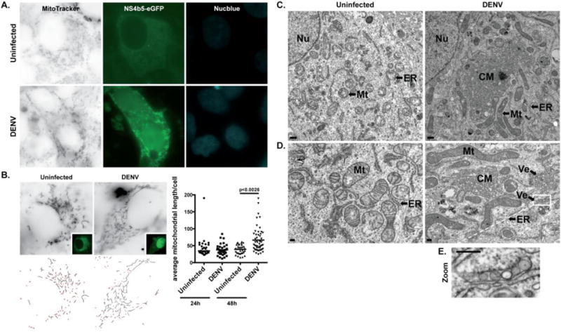
A. Immunofluorescence analysis of mitochondrial morphology in uninfected (cytoplasmic GFP) or DENV-infected (cytoplasmic and nuclear GFP) Huh7 cells. Cells were transfected with pNS4b5-eGFP reporter and infected with DENV (MOI of 0.1) for 48h. Cells were subsequently stained with MitoTracker Deep Red FM prior to imaging. Images were taken at 100× magnification and are representative of at least three independent experiments. B. Quantitative analysis of mitochondrial lengths in uninfected and infected Vero cells after 24h and 48h of infection. Vero cells transfected with pNS4b5-eGFP reporter were infected with DENV (MOI of 1) for 24h and 48h and mitochondria were stained with MitoTracker Deep Red FM. The average mitochondrial lengths for cells within each condition were calculated using a macro developed in ImageJ (NIH). Statistical analysis was done on the average mitochondrial lengths of each cell as described. The results reflect three independent experiments. C. and D. Ultra-thin sections TEM images of uninfected and DENV-infected (MOI of 1) resin-embedded Huh7 cells at different magnifications (C., ×7900, scale bar: 500 nm; D., ×19000, scale bar: 200 nm). Uninfected Huh7 cells exhibit typical ultrastructural morphology of non-infected cells. DENV-infected cells display elongated mitochondria (Mt), along with previously described convoluted membranes (CM) and invaginated vesicles (Ve) in proximity to the endoplasmic reticulum (ER) membranes. Nucleus (Nu) appears on lower magnification images (C.). E. Magnified image of box area illustrates invaginated vesicles (Ve) present in DENV-infected cells.