Abstract
Translocator Protein (18kDa, TSPO) is a mitochondrial outer membrane transmembrane protein. Its expression is elevated during inflammation and injury. However, the function of TSPO in vivo is still controversial. Here, we constructed a TSPO global knockout (KO) mouse with a Cre-LoxP system that abolished TSPO protein expression in all tissues and showed normal phenotypes in the physiological condition. The birth rates of TSPO heterozygote (Het) x Het or KO x KO breeding were consistent with Mendel’s Law, suggesting a normal viability of TSPO KO mice at birth. RNA-seq analysis showed no significant difference in the gene expression profile of lung tissues from TSPO KO mice compared with wild type mice, including the genes associated with bronchial alveoli immune homeostasis. The alveolar macrophage population was not affected by TSPO deletion in the physiological condition. Our findings contradict the results of Papadopoulos, but confirmed Selvaraj’s findings. This study confirms TSPO deficiency does not affect viability and bronchial alveolar immune homeostasis.
Introduction
Translocator Protein (18kDa) (TSPO), previously known as Peripheral Benzodiazepine Receptor (PBR), is an outer mitochondrial membrane protein with five transmembrane domains[1–3]. TSPO is widely expressed in different tissues including the adrenal cortex, white adipose tissue, brown adipose tissue, lung, liver, spleen and thymus[4,5]. However, its precise function remains unclear. Several inflammatory diseases are associated with elevated TSPO expression[6–8]. Increased uptake of radioisotope labeled high affinity ligands of TSPO have been used clinically as biomarkers to monitor inflammatory status in neurodegenerative diseases, brain injury and cancer[6,8–10]. Before TSPO knockout (KO) mice were constructed, knowledge regarding TSPO function was acquired from studies based on the hypothesis that TSPO is activated by ligands[8]. However, functions of TSPO in vivo are not clear.
TSPO deficiency was thought to be lethal during embryonic development given its presumed crucial roles in steroid biosynthesis, mitochondria functions and secondary signal transduction[8,11] However, Selvaraj and colleagues reported that TSPO KO mice survived with normal phenotypes[12]. Two independent research groups reported that TSPO KO mice survived to adulthood without overt phenotypes, developmental defects or cholesterol metabolism disorders[4,13]. In addition, Papadopoulos and colleagues reported that TSPO deficiency led to reduced viability, but Selvaraj suggested conflicting results[14]. These studies address the questions regarding what roles TSPO play during development.
In this study, we generated TSPO floxed mice using a Cre-LoxP system[15,16], with whole body deletion of TSPO. Consistent with previous studies[4,12,17], TSPO KO mice survived until adulthood with a birth rate consistent with Mendel’s Law. We know lung tissue is quiescent with unique immune homeostasis and high expression of TSPO, especially in bronchial alveolar epithelial cells and alveolar macrophages[7]. Our Digital Gene Expression Profiling (DGE) analysis showed no significant difference in the transcriptome profile of lung tissue between TSPO KO mice and wild type (WT) mice. This suggests that TSPO KO mice have normal gene expression profiles and normal bronchial alveolar immune homeostasis.
Reagents and Methods
Mice
All animal procedures were approved by the Animal Care and Use Committee of Institute of Basic Medical Sciences, Chinese Academy of Medical Sciences (IBMS, CAMS). TSPO Floxed mice, Flp transgenic mice and Protamine-Cre transgenic mice were C57BL/6J background and mice were maintained in SPF conditions at Experimental Animals Center of IBMS-CAMS. Mice were euthanized by Carbon Dioxide, for tissue isolation, mice were anesthetized by sodium pentobarbital.
Genotyping
TSPO floxed and KO pups were labeled by cutting toes 7 to 10 days after birth, one toe or tail tip was selected for genotyping. Genomic DNA was isolated according to the Dirty DNA Isolation Protocol of the Jackson Laboratory[18]. Briefly, samples were digested 90min at 98°C, placed at 15°C with 75μl NaOH-EDTA solution (25mM NaOH, 0.2mM EDTA), then mixed with 75μl 40mM Tris-HCl (pH5.5) Genomic DNA was used for genotyping. Standard PCR (Taq Master Mix, Quick Taq HS DyeMix, TOYOBO) was used, annealing temperature was 60°C with 35 cycles. The oligonucleotides for mouse genotyping were Flp forward (F) = 5’-tac aag tgg atc gat cct acc cct tgc g-3’; Flp reverse (R) = 5’-tcc cag gtc caa ctg cag ccc aag ctt cc-3’; Protamine-Cre F = 5’-CAT GTT CAG GGA TCG CCA GGC GTT T-3’; Protamine-Cre R = 5’-GTG CTA ACC AGC GTT TTC GTT CTG CCA A-3’; KO Forward (P1) = 5’-GAT GGA GAA ACT GAG TCC CAG TCA GGG T-3’; KO Reverse (P2) = 5’-GCT CTG CCC TAA TCA CAA AGT TTC ACA C-3’; KO Reverse (P3) = 5’-TTA AGG AGA GGT TTT GTC CTT GTG TC-3’.
Antibodies
Anti-mouse TSPO Antibody (#9530) for Western blot was purchased from Cell Signaling Technology® (CST, Danvers, Massachusetts); Anti-PBR (TSPO) RabMAb® [EPR5384] for WB and IHC was purchased from Abcam (Cambridge, UK); Mouse Anti-GAPDH antibody was purchased from BOSTER (BM1623, Wuhan, P.R.China); Alexa Fluor® 488 anti-mouse F4/80 Antibody (BM8) and Alexa Fluor® 647 anti-mouse CD206 (MMR) Antibody (C068C2) were purchased from BioLegend® (San Diego, California, U.S.); HRP anti-mouse/rabbit IgG secondary antibodies for WB and DAB Kit for IHC were purchased from ZSGB-BIO (Beijing, P.R.China); Mouse on Mouse (M.O.M.™) ImmPRESS™ HRP (Peroxidase) Polymer Kit and ImmPRESS™ HRP Anti-Rabbit IgG (Peroxidase) Polymer Detection Kit for IHC were purchased from Vector Laboratories, Inc.(Burlingame, Ca, U.S.).
Western blotting
TSPO expression of different tissues was examined by Western blot as previously described[19]. Tissues were homogenized in EDTA-free RIPA buffer containing protease inhibitor (Thermo Scientific) on ice, lysates were separated through 12% SDS-PAGE and blotted onto NC membrane. After incubating TSPO antibody (CST) for 36h-48h at 4°C and HRP-secondary antibody for 1h at room temperature, signal was exposed with Supersignal West Pico Chemiluminescent Substrate (Thermo Scientific) and CliNX chemiscope 3400 (CliNX Science Instruments Co., Ltd).
H&E staining and immunohistochemistry (IHC)
TSPO WT and KO (6 weeks, male) were anesthetized with 2% sodium pentobarbital (100mg/kg), and then fixed by cardiac perfusion with 4% PFA and processed for H&E staining and IHC as described previously[4]. Briefly, tissue sections were blocked with 5% goat serum and incubated with Anti-PBR (TSPO) RabMAb® (Abcam, 1:1500) at 4°C overnight and ImmPRESS™ HRP Anti-Rabbit IgG (Peroxidase) Polymer Detection Kit at room temperature for 45 minutes. HRP was detected with DAB Kit (ZSGB-BIO). Nikon microscope was used for whole viewed section scan.
RNA-seq and differential analysis
Total RNAs from lung tissues of 9 TSPO WT and 9 TSPO KO mice were isolated, 3 samples of every group were combined after RNA extraction for quality control and RNA-seq loading samples. RNA-seq was performed as DGE on an Illumia HiSeq platform and 50 bp paired-end reads were generated (RiboBio Co. Ltd.). The NCBI Gene Expression Omnibus (GEO) of the deep sequencing data were submitted under accession umber GSE84942. High-quality reads were aligned to the mouse reference genome (mm10)[20]. Significant differences were analyzed by negative binomial distribution test after read-counts normalization[21], fold change and adjusted P value (Q value) by the Benjamini-Hocherg false discovery rate (FDR) procedure (Q value) were considered as factors for differential analysis[22]. Genontology (GO) and Kyoto Encyclopedia of Genes and Genomes (KEGG) pathway analysis were performed as previously described[22–24]. DAVID (NIAID, NIH) Database was employed for gene annotation analysis[25,26]. TSPO WT and KO locus (mm10) of RNAseq were analyzed by IGV[27,28] software. Heat maps of interaction protein genes and bronchoalveolar immune microenvironment associated genes were analyzed by Multiple Array Viewer (MeV) v4.9 software[29].
Bronchoalveolar lavage fluid (BALF)
Mice were anesthetized, trachea was exposed, BALF and bronchovesicular cells were collected by lung lavage twice with 1ml normal saline containing 0.05% EDTA. BALF was centrifuged at 4,500 rpm, the first lavage supernatant was used to detect cytokines and mixed cells for flow cytometry analysis.
Flow cytometry
Flow cytometry of alveolar cells was preformed as previously described[30]. BALF cells were resuspended in wash buffer (1% BSA-PBS), fluorescently labeled antibodies were incubated at 4°C in dark for 20 minutes, and washed twice with ice wash buffer. Cells were detected by BD C6 Flow Cytometry and the data were analyzed by C6 software.
Statistical analysis
Results are presented as mean ± S.E.M. unless otherwise specified. Statistical significance was determined by Student’s t-tests; Birth rate difference compared with Mendel’s law was detected using Chi-squared test. Differences of statistical analysis at P < 0.05 were considered significant.
Results
Generation of TSPO KO mice
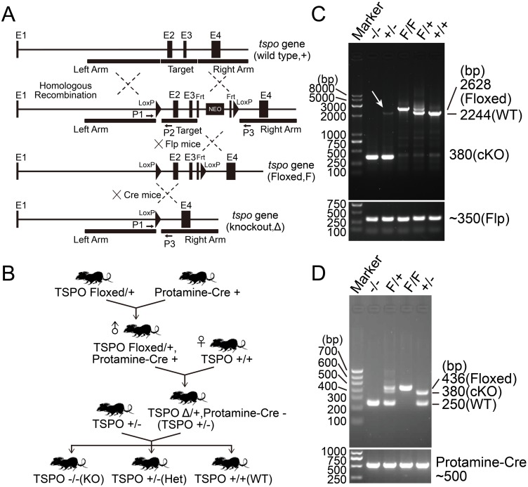
Given previous evidence that deficiency could be lethal during embryonic development[11], TSPO floxed mice for conditional deletion were generated by targeting the transmembrane domains contained within exon 2 and exon 3 of the mouse genomic locus. TSPO targeting vector was constructed by introducing two LoxP sites between exon 2 and exon 3 (Target region) and a NEO selection cassette floxed by Frt sites (Fig 1A). TSPO was genomically deleted by breeding with the male germ-line protamine-Cre deleted mice. After two rounds of breeding, heterozygote mice were generated and then intercrossed to generate homozygote TSPO KO mice (Fig 1B). TSPO KO mice were confirmed by PCR (Fig 1C and 1D). The homozygote TSPO KO mice were viable to adulthood, consistent with previous findings[4,12].
Fig 1. Generation of TSPO KO mice.
(A) Schematic of generation of TSPO Floxed mice and conditional KO mice by Cre-LoxP System. Conditional TSPO KO mice were generated by targeting exon 2 and exon 3 of the mouse genomic locus, a NEO selection cassette Floxed by Frt sites. (B) Schematic of crossing program for TSPO global knockout mice, Protamine-Cre is a transgenic mouse where Cre recombinase expression is driven by sperm specific protamine promotor. (C) Genotyping of TSPO Floxed mice and KO mice by a TSPO genotyping primer pair 1 & 3, and Flp primers. (D) Genotyping of TSPO Floxed mice and KO mice by three TSPO genotyping primers 1, 2 and 3, and Protamine-Cre primers.
TSPO KO mice have normal viability
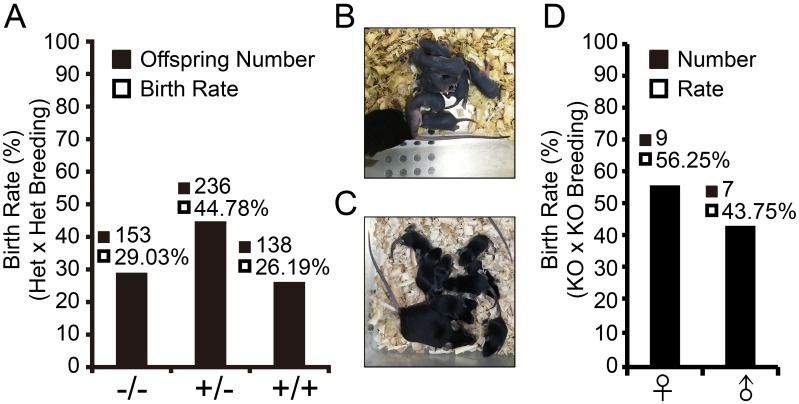
To determine the viability of TSPO KO mice, TSPO Het x Het mouse breeding was set up to examine the birth rate of each genotype (WT, Het and KO). 527 offspring were born during more than 2 years of Het x Het breeding and consisted of 138 WT (26.19%), 236 Het (44.78%) and 153 KO (29.03%) mice (Fig 2A). These numbers were consistent with Mendel’s Law: WT 25%, Het 50%, KO 25% and 50% female, 50% male. No significant difference was observed in the birth rates by Chi-Squared Tests (P<0.05). Thus, TSPO knockout did not affect the viability of mice. To further verify the viability rate of TSPO KO mice, TSPO KO x KO mouse breeding was also set up. Two litters produced 16 pups with normal physical condition (Fig 2B and 2C) and gender birth rate (Fig 2D).
Fig 2. TSPO deletion does not affect viability by Het x Het and KO x KO breeding.
(A) Birth rate of offspring of TSPO Het x Het breeding. (B) One litter Pups born 4 days from TSPO KO x KO breeding. (C) One litter pups born 9 days from TSPO KO x KO breeding. (D) Birth rate of offspring in TSPO KO x KO breeding. Chi-Squared Test was used to determine significant difference compared with Mendel’s Law.
Global deletion of TSPO did not affect the morphology of main organs and tissues
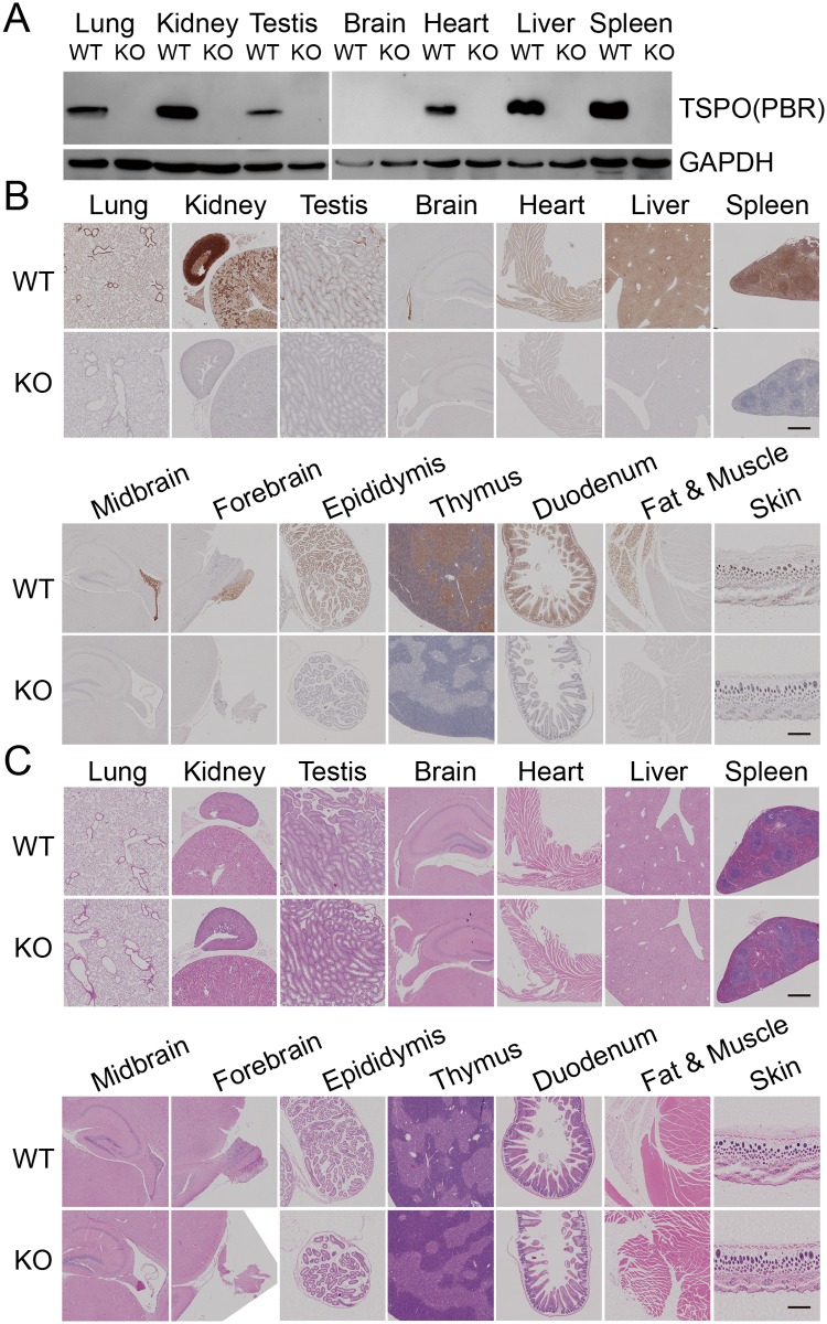
Previous studies demonstrated that TSPO KO mice had normal phenotypes[4,12,13,17]. To further characterize the phenotypes of TSPO KO mice, we first examined the expression of TSPO protein in major organs and tissues from TSPO WT and KO mice by Western blotting and IHC. The results show high levels of TSPO expression in the bronchial alveolar epithelium, alveolar macrophages, heart muscle, liver, spleen, thymus, kidney, testicular/epididymal mesenchymal, fat, skin, duodenum, ependyma and olfactory bulb. In comparison, we found low levels of TSPO expression in lung parenchyma, brain cortex, hippocampus, midbrain and leg muscle tissue (Fig 3A and 3B). The expression of TSPO protein was completely deleted in all tissues in KO mice (Fig 3A and 3B). To determine whether global deletion of TSPO results in the morphological changes in the main organs or tissues, H&E staining of tissue sections from WT and TSPO KO mice was performed. No distinguishable morphological changes were observed in the main tissues from KO mice compared with WT mice (Fig 3C). These results suggest that whole body deletion of TSPO does not affect the development of major organs and tissues in TSPO KO mice.
Fig 3. TSPO expression was abolished in global KO mice without pathological changes.
TSPO expression in different tissues from WT and KO mice were detected by western blotting (A) and IHC (B); (C) H&E staining of different tissues from WT and TSPO KO mice. Scale Bars, 100μm.
TSPO deletion did not affect gene expression profile in lungs of TSPO KO mice
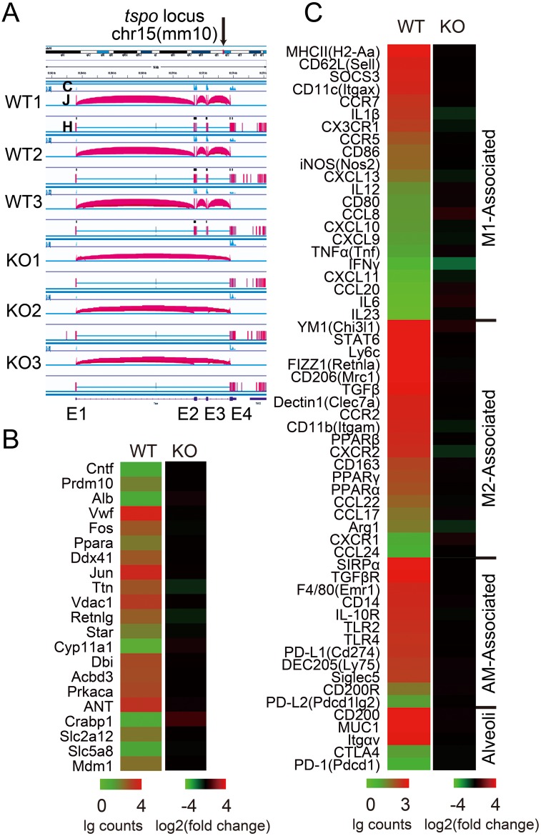
To ascertain which genes were impacted after TSPO deletion, transcriptome profiles of WT and KO lung tissues were analyzed using RNA-seq. The mouse TSPO gene is located at chr15:83,561,573–83,576,203 (mm10). Fig 4A shows the locus of samples for RNA-seq by IGV software. TSPO KO mouse model was constructed by deleting exon 2 and 3 (Fig 1A). RNA-seq data confirmed the deletion of this region (Fig 4A). TSPO interacting proteins reported in the literatures[31,32] and STRING (functional protein association networks) database (Version 10.0)[33] were analyzed and are presented as a heat map (Fig 4B). TSPO was the only differentially expressed gene between TSPO WT and KO mice (data not shown). We also focused on pulmonary alveolar epithelium and macrophage related genes, which are crucial regulators of the steady state of the alveolar immune microenvironment (Fig 4C). TSPO KO mice showed no difference compared with WT mice, indicating TSPO deficiency does not affect the expression of TSPO interacting proteins and bronchial alveolar immune microenvironment homeostasis (Fig 4B and 4C).
Fig 4. TSPO KO did not affect gene expression profiles.
(A) TSPO gene locus of WT and KO mice lung tissue samples used for RNA-seq, results visualized by Integrative Genomics Viewer (IGV) software (NIH), C: coverage of reads, J: junctions, H: hits; (B) Expression level of potential TSPO interaction proteins reported by literatures and SRING database (Version 10.0); (C) Expression profile of bronchoalveolar immune microenvironment associated genes; (B&C) Significant difference determined by negative binomial distribution test.
TSPO KO mice showed normal alveolar macrophage population
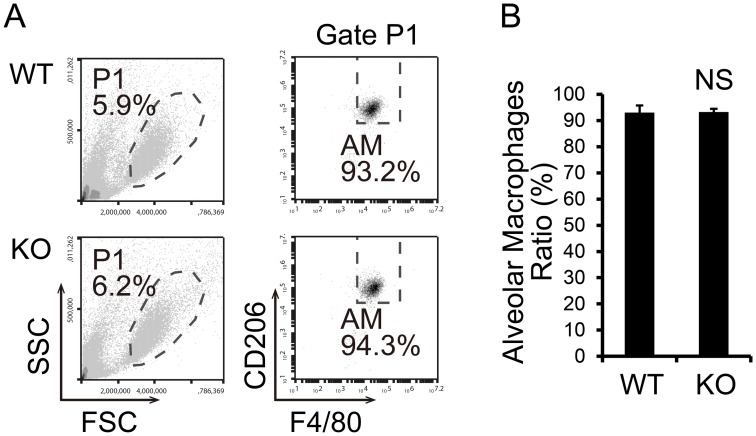
Alveolar macrophages are a specific macrophage subtype that reside in the alveolar duct in close contact with the respiratory epithelium. Mouse alveolar macrophages are classified as M2 macrophages and typically express F4/80, CD206, IL-10 receptor and TGFβ receptor[34]. We analyzed the alveolar macrophage population (F4/80+, CD206+) in bronchoalveolar lavage fluid (BALF) from WT and TSPO KO mice. The data show normal cell subtype populations in BALF and mainly alveolar macrophages in TSPO KO mice (Fig 5).
Fig 5. TSPO KO mice show normal alveolar macrophage population.
(A) FACS analysis of BALF and alveolar macrophages was identified as F4/80+, CD206+. (B) The percentages of alveolar macrophages in BALF from WT and TSPO KO mice are shown as the mean ± S.E.M. from four animals in each group (n = 4). NS, not significant by Student’s t-test.
Discussion
TSPO contains five transmembrane domains and is located at the outer mitochondrial membrane. Hundreds of previous studies demonstrated that TSPO possesses high affinity to cholesterol and plays a crucial role in the translocation of cholesterol from the cytosol to the mitochondrion[8,35–38], and is involved in the rate-limiting step in steroidogenesis[8]. Interestingly, recent in vivo studies based on knockout mouse models and in vitro studies based on deletion in steroidogenic cells have refuted the links between TSPO and cholesterol transfer[12,39]. The link between TSPO and mitochondrial energy homeostasis is inconsistent and perhaps dependent on cell type. Studies have reported that TSPO deletion results in a decreased oxygen consumption rate (OCR) in microglia[4] and fibroblasts[40], but no change in OCR was observed in hepatocytes[13] and Leydig cells[41]. An increase in mitochondrial fatty acid oxidation was observed in steroidogenic cells and tissues[41]. But TSPO is not required for steroid hormone biosynthesis as previously suggested[4,5,12,17,40]. The loss of TSPO in experimental autoimmune encephalomyelitis (EAE) mice, an animal model of multiple sclerosis (MS), caused mild astrogliosis and less EAE clinical scoring[42]. These studies demonstrated that TSPO KO mice exhibit defective phenotypes under pathological conditions. Inflammation and injury-derived TSPO upregulation was studied in multiple scenarios, including as an anti-inflammatory drug target, and as a positron emission tomography (PET) imaging radioligand target. The ligands of TSPO have been evaluated as protective agents to evaluate the inflammatory status in the brains of patients with Alzheimer’s Disease (PK11195 and Ro5-4864)[43], anxiety (XBD-173)[44], MS (PK11195)[45] and acute lung injury (ALI)[7].
In this study, we created TSPO knockout mice with the Cre-LoxP system and characterized the phenotypes under normal conditions to explore systemic physiological functions and networks of TSPO in vivo. In our study, the viability of TSPO KO mice was not affected in both Het x Het breeding and KO x KO breeding. Our results are consistent with Selvaraj’s findings[14], but contradict Papadopoulos[46]. RNA-seq analysis showed normal gene expression profile in lung tissue from TSPO KO mice. RNA-seq analysis showed that nearly all TSPO interacting proteins listed in the databases and published papers had normal mRNA levels in TSPO KO mice. These results indicate that TSPO does not play an important role in physiological conditions.
Several studies using knockout models both in vitro and in vivo have indicated that TSPO is not necessary for cellular and organismal survival. Recent evidences also suggest a strong disparity between pharmacological and genetic dissecting TSPO function. Several recent reviews have dissected these phenomena and explored possible functions for TSPO and its importance in cellular functions[47–51]. However, TSPO does play critical roles in biology, including endoplasmic reticulum associated protein degradation (ERAD), reactive oxygen species (ROS) production, cholesterol efflux, protoporphyrin IX (PPIX) synthesis, and even cytokine production, apoptosis and autophagy[52]. It is important to highlight two issues regarding TSPO. One is explaining why TSPO has a high affinity to cholesterol but does not participate in its transfer. The second is understanding why TSPO is overexpressed in vivo. A re-examination of the characteristics of TSPO with a transgenic mouse model is imperative, especially for drug target mechanism studies[8,44]. In future studies, using the “apparently healthy” knockout mouse model, we aim to uncover new features of TSPO that play roles in inflammation and neurodegenerative diseases.
Additional Information
Deep sequencing data for the RNA-Seq experiments have been deposited in the NCBI Gene Expression Omnibus accession number GSE84942.
Acknowledgments
This study was supported by grants from National Natural Science Foundation of China to JZ (31471016), CAMS Initiative for Innovative Medicine to JZ (2016-I2M-1-008), PUMC Scholarship for Young Scientists to HW (3332015109) and CAMS Central Public Welfare Scientific Research Institute Basal Research Expenses to HW (2016ZX310182-3). Beijing Thorgene Medical Laboratory provided support in the form of salary for FL, but did not have any additional roles in the study design, data collection and analysis, decision to publish, or preparation of the manuscript. We thank Dr. Austin Cape at ASJ Editors for careful review and feedback.
Data Availability
Deep sequencing data for the RNA-Seq experiments have been deposited in the NCBI Gene Expression Omnibus accession number GSE84942.
Funding Statement
This study was supported by grants from National Natural Science Foundation of China to JZ (31471016), CAMS Initiative for Innovative Medicine to JZ (2016-I2M-1-008), PUMC Scholarship for Young Scientists to HW (3332015109) and CAMS Central Public Welfare Scientific Research Institute Basal Research Expenses to HW (2016ZX310182-3). Beijing Thorgene Medical Laboratory provided support in the form of salaries for author FL, but did not have any additional role in the study design, data collection and analysis, decision to publish, or preparation of the manuscript. The specific roles of this author are articulated in the ‘Author Contributions’ section. The funders had no role in study design, data collection and analysis, decision to publish, or preparation of the manuscript.
References
- 1.Anholt RR, Pedersen PL, De Souza EB, Snyder SH (1986) The peripheral-type benzodiazepine receptor. Localization to the mitochondrial outer membrane. J Biol Chem 261: 576–583. [PubMed] [Google Scholar]
- 2.Murail S, Robert JC, Coic YM, Neumann JM, Ostuni MA, Yao ZX, et al. (2008) Secondary and tertiary structures of the transmembrane domains of the translocator protein TSPO determined by NMR. Stabilization of the TSPO tertiary fold upon ligand binding. Biochim Biophys Acta 1778: 1375–1381. 10.1016/j.bbamem.2008.03.012 [DOI] [PubMed] [Google Scholar]
- 3.Gavish M, Bachman I, Shoukrun R, Katz Y, Veenman L, Weisinger G, et al. (1999) Enigma of the peripheral benzodiazepine receptor. Pharmacol Rev 51: 629–650. [PubMed] [Google Scholar]
- 4.Banati RB, Middleton RJ, Chan R, Hatty CR, Wai-Ying Kam W, Quin C, et al. (2014) Positron emission tomography and functional characterization of a complete PBR/TSPO knockout. Nat Commun 5: 5452 10.1038/ncomms6452 [DOI] [PMC free article] [PubMed] [Google Scholar]
- 5.Stocco DM (2014) The Role of PBR/TSPO in Steroid Biosynthesis Challenged. Endocrinology 155: 6–9. 10.1210/en.2013-2041 [DOI] [PMC free article] [PubMed] [Google Scholar]
- 6.Karlstetter M, Nothdurfter C, Aslanidis A, Moeller K, Horn F, Scholz R, et al. (2014) Translocator protein (18 kDa) (TSPO) is expressed in reactive retinal microglia and modulates microglial inflammation and phagocytosis. J Neuroimmunol 11: 3. [DOI] [PMC free article] [PubMed] [Google Scholar]
- 7.Hatori A, Yui J, Yamasaki T, Xie L, Kumata K, Fujinaga M, et al. (2012) PET imaging of lung inflammation with [18F]FEDAC, a radioligand for translocator protein (18 kDa). PLoS One 7: e45065 10.1371/journal.pone.0045065 [DOI] [PMC free article] [PubMed] [Google Scholar]
- 8.Rupprecht R, Papadopoulos V, Rammes G, Baghai TC, Fan J, Akula N, et al. (2010) Translocator protein (18 kDa)(TSPO) as a therapeutic target for neurological and psychiatric disorders. Nat Rev Drug Discov 9: 971–988. 10.1038/nrd3295 [DOI] [PubMed] [Google Scholar]
- 9.Liu GJ, Middleton RJ, Hatty CR, Kam WW, Chan R, Pham T, et al. (2014) The 18 kDa Translocator Protein, Microglia and Neuroinflammation. Brain Pathol 24: 631–653. 10.1111/bpa.12196 [DOI] [PMC free article] [PubMed] [Google Scholar]
- 10.Mirzaei N, Tang SP, Ashworth S, Coello C, Plisson C, Passchier J, et al. (2016) In vivo imaging of microglial activation by positron emission tomography with [11C] PBR28 in the 5XFAD model of Alzheimer's disease. Glia 64: 993–1006. 10.1002/glia.22978 [DOI] [PubMed] [Google Scholar]
- 11.Papadopoulos V, Amri H, Boujrad N, Cascio C, Culty M, Garnier M, et al. (1997) Peripheral benzodiazepine receptor in cholesterol transport and steroidogenesis. Steroids 62: 21–28. [DOI] [PubMed] [Google Scholar]
- 12.Morohaku K, Pelton SH, Daugherty DJ, Butler WR, Deng W, Selvaraj V (2014) Translocator Protein/Peripheral Benzodiazepine Receptor Is Not Required for Steroid Hormone Biosynthesis. Endocrinology 155: 89–97. 10.1210/en.2013-1556 [DOI] [PMC free article] [PubMed] [Google Scholar]
- 13.Š J., Blachly-Dyson E, Sewell R, Carpi A, Menabò R, Di-Lisa F, et al. (2014) Regulation of the mitochondrial permeability transition pore by the outer membrane does not involve the peripheral benzodiazepine receptor (Translocator Protein of 18 kDa (TSPO)). J Biol Chem 289: 13769–13781. 10.1074/jbc.M114.549634 [DOI] [PMC free article] [PubMed] [Google Scholar]
- 14.Selvaraj V, Tu LN, Stocco DM (2016) Crucial role reported for TSPO in viability and steroidogenesis is a misconception. A commentary on: Conditional steroidogenic cell-targeted deletion of TSPO unveils a crucial role in viability and hormone-dependent steroid formation by Fan, J., Campioli, E., Midzak, A., Culty, M., and Papadopoulos, V.(2015). Proc. Natl. Acad. Sci. USA. 112, 7261–7266. Front Endocrinol 7: 91. [DOI] [PMC free article] [PubMed] [Google Scholar]
- 15.O'Gorman S, Dagenais NA, Qian M, Marchuk Y (1997) Protamine-Cre recombinase transgenes efficiently recombine target sequences in the male germ line of mice, but not in embryonic stem cells. Proc Natl Acad Sci U S A 94: 14602–14607. [DOI] [PMC free article] [PubMed] [Google Scholar]
- 16.Seidler B, Schmidt A, Mayr U, Nakhai H, Schmid RM, Schneider G, et al. (2008) A Cre-loxP-based mouse model for conditional somatic gene expression and knockdown in vivo by using avian retroviral vectors. Proc Natl Acad Sci U S A 105: 10137–10142. 10.1073/pnas.0800487105 [DOI] [PMC free article] [PubMed] [Google Scholar]
- 17.Tu LN, Morohaku K, Manna PR, Pelton SH, Butler WR, Stocco DM, et al. (2014) Peripheral Benzodiazepine Receptor/Translocator Protein Global Knockout Mice are Viable with no Effects on Steroid Hormone Biosynthesis. J Biol Chem 289: 27444–27454. 10.1074/jbc.M114.578286 [DOI] [PMC free article] [PubMed] [Google Scholar]
- 18.https://www.jax.org/jax-mice-and-services/customer-support/technical-support/genotyping-resources/dna-isolation-protocols#. The Jackson Laboratory.
- 19.Yin S, Zhang J, Mao Y, Hu Y, Cui L, Kang N, et al. (2013) Vav1-phospholipase C-γ1 (Vav1-PLC-γ1) Pathway Initiated by T Cell Antigen Receptor (TCRγδ) Activation Is Required to Overcome Inhibition by Ubiquitin Ligase Cbl-b during γδT Cell Cytotoxicity. J Biol Chem 288: 26448–26462. 10.1074/jbc.M113.484600 [DOI] [PMC free article] [PubMed] [Google Scholar]
- 20.Anders S, Huber W (2012) Differential expression of RNA-Seq data at the gene level–the DESeq package. Heidelberg, Germany: European Molecular Biology Laboratory (EMBL). [Google Scholar]
- 21.Anders S (2010) Analysing RNA-Seq data with the DESeq package. Mol Biol 43: 1–17. [Google Scholar]
- 22.Wang W, Gao Z, Wang H, Li T, He W, Lv W, et al. (2016) Transcriptome Analysis Reveals Distinct Gene Expression Profiles in Eosinophilic and Noneosinophilic Chronic Rhinosinusitis with Nasal Polyps. Sci Rep 6: 26604 10.1038/srep26604 [DOI] [PMC free article] [PubMed] [Google Scholar]
- 23.Ashburner M, Ball CA, Blake JA, Botstein D, Butler H, Cherry JM, et al. (2000) Gene ontology: tool for the unification of biology. The Gene Ontology Consortium. Nat Genet 25: 25–29. [DOI] [PMC free article] [PubMed] [Google Scholar]
- 24.Kanehisa M, Goto S (2000) KEGG: kyoto encyclopedia of genes and genomes. Nucleic Acids Res 28: 27–30. [DOI] [PMC free article] [PubMed] [Google Scholar]
- 25.Huang DW, Sherman BT, Lempicki RA (2009) Systematic and integrative analysis of large gene lists using DAVID bioinformatics resources. Nat Protoc 4: 44–57. 10.1038/nprot.2008.211 [DOI] [PubMed] [Google Scholar]
- 26.Huang DW, Sherman BT, Lempicki RA (2009) Bioinformatics enrichment tools: paths toward the comprehensive functional analysis of large gene lists. Nucleic Acids Res 37: 1–13. 10.1093/nar/gkn923 [DOI] [PMC free article] [PubMed] [Google Scholar]
- 27.Thorvaldsdottir H, Robinson JT, Mesirov JP (2013) Integrative Genomics Viewer (IGV): high-performance genomics data visualization and exploration. Brief Bioinform 14: 178–192. 10.1093/bib/bbs017 [DOI] [PMC free article] [PubMed] [Google Scholar]
- 28.Robinson JT, Thorvaldsdottir H, Winckler W, Guttman M, Lander ES, Getz G, et al. (2011) Integrative genomics viewer. Nat Biotechnol 29: 24–26. 10.1038/nbt.1754 [DOI] [PMC free article] [PubMed] [Google Scholar]
- 29.Eisen MB, Spellman PT, Brown PO, Botstein D (1998) Cluster analysis and display of genome-wide expression patterns. Proc Natl Acad Sci U S A 95: 14863–14868. [DOI] [PMC free article] [PubMed] [Google Scholar]
- 30.Yin S, Mao Y, Li X, Yue C, Zhou C, Huang L, et al. (2015) Hyperactivation and in situ recruitment of inflammatory Vδ2 T cells contributes to disease pathogenesis in systemic lupus erythematosus. Sci Rep 5: 14432 10.1038/srep14432 [DOI] [PMC free article] [PubMed] [Google Scholar]
- 31.Huttlin EL, Ting L, Bruckner RJ, Gebreab F, Gygi MP, Szpyt J, et al. (2015) The BioPlex Network: A Systematic Exploration of the Human Interactome. Cell 162: 425–440. 10.1016/j.cell.2015.06.043 [DOI] [PMC free article] [PubMed] [Google Scholar]
- 32.Liu J, Rone MB, Papadopoulos V (2006) Protein-protein interactions mediate mitochondrial cholesterol transport and steroid biosynthesis. J Biol Chem 281: 38879–38893. 10.1074/jbc.M608820200 [DOI] [PubMed] [Google Scholar]
- 33.Szklarczyk D, Franceschini A, Wyder S, Forslund K, Heller D, Huerta-Cepas J, et al. (2015) STRING v10: protein-protein interaction networks, integrated over the tree of life. Nucleic Acids Res 43: D447–452. 10.1093/nar/gku1003 [DOI] [PMC free article] [PubMed] [Google Scholar]
- 34.Hussell T, Bell TJ (2014) Alveolar macrophages: plasticity in a tissue-specific context. Nat Rev Immunol 14: 81–93. 10.1038/nri3600 [DOI] [PubMed] [Google Scholar]
- 35.Papadopoulos V, Baraldi M, Guilarte TR, Knudsen TB, Lacapère JJ, Lindemann P, et al. (2006) Translocator protein (18kDa): new nomenclature for the peripheral-type benzodiazepine receptor based on its structure and molecular function. Trends Pharmacol Sci 27: 402–409. 10.1016/j.tips.2006.06.005 [DOI] [PubMed] [Google Scholar]
- 36.Korkhov VM, Sachse C, Short JM, Tate CG (2010) Three-dimensional structure of TspO by electron cryomicroscopy of helical crystals. Structure 18: 677–687. 10.1016/j.str.2010.03.001 [DOI] [PMC free article] [PubMed] [Google Scholar]
- 37.Li F, Liu J, Zheng Y, Garavito RM, Ferguson-Miller S (2015) Protein structure. Crystal structures of translocator protein (TSPO) and mutant mimic of a human polymorphism. Science 347: 555–558. [DOI] [PMC free article] [PubMed] [Google Scholar]
- 38.Guo Y, Kalathur RC, Liu Q, Kloss B, Bruni R, Ginter C, et al. (2015) Protein structure. Structure and activity of tryptophan-rich TSPO proteins. Science 347: 551–555. [DOI] [PMC free article] [PubMed] [Google Scholar]
- 39.Tu LN, Zhao AH, Stocco DM, Selvaraj V (2015) PK11195 effect on steroidogenesis is not mediated through the translocator protein (TSPO). Endocrinology 156: 1033–1039. 10.1210/en.2014-1707 [DOI] [PMC free article] [PubMed] [Google Scholar]
- 40.Zhao AH, Tu LN, Mukai C, Sirivelu MP, Pillai VV, Morohaku K, et al. (2016) Mitochondrial Translocator Protein (TSPO) Function Is Not Essential for Heme Biosynthesis. J Biol Chem 291: 1591–1603. 10.1074/jbc.M115.686360 [DOI] [PMC free article] [PubMed] [Google Scholar]
- 41.Tu LN, Zhao AH, Hussein M, Stocco DM, Selvaraj V (2016) Translocator Protein (TSPO) Affects Mitochondrial Fatty Acid Oxidation in Steroidogenic Cells. Endocrinology 157: 1110–1121. 10.1210/en.2015-1795 [DOI] [PMC free article] [PubMed] [Google Scholar]
- 42.Daugherty DJ, Chechneva O, Mayrhofer F, Deng W (2016) The hGFAP-driven conditional TSPO knockout is protective in a mouse model of multiple sclerosis. Sci Rep 6: 22556 10.1038/srep22556 [DOI] [PMC free article] [PubMed] [Google Scholar]
- 43.Barron AM, Garcia-Segura LM, Caruso D, Jayaraman A, Lee JW, Melcangi RC, et al. (2013) Ligand for translocator protein reverses pathology in a mouse model of Alzheimer's disease. J Neurosci 33: 8891–8897. 10.1523/JNEUROSCI.1350-13.2013 [DOI] [PMC free article] [PubMed] [Google Scholar]
- 44.Rupprecht R, Rammes G, Eser D, Baghai TC, Schule C, Nothdurfter C, et al. (2009) Translocator Protein (18 kD) as Target for Anxiolytics Without Benzodiazepine-Like Side Effects. Science 325: 490–493. 10.1126/science.1175055 [DOI] [PubMed] [Google Scholar]
- 45.Daugherty DJ, Selvaraj V, Chechneva OV, Liu XB, Pleasure DE, Deng WB (2013) A TSPO ligand is protective in a mouse model of multiple sclerosis. EMBO Mol Med 5: 891–903. 10.1002/emmm.201202124 [DOI] [PMC free article] [PubMed] [Google Scholar]
- 46.Fan J, Campioli E, Midzak A, Culty M, Papadopoulos V (2015) Conditional steroidogenic cell-targeted deletion of TSPO unveils a crucial role in viability and hormone-dependent steroid formation. Proc Natl Acad Sci U S A 112: 7261–7266. 10.1073/pnas.1502670112 [DOI] [PMC free article] [PubMed] [Google Scholar]
- 47.Selvaraj V, Stocco DM, Tu LN (2015) Minireview: Translocator Protein (TSPO) and Steroidogenesis: A Reappraisal. Mol Endocrinol 29: 490–501. 10.1210/me.2015-1033 [DOI] [PMC free article] [PubMed] [Google Scholar]
- 48.Hatty CR, Banati RB (2015) Protein-ligand and membrane-ligand interactions in pharmacology: the case of the translocator protein (TSPO). Pharmacol Res 100: 58–63. 10.1016/j.phrs.2015.07.029 [DOI] [PubMed] [Google Scholar]
- 49.Guilarte TR, Loth MK, Guariglia SR (2016) TSPO Finds NOX2 in Microglia for Redox Homeostasis. Trends Pharmacol Sci 37: 334–343. [DOI] [PMC free article] [PubMed] [Google Scholar]
- 50.Selvaraj V, Tu LN (2016) Current status and future perspectives: TSPO in steroid neuroendocrinology. J Endocrinol 231: R1–R30. 10.1530/JOE-16-0241 [DOI] [PubMed] [Google Scholar]
- 51.Middleton RJ, Liu GJ, Banati RB (2015) Guwiyang Wurra—'Fire Mouse': a global gene knockout model for TSPO/PBR drug development, loss-of-function and mechanisms of compensation studies. Biochem Soc Trans 43: 553–558. 10.1042/BST20150039 [DOI] [PubMed] [Google Scholar]
- 52.Selvaraj V, Stocco DM (2015) The changing landscape in translocator protein (TSPO) function. Trends Endocrinol Metab 26: 341–348. 10.1016/j.tem.2015.02.007 [DOI] [PMC free article] [PubMed] [Google Scholar]
Associated Data
This section collects any data citations, data availability statements, or supplementary materials included in this article.
Data Availability Statement
Deep sequencing data for the RNA-Seq experiments have been deposited in the NCBI Gene Expression Omnibus accession number GSE84942.


