

Abstract
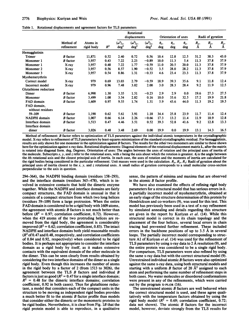
The extent to which the librations of rigid molecules can model the crystallographic temperature factor profiles of proteins has been examined. For all proteins considered, including influenza virus hemagglutinin, glutathione reductase, myohemerythrin, myoglobin, and streptavidin, a simple 10-parameter model [V. Schomaker and K. N. Trueblood (1968) Acta Crystallogr. Sect. B 24, 63-76] is found to reproduce qualitatively the patterns of maxima and minima in the isotropic backbone meansquare displacements. Large deviations between the rigid molecule and individual atomic temperature factors are found to be correlated with a region in hemagglutinin for which the refined structural model is unsatisfactory and with errors in the structure in a partially incorrect model of myohemerythrin. For the high-resolution glutathione reductase structure, better results are obtained on treating each of the compact domains in the structure as independent rigid bodies. The method allows for the refinement of reliable temperature factors with the introduction of minimal parameters and may prove useful for the evaluation of models in the early stages of x-ray structure refinement. While these results by themselves do not establish the nature of the underlying displacements, the success of the rigid protein model in reproducing qualitative features of temperature factor profiles suggests that rigid body refinement results should be considered in any interpretation of crystallographic thermal parameters.
Full text
PDF

Selected References
These references are in PubMed. This may not be the complete list of references from this article.
- Caspar D. L., Clarage J., Salunke D. M., Clarage M. Liquid-like movements in crystalline insulin. Nature. 1988 Apr 14;332(6165):659–662. doi: 10.1038/332659a0. [DOI] [PubMed] [Google Scholar]
- Diamond R. On the use of normal modes in thermal parameter refinement: theory and application to the bovine pancreatic trypsin inhibitor. Acta Crystallogr A. 1990 Jun 1;46(Pt 6):425–435. doi: 10.1107/s0108767390002082. [DOI] [PubMed] [Google Scholar]
- Doucet J., Benoit J. P. Molecular dynamics studied by analysis of the X-ray diffuse scattering from lysozyme crystals. Nature. 1987 Feb 12;325(6105):643–646. doi: 10.1038/325643a0. [DOI] [PubMed] [Google Scholar]
- Hendrickson W. A., Pähler A., Smith J. L., Satow Y., Merritt E. A., Phizackerley R. P. Crystal structure of core streptavidin determined from multiwavelength anomalous diffraction of synchrotron radiation. Proc Natl Acad Sci U S A. 1989 Apr;86(7):2190–2194. doi: 10.1073/pnas.86.7.2190. [DOI] [PMC free article] [PubMed] [Google Scholar]
- Holbrook S. R., Wang A. H., Rich A., Kim S. H. Local mobility of nucleic acids as determined from crystallographic data. III. A daunomycin-DNA complex. J Mol Biol. 1988 Jan 20;199(2):349–357. doi: 10.1016/0022-2836(88)90318-x. [DOI] [PubMed] [Google Scholar]
- Howlin B., Moss D. S., Harris G. W. Segmented anisotropic refinement of bovine ribonuclease A by the application of the rigid-body TLS model. Acta Crystallogr A. 1989 Dec 1;45(Pt 12):851–861. doi: 10.1107/s0108767389009177. [DOI] [PubMed] [Google Scholar]
- Karplus P. A., Schulz G. E. Refined structure of glutathione reductase at 1.54 A resolution. J Mol Biol. 1987 Jun 5;195(3):701–729. doi: 10.1016/0022-2836(87)90191-4. [DOI] [PubMed] [Google Scholar]
- Kuriyan J., Brünger A. T., Karplus M., Hendrickson W. A. X-ray refinement of protein structures by simulated annealing: test of the method on myohemerythrin. Acta Crystallogr A. 1989 Jun 1;45(Pt 6):396–409. doi: 10.1107/s0108767389000437. [DOI] [PubMed] [Google Scholar]
- Kuriyan J., Wilz S., Karplus M., Petsko G. A. X-ray structure and refinement of carbon-monoxy (Fe II)-myoglobin at 1.5 A resolution. J Mol Biol. 1986 Nov 5;192(1):133–154. doi: 10.1016/0022-2836(86)90470-5. [DOI] [PubMed] [Google Scholar]
- Sheriff S., Hendrickson W. A., Smith J. L. Structure of myohemerythrin in the azidomet state at 1.7/1.3 A resolution. J Mol Biol. 1987 Sep 20;197(2):273–296. doi: 10.1016/0022-2836(87)90124-0. [DOI] [PubMed] [Google Scholar]
- Sternberg M. J., Grace D. E., Phillips D. C. Dynamic information from protein crystallography. An analysis of temperature factors from refinement of the hen egg-white lysozyme structure. J Mol Biol. 1979 May 25;130(3):231–252. doi: 10.1016/0022-2836(79)90539-4. [DOI] [PubMed] [Google Scholar]
- Weis W. I., Brünger A. T., Skehel J. J., Wiley D. C. Refinement of the influenza virus hemagglutinin by simulated annealing. J Mol Biol. 1990 Apr 20;212(4):737–761. doi: 10.1016/0022-2836(90)90234-D. [DOI] [PubMed] [Google Scholar]
- Wilson I. A., Skehel J. J., Wiley D. C. Structure of the haemagglutinin membrane glycoprotein of influenza virus at 3 A resolution. Nature. 1981 Jan 29;289(5796):366–373. doi: 10.1038/289366a0. [DOI] [PubMed] [Google Scholar]