Figure 6.
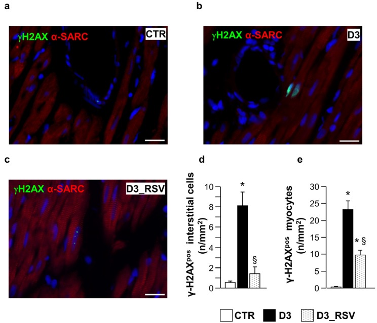
RSV treatment reduced diabetes-induced DNA damage. Panels a–c: detection of DNA double strand breaks in sections of the LV myocardium from control (a), untreated (b) and RSV-treated (c) diabetic rats, by immunofluorescence. Nuclear labeling by antibodies against gamma-Histone2 AX (green, γH2AX) is documented in cardiomyocytes recognized by the red fluorescence of α-sarcomeric actin (red). Two cardiomyocytes show diffuse nuclear fluorescence in the D3 heart while dot-like signals are present in a cardiomyocyte from RSV-treated myocardium. Blue fluorescence corresponds to DAPI staining of nuclei. Scale bars: 20 µm; In (d–e), bar graphs illustrating the density of γH2AX positive interstitial cells (d) and cardiomyocytes (e), in control (CTR), untreated (D3) and RSV-treated (D3_RSV) diabetic myocardium. Data are reported as mean ± SEM * p < 0.05 vs. CTR; § p < 0.05 significant differences between D3 and D3_RSV (one-way ANOVA, Games-Howell post-hoc test).

