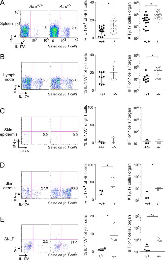
Fig. 2. Increased frequency and number of Tγδ17 cells in peripheral tissues of Aire-deficient mice.
Designated organs of Aire+/+ and Aire−/− littermates were excised at 3-9 weeks of age and examined by flow cytometry, focusing on Tγδ17 cells. Left panels: typical cytofluorometric profiles. Numbers refer to fraction of total γδ T cells in that gate. SI-LP = small-intestinal lamina propria. Center panels: summary data for fractional representation (n=3-16). Right panels: corresponding summary data for number per organ. Statistics as per Fig. 1. Dot plot scales for this figure are all the same.

