CELL BIOLOGY Correction for “Drosophila Mgr, a Prefoldin subunit cooperating with von Hippel Lindau to regulate tubulin stability,” by Nathalie Delgehyr, Uta Wieland, Hélène Rangone, Xavier Pinson, Guojie Mao, Nikola S. Dzhindzhev, Doris McLean, Maria G. Riparbelli, Salud Llamazares, Giuliano Callaini, Cayetano Gonzalez, and David M. Glover, which appeared in issue 15, April 10, 2012, of Proc Natl Acad Sci USA (109:5729–5734; first published March 26, 2012; 10.1073/pnas.1108537109).
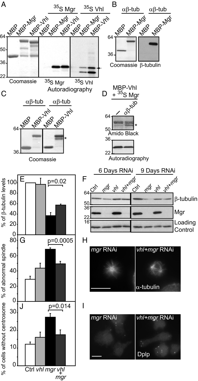
The authors wish to note, “In Fig. 4A in this article, the two last panels were identical due to incorrect handling during the final assembly of the figure. The description of the results is accurate and these changes have no bearing on the experimental results or the conclusions. The figure legend has been corrected to indicate 35S-Vhl and its likely degradation product. We apologize for any inconvenience that this error has caused.” The corrected Fig. 4 and its corrected legend appear below.
Fig. 4.
Mgr and Vhl cooperate in regulating tubulin destruction. (A) MBP, MBP-Mgr, and MBP-Vhl, affinity purified from Escherichia coli extracts (Coomassie stain) tested for binding 35S-Methionine labeled Mgr and Vhl synthesized by coupled transcription-translation in vitro. The arrow indicates 35S-Vhl, the second band being likely a degradation product (autoradiography). (B) MBP and MBP-Mgr, affinity-purified from E. coli extracts (Coomassie stain) tested for binding purified αβ-tubulin (Western blot). (C) MBP and MBP-Vhl, affinity-purified from E. coli extracts (Coomassie stain, Right) tested for binding-purified αβ-tubulin (Coomassie stain, Left). (D) MBP-Vhl, affinity purified from E. coli extracts, and tested for binding 35S-Mgr (as in A). Excess of purified αβ-tubulin is insufficient to release the Vhl:Mgr interaction. (E–J) DMEL-2 cells treated with Control, mgr, Vhl, or mgr and Vhl dsRNA for 6 or 9 d. (E) Levels of β-tubulin in three independent experiments 9 d after transfection. (F) Western blot of β-tubulin and Mgr after such treatment. H2A is used as loading control (Ctrl). (G) Percentage of prometaphase and metaphase cells with monopolar or disorganized spindles after indicated dsRNA treatment. Error bars = SEMs of three independent experiments. n > 300 metaphase cells; (H) Mitotic cells immunostained to reveal microtubules (α-tubulin). (Scale bar, 10 μm.) (I) Percentage of cells without centrosome 9 d after indicated transfections. Error bars = SEM of three independent experiments. n > 600 cells. (J) Cells immunostained to reveal centrosomes (Dplp). (Scale bar, 10 μm.) All P values are from Student t tests.


