Figure 2.
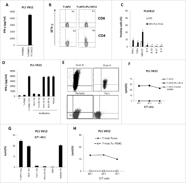
Generation of one cytotoxic CD4+ T cell line against the BCR peptide from a PL patient. (A) IFNγ ELISA assay of autologous CD4+ T cells stimulated with autologous PBMCs, pulsed or nonpulsed with patient-derived peptide. (B, C) Intracellular staining of cytokines secretion by autologous cytotoxic CD4+ T cells versus APCs, pulsed or nonpulsed with peptide. (D) Blocking of IFNγ production by autologous BCR peptide-specific CD4+ T cells with HLA antibodies. (E) Intracellular staining of Granzyme A, B, Perforin, and FasL of autologous cytotoxic CD4+ T cells. (F) Cytotoxicity of autologous CD4+ T cells against APCs pulsed with PL1VK12, control peptide, or APCs alone, and (G) in the presence or absence of HLA class I and II, HLA DR antibodies, and CMA inhibitor. (H) Cytotoxicity of autologous CD4+ T cells against autologous tumor and tumor-free PBMCs as negative controls. PL, plasma cell leukemia.

