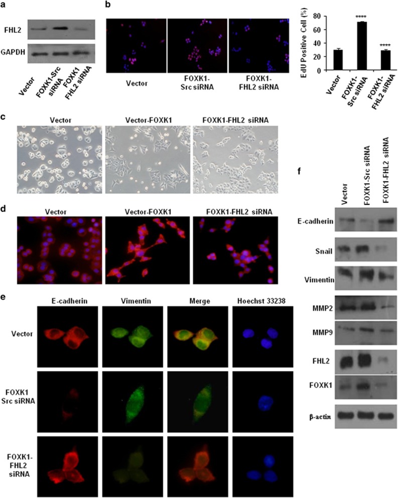
Figure 4.
FOXK1 and FHL2 promote the proliferation and EMT in CRC cell. (a) Expression levels of FHL2 were detected by western blot analysis in SW480 cells, which were transfected with FOXK1 overexpressing plasmids, followed by transfection with FHL2 siRNA or Scr siRNA as a negative control. (b) SW480 stable transfectants of FOXK1, by transfection with FHL2 siRNA or Scr siRNA for 48 h, were subjected to the EdU incorporation assay; ****P<0.001. (c) The aberrant morphology of stably expressing FOXK1 transfected with FHL2 siRNA or src siRNA in SW480 cells, analysed by phase-contrast microscopy. (d) SW480 cells stained with rhodamine-phallotoxin for 48 h to identify F-actin filaments were visualized under fluorescent microscopy. (e) Immunofluorescence and microscopic visualization of E-cadherin (red) and vimentin (green) staining in Vector, FOXK1 src siRNA and FOXK1–FHL2-siRNA cells. (f) EMT biomarkers, including E-cadherin, vimentin, Snail, MMP2, MMP9, FOXK1 and FHL2, were detected by western blot 48 h after transfection. All the experiments were repeated three to four times with similar findings. Scale bars represent 100 μm in b, 20 μm in c and d, 10 μm in e.

