Abstract
BACKGROUND:
Pulmonary aspiration of gastric contents during tracheal intubation is a life-threatening complication in emergency patients. Rapid sequence intubation is commonly performed to prevent aspiration but is not associated with low risk of intubation related complications. Although it has been considered that aspiration can be prevented in the lateral position, few studies have evaluated the ability to prevent aspiration. Moreover, this position is not always a favorable position for tracheal intubation. If aspiration can be prevented in a clinically relevant semi-lateral position, it may be advantageous. We assessed the ability to prevent aspiration in the lateral position and various degrees of the semi-lateral position using a vomiting–regurgitation manikin model.
METHODS:
A manikin’s head was placed in the neutral, simple extension, or sniffing position. The amount of aspirated saline into the bronchi during simulated vomiting was measured at semi-lateral position angles of 0º to 90º in 10º increments. The difference in the vertical height between the mouth corner and the inferior border of the vocal cord was measured radiologically at each semi-lateral position in the three head-neck positions.
RESULTS:
Pulmonary aspiration was prevented at the ≥70º, ≥80º, and 90º semi-lateral positions in the neutral, simple extension, and sniffing positions, respectively. The mouth was lower than the vocal cord in the semi-lateral position in which aspiration was prevented.
CONCLUSION:
The lateral or excessive semi-lateral position was necessary to protect the lung from aspiration in the head-neck positions commonly used for tracheal intubation. Prevention of aspiration was difficult within clinically relevant semi-lateral positions.
KEY WORDS: Pulmonary aspiration, Lateral position, Semi-lateral position
INTRODUCTION
Special attention should be paid to tracheal intubation in emergency patients who are at high risk for aspiration of gastric contents.[1,2] Rapid sequence intubation (RSI) is the standard for definitive airway management in these patients.[3,4] Even with RSI, aspiration of gastric contents is not avoided when vomiting or regurgitation occurs at an interval before tracheal intubation.[5] This problem remains unsolved despite the fact that aspiration is a life-threatening complication associated with tracheal intubation in emergency patients.
When vomiting and regurgitation occur, gastric contents flow from the esophagus into the pharynx. These contents are then expelled from the mouth or enter the larynx and trachea. The direction of their flow depends on the differences in the vertical height of the mouth and larynx.[6] If the mouth is placed more inferiorly than the larynx in the supine position, gastric contents do not enter the trachea; this is made possible by the use of the head-down tilt. Thus, some emergency physicians and anesthesiologists argue in favor of the head-down position during attempt in tracheal intubation in patients at risk of aspiration of gastric contents.[1,7,8] When the patient lay supine in the head-neck positions commonly used for tracheal intubation, the head-down tilt required to protect the trachea and lung from aspiration is not within a clinically relevant range.[6] Thus it is difficult to prevent aspiration of the gastric contents in the supine position.
In contrast, it has been considered that aspiration can be prevented in the lateral position because of the equal height of the mouth and larynx. This position has been recommended for tracheal intubation in patients at risk of aspiration.[9] However, to our knowledge, no studies have evaluated the ability to prevent aspiration in this position. Therefore, the first aim of this study was to assess the ability to prevent aspiration in the lateral position using three head-neck positions for tracheal intubation: the neutral, simple extension, and sniffing positions.
Tracheal intubation with a conventional laryngoscope in the lateral position is more difficult than that in the supine position.[10] Moreover, many emergency physicians and anesthesiologists may be unfamiliar with tracheal intubation in this position.[11] The lateral position is not always a favorable position for airway management. Aspiration of gastric contents into the airways of mechanically ventilated patients is an important cause of ventilator-associated pneumonia.[12] A previous study demonstrated that the 45º semi-lateral position decreases the incidence of aspiration and the risk of ventilator-associated pneumonia.[13] If aspiration is prevented during intubation attempt in a clinically relevant semi-lateral position, this position may be advantageous for tracheal intubation. Our second aim was to determine the optimal degree of the semi-lateral position required to prevent aspiration in the three head-neck positions for tracheal intubation.
METHODS
This was an observational study designed to assess the ability to prevent aspiration in the lateral and semi-lateral positions. The Institutional Ethical Committee determined this study was exempt because it did not involve human subjects.
Simulated pulmonary aspiration caused by vomiting-regurgitation using the Airway Trainer Manikin (Laerdal Medical, Stavanger, Norway). This manikin had the life-like and watertight structure of the upper airway and separation of the esophagus and trachea.[6] The manikin lay supine on the table without a pillow, and its head was neither extended nor flexed (neutral position). The esophagus and bronchi were connected to flexible tubes of 2.5 and 1.5 cm in diameter, respectively. The distal end of the esophageal tube was attached to a reservoir over the head, and the tube was clamped. The reservoir was filled with 1.0 L of normal saline colored with povidone iodine, which was the normal gastric content volume. The height of the saline was maintained at 130 cm above the center of the esophageal tube. After removing the clamp, the saline that entered the bronchial tube was collected in a beaker. After removing residual saline from the upper airway, trachea, and tubes, the table was rotated counterclockwise to an angle of 10º to 90º (10º–80º, semi-lateral position; 90º, lateral position) in 10º increments using a goniometer. At each semi-lateral or lateral position, the release of colored saline from the esophagus was repeated. The head-neck position was then changed from the neutral position to the simple extension position (in which the head was maximally extended without a pillow) and sniffing position (in which the head was maximally extended with a 7-cm-high pillow).[14,15] The order of the two positions was randomly determined using a sealed envelope technique. The release of colored saline from the esophageal tube was repeated in the same fashion as in the neutral position. Measurements were repeated 10 times at each semi-lateral position in each head-neck position. Two radiopaque marks were placed on the manikin; one at the left corner of the mouth and the other at the inferior border of the left vocal cord. Lateral cervical radiographs were taken in the supine, 10º to 80º left semi-lateral, and left lateral positions in the three head-neck positions. The researcher, who was blinded to the table rotation angle and head-neck position being used, measured the volume of saline that entered the bronchial tube and the difference in the vertical height between the mouth corner and the inferior border of the left vocal cord on each lateral cervical radiograph.
RESULTS
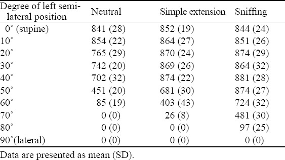
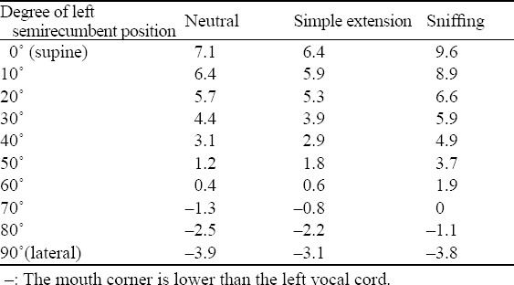
The aspiration of colored saline into the manikin bronchi after simulated vomiting and regurgitation at various degrees of the semi-lateral position in each head-neck position was shown in Table 1. To avoid pulmonary aspiration, the ≥70º semi-lateral, ≥80º semi-lateral, and lateral positions were needed in the neutral, simple extension, and sniffing positions, respectively. Radiological examination showed that the corner of the mouth was lower than the inferior border of the vocal cord in the ≥70º semi-lateral position in the three head-neck positions (Table 2). The vertical height of the three positions became equal at the 60º to 70º semi-lateral position in the three head-neck positions. Prevention of aspiration was impossible at the ≤60º, ≤70º, and ≤80º semi-lateral positions in the neutral, simple extension, and sniffing positions, respectively.
Table 1.
Volume (mL) of aspirated saline in the manikin bronchi after simulated vomiting and regurgitation in various degrees of left semi-lateral position in each head-neck position

Table 2.
Difference in vertical height (cm) between mouth corner and inferior border of left vocal cord in various degrees of left semi-lateral position in each head-neck position

DISCUSSION
This study showed that aspiration of gastric contents was prevented in the lateral position even when the manikin’s head was placed in the positions for tracheal intubation. The larynx is a midline structure, and the glottic opening is located in the center of the larynx. Thus, the corner of the mouth is always located more inferiorly than the glottic opening in the lateral position, which was demonstrated in our radiological study (Table 2). A difference in the vertical height between these tissues is an important determinant for the occurrence of pulmonary aspiration and is a possible reason for prevention of aspiration in the lateral position.[6] The findings in this manikin study should be clinically applicable.
The laryngoscopist often tries to place the head in the sniffing position when the glottis is poorly visualized during direct laryngoscopy.[16] When the sniffing position is achieved in the supine position, the difference in the vertical height between the mouth and larynx markedly increases secondary to neck flexion. Thus, excessive head-down tilt is needed to avoid pulmonary aspiration.[6] In contrast, the vertical heights of the mouth and larynx should be unchanged in the lateral position, even when placing the head in the sniffing position (Table 2). The validity of the lateral position in terms of preventing aspiration is not lost even in the sniffing position. This may be another advantage of the lateral position.
An excessive semi-lateral position was necessary to protect the trachea and lung from aspiration in the head-neck positions that we assessed. Our radiological study showed that the corner of the mouth was level with the glottic opening at a table rotation angle of 60º to 70º (Table 2), which supported the results in this manikin model for aspiration. In general, a means for preventing the patient from sliding is needed when the table is rotated or tilted to 25º.[17] Thus, 15º to 20º is a reasonable upper limit. These findings indicate that prevention of aspiration is difficult in clinically relevant semi-lateral positions. Laboratory studies have suggested that the lateral Trendelenburg position is effective in preventing ventilator-associated pneumonia.[18,19] Pulmonary aspiration may be prevented during intubation attempt by the use of a combination of the head-down tilt and the semi-lateral position. Further investigations are needed.
The degree of aspiration was well correlated with the difference in the vertical height between the mouth corner and the larynx measured radiologically. Previous investigations have demonstrated that the lateral and 45º semi-lateral position decreases the incidence of aspiration and the risk of ventilator-associated pneumonia.[13,18,19] Measuring the difference in the height between these tissues may be effective in deciding the degree of the semi-lateral position required to prevent ventilator-associated pneumonia.
Implementing the lateral position may be only a method for preventing aspiration during tracheal intubation. Tracheal intubation with a conventional laryngoscope is difficult in this position, and many experienced anesthesiologists and emergency physicians are unfamiliar with it.[10,11] Therefore, airway management maneuvers should be devised. There is evidence regarding the usefulness of the laryngeal mask airway, illuminating stylet, and a combination of both devices for tracheal intubation in the lateral position.[20–22] The Airway Scope (HOYA, Tokyo, Japan) has potential as a useful airway device to secure the airway in the lateral position.[23] The choice of the lateral position during intubation attempt in emergency patients should be weighed in light of the severity of the risk for pulmonary aspiration, degree of difficulty in the airway, and the skill of the laryngoscopist.
There are some potential limitations of our experimental design. First, the configuration of the upper airway of the manikin that we used and its changes with the different head-neck positions may differ from those of patients. However, a human study of pulmonary aspiration by vomiting or regurgitation was not feasible. We examined the similarity of a number of manikins to the human airway in terms of, their watertight properties and the ease with which they can be placed in the simple extension position. We found that the Airway Trainer Manikin was the best at meeting these demands.[6] Second, this study was performed in the left semi-lateral or left lateral position because it is the first choice for managing the airway in the lateral position. Direct laryngoscopy is considered to be easy in the left lateral position because there is no need to shift the tongue to the opposite side.[10,11,23] Finally, many factors are involved in pulmonary aspiration during vomiting or regurgitation, including the type of aspirate and esophageal pressure. We did not examine aspiration of particulate matter, which sometimes produces airway obstruction and may change the direction of the flow of gastric contents. Esophageal pressure during vomiting or regurgitation was postulated to be 60 to 105 cmH2O, although it has not been extensively studied.[24,25] To simulate a more clinically urgent situation in terms of the occurrence of pulmonary aspiration, we used an esophageal pressure of 130 cmH2O, which was equal to that used by Bercker et al.[26]
In summary, prevention of aspiration of gastric contents was difficult in the semi-lateral position within clinically relevant ranges when placing the head in the neutral, simple extension, and sniffing positions. The lateral or excessive semi-lateral position was necessary to protect the trachea and lung from aspiration. Because the lateral position is not always a favorable position for tracheal intubation, the choice of this position should be weighed in light of the severity of the risk for pulmonary aspiration, degree of airway difficulty, and skill of the laryngoscopist.
Footnotes
Funding: None.
Ethical approval: The Institutional Ethical Committee determined this study was exempt because it did not involve human subjects.
Conflicts of interest: The authors declare there is no competing interest related to the study, authors, other individuals or organizations.
Contributors: Takenaka I participated in study design, conduct of the study, data analysis, and manuscript preparation. Aoyama K participated in manuscript preparation.
REFERENCES
- 1.Sellick BA. Cricoid pressure to control regurgitation of stomach contents during induction of anaesthesia. Lancet. 1961;2:404–406. doi: 10.1016/s0140-6736(61)92485-0. [DOI] [PubMed] [Google Scholar]
- 2.Green SM, Krauss B. Pulmonary aspiration risk during emergency department procedural sedation--an examination of the role of fasting and sedation depth. Acad Emerg Med. 2002;9:35–42. doi: 10.1197/aemj.9.1.35. [DOI] [PubMed] [Google Scholar]
- 3.American College of Emergency Physicians. Rapid-sequence intubation. Ann Emerg Med. 1997;29:573. [PubMed] [Google Scholar]
- 4.Gebremedhn EG, Gebeyehu KD, Ayana HA, Oumer KE, Ayalew HN. Techniques of rapid sequence induction and intubation at a university teaching hospital. World J Emerg Med. 2014;5:107–111. doi: 10.5847/wjem.j.issn.1920-8642.2014.02.005. [DOI] [PMC free article] [PubMed] [Google Scholar]
- 5.Tayal VS, Riggs RW, Marx JA, Tomaszewski CA, Schneider RE. Rapid-sequence intubation at an emergency medicine residency: success rate and adverse events during a two-year period. Acad Emerg Med. 1999;6:31–37. doi: 10.1111/j.1553-2712.1999.tb00091.x. [DOI] [PubMed] [Google Scholar]
- 6.Takenaka I, Aoyama K, Iwagaki T. Combining head-neck position and head-down tilt to prevent pulmonary aspiration of gastric contents during induction of anaesthesia: a volunteer and manikin study. Eur J Anaesthesiol. 2012;29:380–385. doi: 10.1097/EJA.0b013e328354a51a. [DOI] [PubMed] [Google Scholar]
- 7.Cameron JL, Zuidema GD. Aspiration pneumonia. Magnitude and frequency of the problem. JAMA. 1972;219:1194–1196. doi: 10.1001/jama.219.9.1194. [DOI] [PubMed] [Google Scholar]
- 8.Cucchiara RF, Faust RJ. Patient positioning. In: Miller RD, editor. Anesthesia. 5th edition. Philadelphia: Churchill Livingston; 2000. pp. 1017–1032. [Google Scholar]
- 9.Kirchner E. Emergencies and aspiration. Can the usual methods for the prophylaxis of aspiration be further developed? Anaesthesist. 1978;27:119–126. [PubMed] [Google Scholar]
- 10.McCaul CL, Harney D, Ryan M, Moran C, Kavanagh BP, Boylan JF. Airway management in the lateral position: a randomized controlled trial. Anesth Analg. 2005;101:1221–1225. doi: 10.1213/01.ane.0000171712.44746.bb. [DOI] [PubMed] [Google Scholar]
- 11.Nathanson MH, Gajraj NM, Newson CD. Tracheal intubation in a manikin: comparison of supine and left lateral positions. Br J Anaesth. 1994;73:690–691. doi: 10.1093/bja/73.5.690. [DOI] [PubMed] [Google Scholar]
- 12.Chastre J, Fagon JY. Ventilator-associated pneumonia. Am J Respir Crit Care Med. 2002;165:867–903. doi: 10.1164/ajrccm.165.7.2105078. [DOI] [PubMed] [Google Scholar]
- 13.Mauri T, Berra L, Kumwilaisak K, Pivi S, Ufberg JW, Kueppers F, et al. Lateral-horizontal patient position and horizontal orientation of the endotracheal tube to prevent aspiration in adult surgical intensive care unit patients: a feasibility study. Respir Care. 2010;55:294–302. [PubMed] [Google Scholar]
- 14.Adnet F, Baillard C, Borron SW, Denantes C, Lefebvre L, Galinski M, et al. Randomized study comparing the “sniffing position” with simple head extension for laryngoscopic view in elective surgery patients. Anesthesiology. 2001;95:836–841. doi: 10.1097/00000542-200110000-00009. [DOI] [PubMed] [Google Scholar]
- 15.Takenaka I, Aoyama K, Iwagaki T, Kadoya T. The sniffing position provides greater occipito-atlanto-axial angulation than simple head extension: a radiological study. Can J Anesth. 2007;54:129–133. doi: 10.1007/BF03022009. [DOI] [PubMed] [Google Scholar]
- 16.Berry JM. Conventional (laryngoscopic) orotracheal and nasotracheal intubation (single lumen tube) In: Hagberg CA, editor. Benumof’s airway management: principles and practice. 2nd edition. Philadelphia: Mosby Elsevier; 2007. pp. 379–392. [Google Scholar]
- 17.Dixon BJ, Dixon JB, Carden JR, Burn AJ, Schachter LM, Playfair JM, et al. Preoxygenation is more effective in the 25 degrees head-up position than in the supine position in severely obese patients: a randomized controlled study. Anesthesiology. 2005;102:1110–1115. doi: 10.1097/00000542-200506000-00009. [DOI] [PubMed] [Google Scholar]
- 18.Panigada M, Berra L, Greco G, Stylianou M, Kolobow T. Bacterial colonization of the respiratory tract following tracheal intubation-effect of gravity: an experimental study. Crit Care Med. 2003;31:729–737. doi: 10.1097/01.CCM.0000049943.01252.E5. [DOI] [PubMed] [Google Scholar]
- 19.Zanella A, Cressoni M, Epp M, Hoffmann V, Stylianou M, Kolobow T. Effects of tracheal orientation on development of ventilator-associated pneumonia: an experimental study. Intensive Care Med. 2012;38:677–685. doi: 10.1007/s00134-012-2495-2. [DOI] [PubMed] [Google Scholar]
- 20.Dimitriou V, Voyagis GS, Latrou C, Brimacombe J. Flexible lightwand-guided intubation using the intubating laryngeal mask airwayTM in the supine, right, and left lateral positions in healthy patients by experienced users. Anesth Analg. 2003;96:896–898. doi: 10.1213/01.ANE.0000048839.12552.50. [DOI] [PubMed] [Google Scholar]
- 21.Komatsu R, Nagata O, Sessler DI, Ozaki M. The intubating laryngeal mask airway facilitates tracheal intubation in the lateral position. Anesth Analg. 2004;98:858–861. doi: 10.1213/01.ane.0000100741.46539.6b. [DOI] [PMC free article] [PubMed] [Google Scholar]
- 22.Biswas BK, Agarwal B, Bhattacharyya P, Badhani UK, Bhattarai B. Intubating laryngeal mask for airway management in lateral decubitus state: Comparative study of right and left lateral positions. Br J Anaesth. 2005;95:715–718. doi: 10.1093/bja/aei226. [DOI] [PubMed] [Google Scholar]
- 23.Takenaka I, Aoyama K, Iwagaki T, Kadoya T. Efficacy of the Airway Scope on tracheal intubation in the lateral position: comparison with the Macintosh laryngoscope. Eur J Anaesthesiol. 2011;28:164–168. doi: 10.1097/EJA.0b013e328340c368. [DOI] [PubMed] [Google Scholar]
- 24.Fanning GL. The efficacy of cricoid pressure in preventing regurgitation of gastric contents. Anesthesiology. 1970;32:553–555. doi: 10.1097/00000542-197006000-00019. [DOI] [PubMed] [Google Scholar]
- 25.Brimacombe J, Keller C. Hypopharyngeal seal pressure during projectile vomiting with the ProSealTM laryngeal mask airway: a case report and laboratory study. Can J Anesth. 2006;53:328. doi: 10.1007/BF03022230. [DOI] [PubMed] [Google Scholar]
- 26.Bercker S, Schmidbauer W, Volk T, Bogusch G, Bubser HP, Hensel M, et al. A comparison of seal in seven supraglottic airway devices using a cadaver model of elevated esophageal pressure. Anesth Analg. 2008;106:445–448. doi: 10.1213/ane.0b013e3181602ae1. [DOI] [PubMed] [Google Scholar]


