Abstract
Transcriptional activators interact with diverse proteins and recruit transcriptional machinery to the activated promoter. Recruitment of the Mediator complex by transcriptional activators is usually the key step in transcriptional activation. However, it is unclear how Mediator recognizes different types of activator proteins. To systematically identify the subunits responsible for the signal- and activator-specific functions of Mediator in Drosophila melanogaster, each Mediator subunit was depleted by RNA interference, and its effect on transcriptional activation of endogenous as well as synthetic promoters was examined. The depletion of some Mediator gene products caused general transcriptional defects, whereas depletion of others caused defects specifically related to activation. In particular, MED16 and MED23 were required for lipopolysaccharide- and heat-shock-specific gene expression, respectively, and their activator-specific functions appeared to result from interaction with specific activators. The corequirement of MED16 for other forms of differentiation-inducing factor-induced transcription was confirmed by microarray analysis of differentiation-inducing factor (DIF)- and MED16-depleted cells individually. These results suggest that distinct Mediator subunits interact with specific activators to coordinate and transfer activator-specific signals to the transcriptional machinery.
Environmental signals induce specific cellular responses. Cell surface receptors and their individual intracellular signaling pathways provide most of the specificity needed to evoke the appropriate cellular responses by activating particular transcription factors (1, 2). For example, heat-shock treatment induces the expression of molecular chaperons (3–5). In response to heat shock, trimerized heat-shock factor (HSF) proteins bind to heat-shock promoters and recruit the Mediator complex to drive the arrested RNA polymerase II into productive elongation. On the other hand, lipopolysaccharide (LPS) treatment of SL2 cells induces a cellular innate immune response mainly manifested by the expression of antimicrobial peptides (AMPs) (6, 7). Nuclear transport and binding of Drosophila NF-κB homologues Relish, differentiation-inducing factor, and dorsal to the AMP promoters in response to LPS induces transcriptional activation of the AMP genes (8, 9). Despite the difference in the environmental triggers and their signaling pathways, both types of activator require the Mediator complex to activate their target genes.
Diverse transcriptional activation processes have been shown to require the Mediator complex. We have reported previously that the MED17 subunit of the Mediator complex is required for transcriptional activation of Drosomycin when Toll is activated (10). MED23 and MED1 have been shown to mediate transcriptional activation of E1A (11, 12) and ligand-bound nuclear receptors (13, 14), respectively, in the mouse. In addition, a recent study revealed that MED15 is a critical mediator of transforming growth factor type β/activin/Nodal signaling by means of SMAD2/3 activators (15), and that MED25 is a specific binding target of the VP16 activator protein (16, 17). All these studies indicate that eukaryotic Mediator plays an important role in gene regulatory pathways. However, it is not known whether the above transcriptional effects result from activator specificity of the Mediator subunits.
To understand the specificity of the Mediator complex in transcriptional activation, we examined the effect of depleting individual Mediator subunits one by one by RNA interference (RNAi) on transcriptional activation in response to natural signals (LPS and heat shock) or to the overexpression of synthetic activator proteins (DIF or HSF). We found that MED16 and MED23, of the 23 Mediator proteins tested, were required for DIF- and HSF-mediated transcriptional activation, respectively. GST pull-down assays revealed that DIF and HSF fusion proteins bound to different Mediator proteins. In particular DIF was found to bind specifically to MED16. Microarray analysis of Dif-, Med16-, and Med23-depleted cells revealed that Med16, but not Med23, is required for transcription of all of the genes activated by DIF upon LPS treatment. We conclude that different transcriptional activator proteins depend on interaction with particular target proteins of the Mediator complex to activate transcription.
Materials and Methods
Plasmids. pG5-E1b-luciferase was constructed by replacing the chloramphenicol acetyltransferase cassette of pG5-E1b-chloramphenicol acetyltransferase (a gift from Michael Green, University of Massachusetts, Amherst, MA) with the luciferase gene. To construct the DNA templates for synthesis of double-stranded RNA (dsRNA), Med1 (+1 to +1,019; these and the numbers in the following genes indicate the nucleotide positions relative to the initiation codon at +1), Med4 (+1 to +827), Med6 (+1 to +603), Med7 (+1 to +709), Med10 (+1 to +402), Med11 (+1 to +531), Med12 (+2,870 to +3,647), Med14 (+1,298 to +2,277), Med16 (+1 to +759), Med17 (+262 to +983), Med18 (+1 to +618), Med20 (+1 to +829), Med21 (+1 to +429), Med22 (+1 to +432), Med23 (+490 to +1,258), Med24 (+1 to +985), Med25 (+1 to +794), Med26 (+1 to +653), Med27 (+1 to +882), Med28 (+1 to +570), Med30 (+1 to +957), Med31 (+1 to +615), CycC (+1 to +588), Rpb2(+540 to +1,463), TATA-binding protein (TBP) (+1 to +1,062), luciferase (+1 to +1,305), and GFP (+1 to +720) were cloned into pBluescript II KS(+) (Stratagene) and designated pdX, where X is the symbol of the gene cloned; the symbols for the Mediator genes are according to the unified nomenclature for Mediator subunits (18).
GST Pull-Down Assays. GST pull-down experiments were carried out as described in ref. 19. The supernatants were incubated with glutathione-Sepharose beads (Amersham Pharmacia) preequilibrated with sonication buffer for 1 h at 4°C, and the beads were washed three times with sonication buffer and once with IP100 buffer (20 mM Hepes·KOH, pH 7.6/10% glycerol/0.1 mM EDTA/100 mM potassium acetate/1 mM DTT/1 mM benzamidine·HCl/0.02% Nonidet P-40). GST pull-down assays were performed by incubating 35S-radiolabled Mediator protein fragments produced in the TNT-coupled transcription–translation system (Promega) with 10 μl of beads retaining 2 μg of each GST fusion protein for 6 h at 4°C. After washing the beads with IP300 buffer (20 mM Hepes·KOH, pH 7.6/10% glycerol/0.1 mM EDTA/300 mM potassium acetate/1 mM DTT/1 mM benzamidine·HCl/0.02% Nonidet P-40), bound proteins were eluted with SDS gel sample buffer and analyzed by autoradiography along with 1/20th of the input proteins.
RNAi Analysis. To produce single-stranded RNAs (ssRNAs) of the genes tested, in both directions, pBluescript II KS(+) plasmids containing a gene of interest were digested with BssHII, and the small fragments containing the T7 and T3 promoter sequences, respectively, at the ends of each template, were purified. By using the purified fragments as templates, ssRNA products were prepared with MEGAscript T7 and T3 transcription kits (Ambion, Austin, TX) and purified on Micro Bio-Spin 6 Chromatography columns (Bio-Rad). The ssRNAs derived from each gene were mixed and annealed to make dsRNAs by incubation at 65°C for 30 min followed by slow cooling to room temperature. The dsRNAs were analyzed by agarose gel (1%) electrophoresis to ensure that most of the RNA migrated as a single band of the expected size.
Drosophila SL2 cells were diluted to a final concentration of 1 × 10⁶ cells per ml in serum-free HyQ-CCM3 medium (HyClone, Logan, UT), and 4 ml of the suspension was plated in 35-mm flasks (Nunc). Transfection of dsRNA (6.25 μg) by using Cellfectin reagent (GIBCO/BRL) was performed according to the manufacturer's protocol. The transfected cells were incubated for 4 days at 25°C before being examined for RNAi.
Quantitative RT-PCR Analysis. Total RNA (5 μg) isolated from the SL2 cells was used for cDNA synthesis by the SuperScript II reverse transcriptase system (Invitrogen). The level of the transcript present in each cDNA sample was measured by real time PCR analysis with specific primers according to the manufacturer's instructions. The PCRs contained 1× SYBR Green mix (Applied Biosystems), 10 pmol of forward and reverse primers, and cDNA corresponding to 0.1 μg of total RNA. The reactions were subjected to 40 PCR cycles (95°C for 15 sec and 50°C for 1 min) in a Bio-Rad iCycler and detection system.
Luciferase Reporter Assays of Gal4 Fusion Activators. For luciferase reporter assay from chromosomal templates, a mix of SL2 cells containing the reporter at various chromosomal locations was isolated. SL2 cells were diluted to a final concentration of 1 × 10⁶ cells per ml in serum-free HyQ-CCM3 medium containing 10 μg/ml gentamycin (Invitrogen). The cells (4 ml) were transfected with pG5-E1b-luciferase (5 μg) and a plasmid bearing the hygromycin resistance gene (1 μg) by using Lipofectin (Invitrogen) and then were incubated at 25°C. After 4 days, the transfected cells were diluted to a final concentration of 4 × 10⁶ cells per ml in HyQ-CCM3 medium with 300 μg/ml hygromycin (Invitrogen) and incubated for a further 4 days. Thereafter, the cells were passaged by a 1:4 dilution with the same medium every 4 days for 45 days. The established cell lines containing the pG5-E1b-luciferase were transfected with expression constructs bearing the Gal4 fusion activator under the control of a metal inducible promoter (pMTG4-AD), along with a lacZ reporter controlled by an actin promoter (pActin-lacZ) after RNAi treatment as described above. For luciferase reporter assay from transiently transfected templates, the RNAi-treated SL2 cells (4 ml) were cotransfected with pG5-E1b-luciferase (5 μg) and pMTG4-AD, along with a pActin-lacZ. In both cases, the Gal4 fusion activators were induced with 0.7 mM Cu2+ for 12 h 24 h after transfection, and whole-cell lysates were prepared as described in Park et al. (10). The activities of firefly luciferase and β-galactosidase in the supernatant were analyzed with the Luciferase Assay system (Promega) and Galacto-Light Plus system (Tropix, Bedford, MA), respectively, according to the manufacturers' instructions.
cDNA Microarray Analysis. The cDNA microarrays used in this study contained 5,929 cDNA elements representing 5,405 different genes (based on data from the National Center for Biotechnology Information UniGene database accessed on August 1, 2003). Information concerning the cDNA elements is available from http://annotation.digital-genomics.co.kr/excel/fly_annotation.xls. All of the microarray experiments were done in duplicate with dye-swapping by using twin array cDNA chips from Digital Genomics (Seoul, Korea) as described in Kim et al. (20). To identify genes whose expression changed significantly in response to the LPS treatment, the Cy5-labeled RNAs from LPS-treated cells (for 1 h) were cohybridized with the Cy3-labeled RNAs from the nontreated cells. Four sets of microarray data (from twin arrays and the dye swap) were analyzed with sam (Significance Analysis of Microarray) in one class response format. Genes with significant expression changes (>1.7 fold) after LPS treatment were selected with 5% of a q value cutoff. To examine the effect of RNAi on LPS-induced RNA levels, total RNAs purified after LPS induction (1 h) from the Dif-, Med16-, or Med23-RNAi cells were cohybridized with the RNAs prepared from the LPS-treated luciferase RNAi cells. The microarray data were analyzed by scatter plot analysis in the brb array tool Version 3.0 software package from the National Cancer Institute.
Results
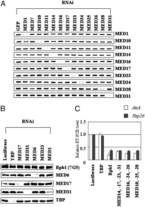
Depletion of Individual Mediator Gene Products by RNAi. To examine the specific requirement for each individual Mediator protein for signal-dependent transcriptional activation, individual Mediator gene products were eliminated by dsRNAi. dsRNA prepared from the 5′ region of each Mediator cDNA was introduced individually into SL2 cells, and the level of the corresponding Mediator mRNA was monitored by quantitative RT-PCR with primers designed to detect only the endogenous form of the corresponding Mediator mRNA (Fig. 1A). Most of the targeted mRNAs disappeared selectively within 2 days after the RNAi treatment. Western blot analysis of the RNAi-treated samples with antibodies against several Mediator subunits confirmed the absence of the corresponding protein. However, knockdown of MED1 by RNAi slightly decreased the level of MED31, indicating a possible connection between MED1 and MED31 proteins. We conclude that RNAi of individual Mediator subunits causes specific loss of that subunit, but that the elimination of certain subunits in the core region of the complex may cause concurrent partial loss of neighboring Mediator subunits, as has been shown in yeast and humans (11, 13).
Fig. 1.
Knockdown of Mediator genes by dsRNAi. (A) Quantitative RT-PCR of the Mediator mRNAs is indicated on the right after RNAi treatment for the genes, which is indicated at the top. (B) dsRNAi specifically depletes the corresponding Mediator protein. Total cell extracts from cells treated with the dsRNAs indicated at the top were analyzed by immunoblotting with antibodies against the proteins indicated on the right. (C) SL2 cells were treated with the mixture of dsRNAs indicated at the bottom. After treatment with an external stimulus (1-h LPS treatment for AttA; 30-min heat shock at 37°C for Hsp26), the levels of AttA and Hsp26 transcripts were determined in triplicate by quantitative RT-PCR. As a control, the level of rp49 transcript in each sample was determined and used to normalize the amount of the amplified RT-PCR product in each assay. The ratio of the mean values in cells treated with the transcription factor dsRNA to those in cells treated with luciferase dsRNA is presented with standard deviation
Signal-Dependent Transcriptional Defects in RNAi-Treated Cells. To understand whether depletion of Mediator causes particular signal-dependent transcriptional defects, transcriptional activation in response to two different environmental signals, LPS and heat shock, was examined for its Mediator requirements. Because of the incomplete nature of the RNAi technique in knocking out the activity of a target gene, it is not easy to observe the effect caused by a complete loss of Mediator function. Therefore, we calibrated the effects of the Mediator knockdowns against a knockdown of the RNA polymerase II subunit, Rpb2, and TBP to compare the magnitude of Mediator RNAi effects to complete ablation of transcription. We exposed each of the knockdown cells to LPS and heat shock and examined the resulting levels of AttA and Hsp26 mRNAs, typical components of the LPS and heat-shock-signaling pathways, respectively. Luciferase dsRNA, as a nonspecific dsRNA control, caused no reduction in either AttA or Hsp26 mRNA. When RNA polymerase II subunit Rpb2 was depleted, the RT-PCR levels of AttA and Hsp26 transcripts were decreased by 65–75% (Fig. 1C). Simultaneous treatment with multiple Mediator dsRNAs in various combinations to remove most Mediator activities decreased the levels of both AttA and Hsp26 transcripts to values comparable with those observed in Rpb2 knockdown cells (Fig. 1C). Because depletion of Rpb2 completely abolishes transcription activity, this roughly 3-fold reduction in semiquantitative RT-PCR levels appears to reflect complete loss of transcriptional activation and shows that Mediator is required for transcriptional activation of these genes. Intriguingly, depletion of TBP caused no detectable defects in transcriptional activation of AttA or Hsp26 (Fig. 1C). It is not clear whether TBP is not involved in the transcription of these genes or whether activation of RNA polymerase complexes bound to the promoter regions of these genes bypasses the requirement for TBP.
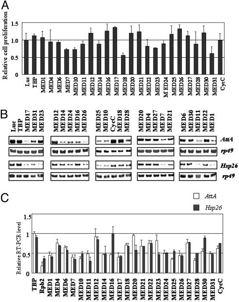
We also checked whether depletion of some Mediator subunits causes a strong physiological defect that might affect the interpretation of a Mediator subunit-specific requirement for transcriptional activation. To this end, we examined the effect of depletion of each Mediator subunit on cell proliferation. When total cell numbers were counted 4 days after the luciferase RNAi treatment, total cell numbers had increased ≈10-fold. There was a similar increase in cell numbers 4 days after incubation with Mediator RNAi (Fig. 2A), and thus no effect on cell proliferation. Only in the case of Med18 and Med31 RNAi was there an ≈40% reduction in proliferation. Having eliminated a general effect on cell proliferation, we searched for those Mediator genes whose depletion caused a >2-fold reduction in the levels of AttA or Hsp26 transcripts measured by RT-PCR (Fig. 2 B and C). Of the 23 Mediator subunits eliminated, 11 (MED1, MED7, MED10, MED11, MED14, MED17, MED24, MED25, MED26, MED28, and MED31) caused a clear-cut defect (>2-fold reduction) in transcriptional activation of both AttA and Hsp26, whereas deficiencies in 10 others (MED4, MED6, MED12, MED18, MED20, MED21, MED22, MED27, MED30, and CycC) had little or no effect (<1.6-fold reduction). Thus, not all of the Mediator proteins are required for transcriptional activation of AttA and Hsp26. The two remaining Mediator subunits proved to be required for particular transcriptional processes: MED16 for transcription of AttA and MED23 for transcription of Hsp26. The requirement of these subunits indicates that different components of the Mediator complex mediate different activation signals, and, in particular, that MED16 and MED23 are coactivators of activators involved in LPS and heat-shock-induced gene expression, respectively.
Fig. 2.
Requirement of each Mediator protein for LPS- and heat-shock-induced transcriptional activation. (A) Effect of Mediator depletion on cell proliferation. The relative fold increases of total cell number 4 days after each Mediator RNAi treatment compared with that of each luciferase RNAi sample are shown. The averages and standard deviations from four independent experiments are shown. (B) Cells treated for 4 days with the dsRNA indicated at the top were given either LPS treatment for 1 h or heat shock for 30 min at 37°C; thereafter the levels of AttA and Hsp26 transcripts together with those of rp49 were determined by quantitative RT-PCR. (C) RNAi-coupled RT-PCR analysis of AttA and Hsp26 transcriptional activation by LPS and heat shock was measured in triplicate, and transcript levels were normalized with the corresponding rp49 transcript level. To show the effect of the Mediator RNAi on LPS-induced (white bars) and heat-shock-induced (black bars) transcription, the ratio of the mean values in the Mediator dsRNA-treated cells to those in cells treated with luciferase dsRNA is presented with standard deviation.
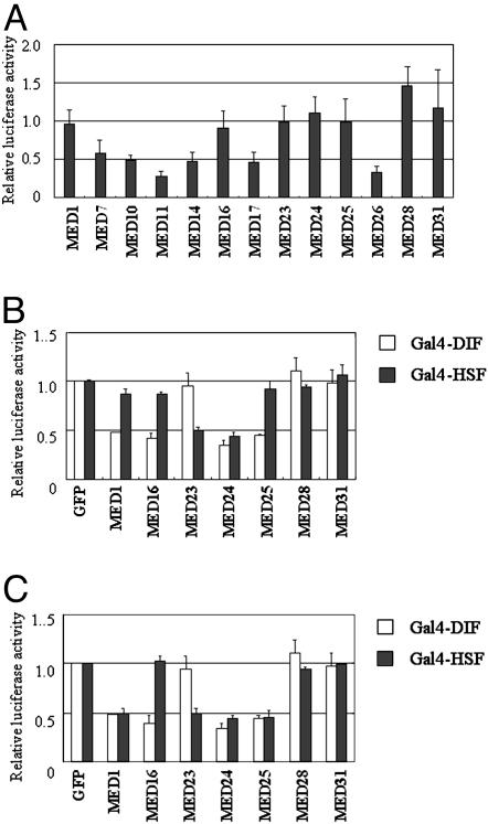
Activator-Specific Requirements for Mediator Protein. Some Mediator proteins, such as the ones that function as structural components needed to maintain the integrity of the Mediator complex, may be required for transcription generally, whereas others may be required specifically for the transfer of activation signals to RNA polymerase II. To determine which Mediator subunits are required generally for transcription, we inquired which of the 13 Mediator subunits that were shown above to be required for AttA or Hsp26 expression were also necessary for the basal level of transcription of a lacZ reporter gene from the actin promoter (Fig. 3A). Depletion of the products of six of these 13 genes, Med7, Med10, Med11, Med14, Med17, and Med26 reduced lacZ expression, and these subunits are presumably needed for general aspects of transcription shared by the various types of endogenous gene promoters and the partial actin promoter. Depletion of the other seven products, on the other hand, had no major effect on lacZ expression, and the fact that they are required for LPS- and heat-shock-induced transcriptional activation in vivo indicates that they are signal- or possibly activator-specific subunits of the Mediator complex.
Fig. 3.
Activator-specific Mediator proteins required for transcriptional activation. (A) Requirement of Mediator subunits for basal transcription. SL2 cells transfected with a lacZ reporter (pActin-lacZ) were treated with the dsRNA indicated at the bottom for 4 days. The ratios of the mean levels of lacZ activity in the Mediator dsRNA-treated cells to those in cells treated with luciferase dsRNA in five independent assays are presented with standard deviation. (B) Requirements of Mediator subunits for activator-specific transcriptional activation from chromosomal templates. After induction of the Gal4-fusion DIF activator (white bars) or Gal4-fusion HSF activator (black bars), luciferase activity induced by the activator proteins was measured in triplicate and normalized by the lacZ activity. The ratios of the mean levels of the normalized luciferase activity to that of the GFP dsRNA-treated cells in four experiments are shown with standard deviation. (C) Requirement of Mediator subunits for activator-specific transcriptional activation from transiently transfected templates. The effect of Mediator depletion indicated at the bottom on transcriptional activation from transiently transfected templates is shown as described for B.
We therefore examined the role of these seven transcriptional activation-specific subunits in combination with defined transcriptional activator proteins consisting of the Gal4 DNA-binding domain fused to the activation domain of DIF or HSF. To this end, we first isolated a mixture of cells containing the luciferase reporter at various chromosomal locations, then examined the effect of Mediator RNAi on their luciferase expression upon induction of the Gal4 fusion activators. Depletion of Med1, Med24, and Med25 caused defects in both Gal4-DIF- and Gal4-HSF-driven transcriptional activation, whereas depletion of Med28 and Med31 had no major effect (Fig. 3B). Depletion of Med16 and Med23 resulted in defective activation by Gal4-DIF and Gal4-HSF, respectively, in agreement with the results obtained above in the analysis of the expression of the endogenous AttA and Hsp26 genes (Fig. 2C). To rule out the possibility that the expression of the activator protein itself was affected by the depletion of the Mediator subunit in each sample, we showed that the levels of Gal4-DIF or Gal4-HSF in all of the Mediator knockdown cells tested were expressed at comparably high levels (Fig. 6, which is published as supporting information on the PNAS web site). This result confirmed that the transcriptional defects are due to the absence of Mediator subunits required for reception of activator signals. We repeated the experiment by using only transiently transfected cells as described in Materials and Methods and obtained a similar result, confirming the specific use of Mediator subunits for distinct transcriptional activation processes (Fig. 3C). However, depletion of MED1 and MED25 did not cause defects in Gal4-HSF-driven transcriptional activation (Fig. 3C), contrary to the previous result from the integrated reporter (Fig. 3B). This discrepancy may result from the different natures of the chromatin structures affecting transcriptional activation by distinct activator proteins. Therefore, MED16 and MED23 may be direct coactivators of DIF and HSF, respectively, and MED1, MED24, and MED25 may function in aspects of activation mechanisms shared by the two transcriptional activator proteins. The absence of Gal4-DIF- and Gal4-HSF-driven transcriptional defects in the MED28- and MED31-deficient cells indicates that they may be required for transcriptional activation of endogenous genes, perhaps by interacting with additional transcriptional activator proteins or coactivator complexes.
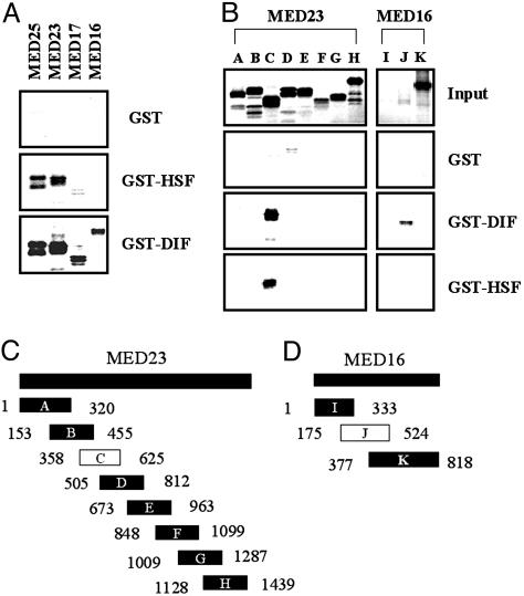
Activator-Specific Binding Targets of Mediator. As pointed out above, the activator-specific requirements for MED16 and MED23 suggest that they may interact directly with DIF and HSF, respectively. Because other Mediator proteins have been shown to bind to activators, all of the Mediator proteins were tested systematically for interaction with activator proteins. The 23 components of Mediator were isotopically labeled by in vitro translation, and their interactions with activator proteins were systematically analyzed by GST pull-down assays with fusions of GST with the HSF and DIF activation domains (Fig. 4 and data not shown). MED17 (with a higher affinity to GST-DIF), MED23, and MED25 turned out to bind strongly to both the DIF and HSF activation domains (Fig. 4A), whereas MED16 interacted only with the DIF activation domain. To map the regions of these Mediator subunits that interact with each of the activation domains, we examined the binding of a series of overlapping fragments of MED16, MED23, and MED25 (Fig. 4 B–D and data not shown). MED23 and MED25 interacted with the two activators by means of region C (Fig. 4C) and the carboxyl-terminal region of MED25 (amino acids 573–863; data not shown), respectively, whereas the J fragment of MED16 interacted only with the DIF activation domain (Fig. 4D). These results, taken together, indicate that a specific interaction between MED16 and DIF is required for DIF-induced transcriptional activation, whereas MED17 and MED25 are required for both DIF- and HSF-induced transcriptional activation. Interestingly, although MED23 is only required for transcriptional activation by HSF, it interacted with DIF and HSF in the in vitro binding assay. This result may be because DIF also interacts with MED16, so that the loss of the MED23 interaction may have only a minor effect on transcriptional activation by DIF compared with its effect on activation by HSF.
Fig. 4.
Activator-specific interaction of Mediator proteins. (A) The Mediator proteins indicated at the top were labeled, and their interactions with the DIF and HSF activation domains (GST-DIF and GST-HSF, respectively) were monitored by GST pull-down assays. As a negative control, GST protein alone (GST) was used. (C and D) A series of overlapping fragments of MED23 (C) and MED16 (D) is shown along with the amino acid positions at each end. The white boxes indicate the fragments that interact with the activation domains.
Requirement for MED16 for DIF-Mediated Transcriptional Activation During LPS Induction. Microarray analysis of LPS-induced changes in the transcriptional profile of SL2 cells identified 92 LPS-responsive genes (>1.7-fold change, q < 0.05 of significance analysis of microarray; see Table 1, which is published as supporting information on the PNAS web site). The binding of LPS to the peptidoglycan-recognition protein receptor activates the inhibitory κB kinase and c-Jun N-terminal kinase (JNK) pathways, and the activated transcription factors downstream of each pathway turn on diverse innate immune responses (21–23). To identify those genes activated by DIF, SL2 cells were depleted of DIF by dsRNAi, and their LPS response was compared by microarray analysis with that of control SL2 cells treated with luciferase dsRNA (Fig. 5A). Depletion of DIF caused up-regulation of most of the LPS-induced genes in the JNK pathway, probably because of release from negative regulation of JNK activity by an NF-κB homologue (24, 25). However, AttA, CG5770, and CG32302 were reduced >2-fold when DIF was depleted, indicating that these three genes are activated by DIF during LPS induction. To find out whether the activator specificity of MED16 and MED23 affects the regulation of these genes, we examined their requirements by using a similar method (Fig. 5 B and C). Intriguingly, a similar microarray analysis with MED16 depleted revealed that only the same three gene transcripts were lowered substantially, whereas MED23 depletion had no discernable effect on any of the LPS-induced transcripts. This result suggests that MED16 is a specific binding partner of the DIF-related transcriptional activator proteins, whereas MED23 and MED25 serve as additional binding partners for other types of transcriptional activator proteins.
Fig. 5.
General requirement of MED16 for DIF-induced transcription. Shown are scatter plot analyses of microarray data. The microarray data represented on the x axes show the log ratios of the RNA levels before and after LPS treatment (1 h). The microarray data on the y axes give the log ratios of the LPS-induced RNA levels in the DIF-depleted (A), MED23-depleted (B), or MED16-depleted (C) cells to those in the control (luciferase dsRNA-treated) cells. Only those genes shown by sam analysis to change their RNA levels significantly (q < 0.05, >1.7-fold) upon LPS treatment are plotted with their values in each microarray experiment. The three genes that showed >2-fold reductions in their RNA levels under DIF-depleted conditions are indicated.
Discussion
We have shown that several Mediator subunits are required for general aspects of transcriptional activation (26, 27), whereas others are only required for the expression of a defined group of genes (28, 29). The modular structure of the Mediator complex suggests that Mediator proteins that have a scaffold function or are associated with the basal transcription machinery are required for most transcriptional processes. On the other hand, those Mediator proteins that exhibit activator-dependent activity may either interact with particular activator proteins or relay the activation signal to the basal transcription machinery (30–32). We have identified a subset of Mediator proteins that bind to DIF and have shown that MED16 is a specific biding partner of the DIF activator. DIF interacts not only with MED16 but also with MED17, MED23, and MED25, and these interactions, apart from the one involving MED23, are essential for DIF-induced transcriptional activation. HSF also interacts with MED17, MED23, and MED25, but not MED16, and each of these interactions is required for HSF-induced transcriptional activation from chromosomal promoters. The presence of multiple activator-binding sites in the Mediator complex appears to reflect the requirement for a strong interaction between activator and Mediator complex for productive transcriptional activation and may also provide the specificity needed for interaction with distinct types of transcriptional activator proteins. In particular, MED16, which interacts with DIF, and MED23, whose interaction with HSF is essential in the absence of MED16, may act as key elements in eliciting activator-specific functions. It is striking that Med16 and Med23 are homologues of yeast sin4 and gal11, respectively. The latter are coregulators involved in carbon source metabolism and yeast cell flocculation and form an activator-binding module. Therefore, the roles of sin4 and gal11 as activator-specific binding targets appear to have been conserved in evolution.
Although we have not demonstrated any physical interaction of MED1 and MED24 with activator proteins (13, 33), both are required for DIF- or HSF-induced transcriptional activation without apparently being required for basal transcription from the actin promoter. These proteins may help the activators to bind strongly to the target proteins identified above or act downstream at the postactivator binding stage. It is also possible that they interact with activator proteins under more physiological conditions. Depletion by RNAi of several Mediator proteins, including MED17, also affected basal transcription from the ubiquitously expressed actin promoter. Our biochemical analysis of the yeast Mediator complex shows that it is required not only for transcriptional activation but also for basal transcription, and the Mediator proteins in the module that interact with RNA polymerase II are responsible for the latter activity (34, 35). Therefore, many of the Mediator proteins involved in basal transcription from the actin promoter may interact with the transcription machinery after the activator proteins have bound to their target sites in the Mediator complex. The defects in transcription from the actin promoter in cells depleted of MED14 and MED17, the Drosophila homologues of the yeast Mediator scaffold proteins RGR1 and SRB4, respectively, also indicate that disintegration of the Mediator complex as the result of depletion of a subunit that function as a scaffold protein may cause transcriptional defects that are detectable by RNAi (1, 29, 36, 37).
Although MED28 and MED31 were absolutely required for LPS- and heat-shock-induced activation of endogenous genes, they were dispensable for Gal4-DIF- and Gal4-HSF-induced transcription of a synthetic promoter. This difference may result from the absence of additional activator proteins that act on the natural promoter or from the different chromosomal context of the transiently transfected template from the endogenous one. If that is the case, MED28 and MED31 may function to integrate these additional signals during the activation process (38–40).
Supplementary Material
Acknowledgments
We thank Dr. Michael Green for plasmids. Microarray analyses were performed by using brb array tools v.3.x developed by Dr. Richard Simon and Amy Peng Lam. This work was supported by a grant from the Creative Research Initiatives Program of the Korean Ministry of Science and Technology (to Y.-J. Kim).
This paper was submitted directly (Track II) to the PNAS office.
Abbreviations: RNAi, RNA interference; dsRNA, double-stranded RNA; LPS, lipopolysaccharide; HSF, heat-shock factor; TBP, TATA-binding protein; DIF, differentiation-inducing factor.
References
- 1.Koh, S. S., Ansari, A. Z., Ptashne, M. & Young, R. A. (1998) Mol. Cell 1, 895-904. [DOI] [PubMed] [Google Scholar]
- 2.Koleske, A. J. & Young, R. A. (1994) Nature 368, 466-469. [DOI] [PubMed] [Google Scholar]
- 3.Wisniewski, J., Orosz, A., Allada, R. & Wu, C. (1996) Nucleic Acids Res. 24, 367-374. [DOI] [PMC free article] [PubMed] [Google Scholar]
- 4.Rasmussen, E. B. & Lis, J. T. (1993) Proc. Natl. Acad. Sci. USA 90, 7923-7927. [DOI] [PMC free article] [PubMed] [Google Scholar]
- 5.Park, J. M., Werner, J., Kim, J. M., Lis, J. T. & Kim, Y. J. (2001) Mol. Cell 8, 9-19. [DOI] [PubMed] [Google Scholar]
- 6.Ip, Y. T., Reach, M., Engstrom, Y., Kadalayil, L., Cai, H., Gonzalez-Crespo, S., Tatei, K. & Levine, M. (1993) Cell 75, 753-763. [DOI] [PubMed] [Google Scholar]
- 7.Kim, Y. S., Ryu, J. H., Han, S. J., Choi, K. H., Nam, K. B., Jang, I. H., Lemaitre, B., Brey, P. T. & Lee, W. J. (2000) J. Biol. Chem. 275, 32721-32727. [DOI] [PubMed] [Google Scholar]
- 8.Khush, R. S., Leulier, F. & Lemaitre, B. (2001) Trends Immunol. 22, 260-264. [DOI] [PubMed] [Google Scholar]
- 9.Dushay, M. S., Roethele, J. B., Chaverri, J. M., Dulek, D. E., Syed, S. K., Kitami, T. & Eldon, E. D. (2000) Gene 246, 49-57. [DOI] [PubMed] [Google Scholar]
- 10.Park, J. M., Kim, J. M., Kim, L. K., Kim, S. N., Kim-Ha, J., Kim, J. H. & Kim, Y. J. (2003) Mol. Cell. Biol. 23, 1358-1367. [DOI] [PMC free article] [PubMed] [Google Scholar]
- 11.Stevens, J. L., Cantin, G. T., Wang, G., Shevchenko, A. & Berk, A. J. (2002) Science 296, 755-758. [DOI] [PubMed] [Google Scholar]
- 12.Cantin, G. T., Stevens, J. L. & Berk, A. J. (2003) Proc. Natl. Acad. Sci. USA 100, 12003-12008. [DOI] [PMC free article] [PubMed] [Google Scholar]
- 13.Ito, M., Okano, H. J., Darnell, R. B. & Roeder, R. G. (2002) EMBO J. 21, 3464-3475. [DOI] [PMC free article] [PubMed] [Google Scholar]
- 14.Ge, K., Guermah, M., Yuan, C. X., Ito, M., Wallberg, A. E., Spiegelman, B. M. & Roeder, R. G. (2002) Nature 417, 563-567. [DOI] [PubMed] [Google Scholar]
- 15.Kato, Y., Habas, R., Katsuyama, Y., Naar, A. M. & He, X. (2002) Nature 418, 641-646. [DOI] [PubMed] [Google Scholar]
- 16.Mittler, G., Stuhler, T., Santolin, L., Uhlmann, T., Kremmer, E., Lottspeich, F., Berti, L. & Meisterernst, M. (2003) EMBO J. 22, 6494-6504. [DOI] [PMC free article] [PubMed] [Google Scholar]
- 17.Yang, F., DeBeaumont, R., Zhou, S. & Naar, A. M. (2004) Proc. Natl. Acad. Sci. USA 101, 2339-2344. [DOI] [PMC free article] [PubMed] [Google Scholar]
- 18.Bourbon, H. M., Aguilera, A., Ansari, A. Z., Asturias, F. J., Berk, A. J., Bjorklund, S., Blackwell, T. K., Borggrefe, T., Carey, M., et al. (2004) Mol. Cell 14, 553-557. [DOI] [PubMed] [Google Scholar]
- 19.Park, J. M., Gim, B. S., Kim, J. M., Yoon, J. H., Kim, H. S., Kang, J. G. & Kim, Y. J. (2001) Mol. Cell. Biol. 21, 2312-2323. [DOI] [PMC free article] [PubMed] [Google Scholar]
- 20.Kim, S. N., Rhee, J. H., Song, Y. H., Park, D. Y., Hwang, M., Kim, J. E., Gim, B. S., Yoon, J. H., Kim, Y. J. & Kim-Ha, J. (2004) Neurobiol. Aging, in press. [DOI] [PubMed]
- 21.Dziarski, R. (2004) Mol. Immunol. 40, 877-886. [DOI] [PubMed] [Google Scholar]
- 22.Hoffmann, J. A. (2003) Nature 426, 33-38. [DOI] [PubMed] [Google Scholar]
- 23.Silverman, N., Zhou, R., Erlich, R. L., Hunter, M., Bernstein, E., Schneider, D. & Maniatis, T. (2003) J. Biol. Chem. 278, 48928-48934. [DOI] [PubMed] [Google Scholar]
- 24.Park, J. M., Brady, M., Ruocco, M., Sun, H., Williams, D., Lee, S., Kato, T., Richards, N., Chan, K., Mercurio, F., et al. (2004) Genes Dev. 18, 584-594. [DOI] [PMC free article] [PubMed] [Google Scholar]
- 25.Boutros, M., Agaisse, H. & Perrimon, N. (2002) Dev. Cell 3, 711-722. [DOI] [PubMed] [Google Scholar]
- 26.Han, S. J., Lee, J. S., Kang, J. S. & Kim, Y. J. (2001) J. Biol. Chem. 276, 37020-37026. [DOI] [PubMed] [Google Scholar]
- 27.Han, S. J., Lee, Y. C., Gim, B. S., Ryu, G. H., Park, S. J., Lane, W. S. & Kim, Y. J. (1999) Mol. Cell. Biol. 19, 979-988. [DOI] [PMC free article] [PubMed] [Google Scholar]
- 28.Li, Y., Bjorklund, S., Jiang, Y. W., Kim, Y. J., Lane, W. S., Stillman, D. J. & Kornberg, R. D. (1995) Proc. Natl. Acad. Sci. USA 92, 10864-10868. [DOI] [PMC free article] [PubMed] [Google Scholar]
- 29.Mizuno, T. & Harashima, S. (2003) Mol. Genet. Genomics 269, 68-77. [DOI] [PubMed] [Google Scholar]
- 30.Baek, H. J., Malik, S., Qin, J. & Roeder, R. G. (2002) Mol. Cell. Biol. 22, 2842-2852. [DOI] [PMC free article] [PubMed] [Google Scholar]
- 31.Hampsey, M. (1998) Microbiol. Mol. Biol. Rev. 62, 465-503. [DOI] [PMC free article] [PubMed] [Google Scholar]
- 32.Roeder, R. G. (1998) Cold Spring Harb. Symp. Quant. Biol. 63, 201-218. [DOI] [PubMed] [Google Scholar]
- 33.Ito, M., Yuan, C. X., Okano, H. J., Darnell, R. B. & Roeder, R. G. (2000) Mol. Cell 5, 683-693. [DOI] [PubMed] [Google Scholar]
- 34.Reeves, W. M. & Hahn, S. (2003) Mol. Cell. Biol. 23, 349-358. [DOI] [PMC free article] [PubMed] [Google Scholar]
- 35.Kang, J. S., Kim, S. H., Hwang, M. S., Han, S. J., Lee, Y. C. & Kim, Y. J. (2001) J. Biol. Chem. 276, 42003-42010. [DOI] [PubMed] [Google Scholar]
- 36.Shim, E. Y., Walker, A. K. & Blackwell, T. K. (2002) J. Biol. Chem. 277, 30413-30416. [DOI] [PubMed] [Google Scholar]
- 37.Boube, M., Faucher, C., Joulia, L., Cribbs, D. L. & Bourbon, H. M. (2000) Genes Dev. 14, 2906-2917. [DOI] [PMC free article] [PubMed] [Google Scholar]
- 38.Naar, A. M., Beaurang, P. A., Zhou, S., Abraham, S., Solomon, W. & Tjian, R. (1999) Nature 398, 828-832. [DOI] [PubMed] [Google Scholar]
- 39.Naar, A. M., Taatjes, D. J., Zhai, W., Nogales, E. & Tjian, R. (2002) Genes Dev. 16, 1339-1344. [DOI] [PMC free article] [PubMed] [Google Scholar]
- 40.Taatjes, D. J., Naar, A. M., Andel, F., III, Nogales, E. & Tjian, R. (2002) Science 295, 1058-1062. [DOI] [PubMed] [Google Scholar]
Associated Data
This section collects any data citations, data availability statements, or supplementary materials included in this article.


