Abstract
Purpose:
To quantitatively investigate the effect of range shifter materials on single-spot characteristics of a proton pencil beam.
Methods:
An analytic approximation for multiple Coulomb scattering (“differential Moliere” formula) was adopted to calculate spot sizes of proton spot scanning beams impinging on a range shifter. The calculations cover a range of delivery parameters: six range shifter materials (acrylonitrile butadiene styrene, Lexan, Lucite, polyethylene, polystyrene, and wax) and water as reference material, proton beam energies ranging from 75 to 200 MeV, range shifter thicknesses of 4.5 and 7.0 g/cm2, and range shifter positions from 5 to 50 cm. The analytic method was validated by comparing calculation results with the measurements reported in the literature.
Results:
Relative to a water-equivalent reference, the spot size distal to a wax or polyethylene range shifter is 15% smaller, while the spot size distal to a range shifter made of Lexan or Lucite is about 6% smaller. The relative spot size variations are nearly independent of beam energy and range shifter thickness and decrease with smaller air gaps.
Conclusions:
Among the six material investigated, wax and polyethylene are desirable range shifter materials when the spot size is kept small. Lexan and Lucite are the desirable range shifter materials when the scattering power is kept similar to water.
Keywords: range shifter material, proton pencil beam, spot size, scattering, bolus
1. INTRODUCTION
Proton beam radiation therapy has expanded rapidly in recent years, with proton pencil beam scanning (PBS) techniques being implemented in many new proton beam therapy centers.1 The pencil beam scanning method allows for the delivery of a more conformal dose to tumor and reduced dose from neutrons than passive scattering protons.2 Current pencil beam scanning systems, however, have a minimum proton energy limitation due to various technical constraints. For example, the Hitachi and IBA systems provide minimum proton energies of 70 MeV.3,4 In order to apply the pencil beam scanning technique to tumors located proximal to the minimum range, a range shifter is needed to degrade the beam energy.3,5,6 A range shifter is a uniform slab of material, usually plastic. Common plastics used include acrylonitrile butadiene styrene (ABS), Lexan, and polyethylene.3,5,6
It is well known that a proton pencil beam is broadened by a range shifter, and the broadening is reduced by moving the range shifter closer to the patient.7 However, it is not always possible to place the range shifter close to the patient, due to the possibility of a collision with the patient or support devices. As an alternative, a bolus may be placed on or very close to the patient.5 In some clinical situations, such as supine craniospinal irradiations, it may be advantageous to integrate the bolus into the patient support device.8 We will not differentiate the range shifter and bolus throughout the paper unless it is necessary.
Several publications have reported on proton range and fluence correction factors for various phantom materials.9–12 These studies focus mainly on the reference dosimetry by accounting for attenuation of primary protons and production of secondary particles due to nonelastic nuclear interactions. None of these publications address PBS spot scattering or specifically address the choice of range shifter material or position. The ideal material for a range shifter minimizes spot growth as the pencil beam propagates through the range shifter. In order to select desirable materials for a range shifter, a thorough investigation of their effect on the pencil beam characteristics is necessary. One evaluation of various range shifter materials was reported by Kanematsu et al.,13 but in the context of carbon-ion radiotherapy.
Commissioning of a range shifter within a treatment planning system (TPS) requires extensive measurements.7 To make efficient use of valuable commissioning time, there is a need for a comprehensive evaluation of range shifter materials, which focus on quantitative effect of range shifter material and position on beam spot characteristics.
The purpose of this paper is to quantitatively evaluate how range shifters of various material compositions affect spot size in different geometries. The results presented here may prove useful in selecting the most appropriate materials for range shifters. The structure of the paper is as follows: in Sec. 2, an analytic spot size formula is introduced and validated by comparison with measurements reported in the literature.5 Then, the analytic tool is applied to various delivery parameter combinations: air gaps ranging from 5 to 50 cm, proton energies from 70 to 200 MeV, and range shifter thicknesses of 7 g/cm2 and 4.5 g/cm2. Results for six commonly used range shifter materials are presented, with water as the reference material. Finally, Secs. 3 and 4 present variations in spot properties due to the various combinations of geometry and material.
2. METHODS AND MATERIALS
2.A. Spot size formula
The principal component of proton beam scattering is due to multiple Coulomb scattering (MCS). Several approximations for MCS have been reported.14–16 Gottschalk et al.14 reported that the Molière theory and Highland approximation agree with measurements to within 1%, on average. Since then, the generalized Highland approximation has been widely used for proton dose calculation algorithms in order to model proton scattering in the patient.17,18 According to Gottschalk,16 an improved differential Molière method including a nonlocal correction was introduced. We applied the analytic differential Molière method to evaluate the spot broadening in a slablike range shifter due to MCS. The advantage of this method is that it does not require beam time for measurements and can be easily expanded to different materials.
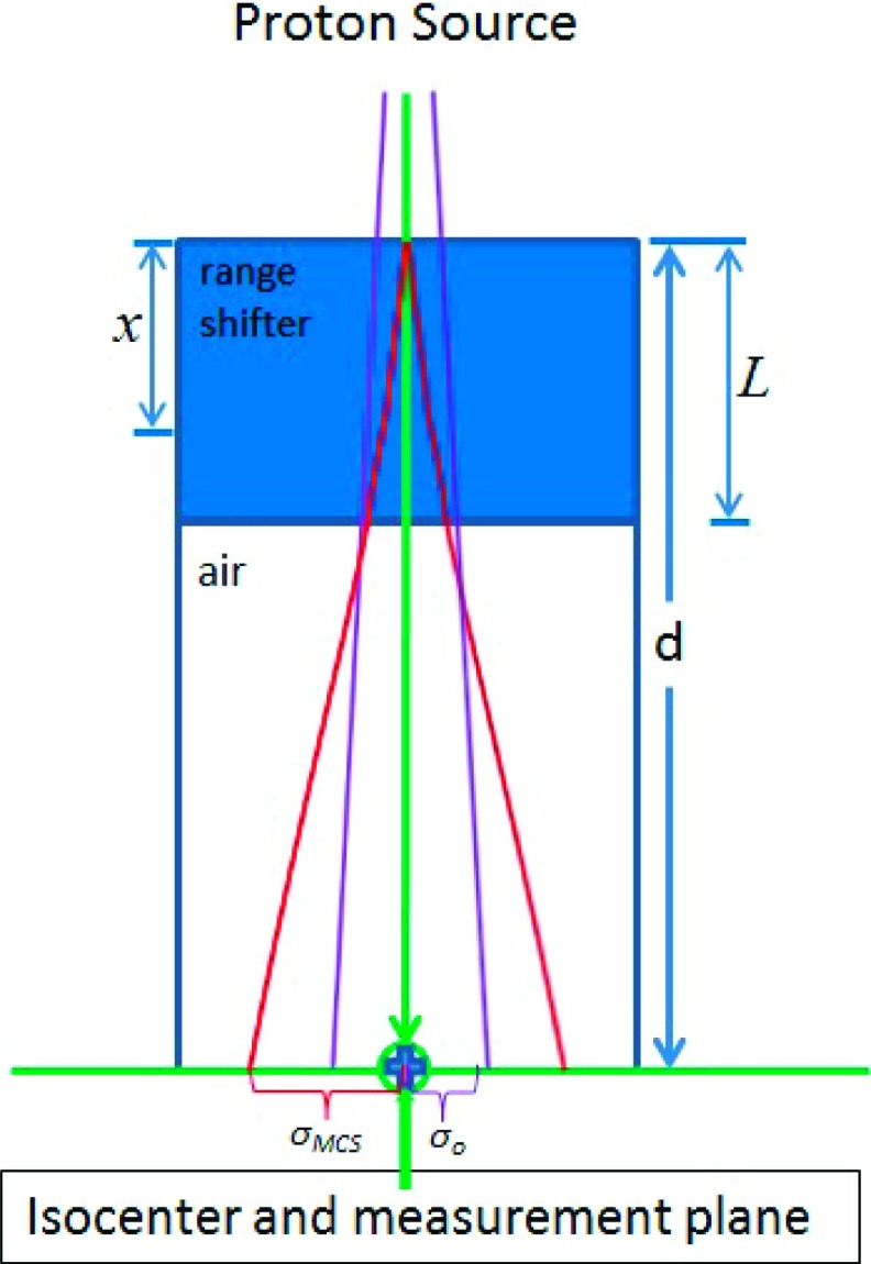
The setup geometry of the study is shown in Fig. 1. The proton beam passes through a slab followed by an air gap before reaching the isocenter. We define the original spot size in air without the range shifter as σo and the spot size with the range shifter as σrs; both quantities are defined at the isocenter. The spot broadening due to scattering in air is included in σo. Spot broadening due to the MCS in the range shifter is accounted for in σMCS. The schematic drawing of σo and σMCS is also shown in Fig. 1. According to Gottschalk et al.,14 the spot size after the range shifter is the quadratic sum of the spot size without range shifter (σo) and the broadening due to scattering by the range shifter (σMCS). Hence, the following formula for σrs holds:
| (1) |
FIG. 1.
Setup geometry of range shifter evaluation for spot sizes. The σo and σMCS are labeled in the figure. The effective source point due to the pure MCS lies inside the slab materials, and σMCS follows an asymptotic envelope. σrs (not shown) is the sum in quadrature of σo and σMCS [Eq. (1)].
The details of the analytic approximation have been described at length in the literature.14–16 Here, we briefly summarize the differential Molière method used in this study for the convenience of the reader. The spot size at the isocenter due to MCS may be obtained through integration of differential scattering along the slab,
| (2) |
In general, T(x) ≡ dθ2/dx is the rate of increase with x of the mean squared projected MCS angle. T(x) is nonlocal, so in addition to material properties and energy at the point of interest, it also depends on how much scattering has already taken place. Using the differential Molière method,16
| (3) |
The factor represents the nonlocal scattering effect, defined as from which the local scattering at the point of interest is related to the initial condition at the entrance point. The terms p0v0 and pv are products of proton momentum and speed at the entrance point and the point of interest, respectively. Es = 15.0 MeV, and XS denotes the scattering length, a quantity related to the radiation length.16 We note that σMCS depends on the incident beam energy, slab thickness, slab density, stopping power, and chemical composition. The nongeometric parameters may be found in International Commission on Radiation Units and Measurement (ICRU) Report 49.19
2.B. Validation of the method with measurements reported in the literature
In order to validate the analytic method described here, it was applied to the experimental setup described in Both et al.5 In that study, the spot sizes were measured at the isocenter for proton beam energies of 110–200 MeV in four scenarios: without a range shifter in the beam line, with a 7.4 g/cm2 Lexan range shifter positioned 34.5 cm upstream of isocenter, with a 5.5 g/cm2 wax-made bolus positioned at 8 and 2 cm upstream of isocenter. The spot size components σMCS due to the MCS in the range shifter or bolus were calculated using Eq. (2). The final spot sizes were calculated by adding the spot sizes σo (without degrader) and σMCS according to Eq. (1). The calculated spot sizes were then compared with the measurements of Both et al.5
2.C. Evaluation of spot sizes for different materials and distances
Next, six materials commonly used as range shifters were evaluated: ABS, Lexan, Lucite, polyethylene, polystyrene, and wax. Water was used as a reference material.5,8 The mass density, physical thickness, and the scattering length of each material are listed in Table I. The range shifters were positioned 50, 40, 30, 20, 10, and 5 cm from the isocenter. Proton pencil beams of energy 100 and 200 MeV for a 7 g/cm2 range shifter and 75 and 200 MeV for a 4.5 g/cm2 range shifter were used for spot size evaluation. Typical in-air spot sizes of σo = 4.5, 3.5, and 2.3 mm for the undegraded beam at the isocenter were used for 75, 100, and 200 MeV proton energies, respectively. In order to study the relative spot size variations due to different range shifter materials (σrs), the relative spot size variations from water, defined as (σwat − σrs)/σwat, were recorded.
TABLE I.
Physical parameters of the range shifter materials, taken from ICRU Report 49 (Ref. 19).
| Element weight | ||||||
|---|---|---|---|---|---|---|
| Material | ρ (g/cm3) | H | C | N | O | Scattering length (g/cm2) |
| Water | 1.0 | 0.1119 | — | — | 0.8881 | 46.88 |
| ABSa | 1.07 | 0.0811 | 0.8526 | 0.0663 | 58.66 | |
| Lexan | 1.20 | 0.0555 | 0.7558 | — | 0.1888 | 55.05 |
| Lucite | 1.19 | 0.0805 | 0.5998 | — | 0.3196 | 53.76 |
| Polyethylene | 0.94 | 0.1437 | 0.8563 | — | — | 61.79 |
| Polystyrene | 1.06 | 0.0774 | 0.9226 | — | — | 59.15 |
| Wax | 0.93 | 0.1486 | 0.8514 | — | — | 61.99 |
ABS is not listed in ICRU 49, in which stopping power is derived from its elements by the Bragg additivity theory.
3. RESULTS
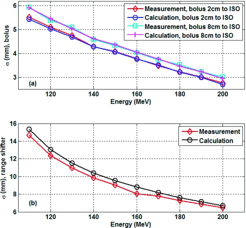
Figure 2 shows the comparison between the analytical method in this work and experimental results reported by Both et al.5 Figure 2(a) is for spot sizes with the wax-made bolus placed 2 and 8 cm proximal to the isocenter, and Fig. 2(b) is for spot sizes with a range shifter made of Lexan placed 34.5 cm proximal to the isocenter. Figure 2 shows that the calculated spot sizes in this study match measurements for both bolus and range shifter conditions, with the maximum deviation being less than 5.5%.
FIG. 2.
Comparison of the calculated spot sizes with measurements for a 5.5 cm WET wax bolus at 2 and 8 cm from the isocenter (a) and a 7.4 cm WET Lexan range shifter at 34.5 cm from the isocenter (b).
Table II shows spot sizes with range shifters of 7 g/cm2 composed of six materials and placed at six positions between 5 and 50 cm from the isocenter for proton beam energies of 100 and 200 MeV. Table III shows spot sizes with a range shifters of 4.5 g/cm2 composed of six materials and placed at six positions from the isocenter for proton beam energies of 75 and 200 MeV. The results in Tables II and III show that the spot sizes with a range shifter made of a material other than water are smaller than those with a range shifter made of water. The spot sizes after a range shifter made of wax or polyethylene are more than 15% smaller than those after a range shifter made of water when they are placed 50 cm proximal to the isocenter. The spot sizes with range shifters made of Lexan and Lucite are less than 6% smaller than those corresponding to water in the same geometry. For ABS and polystyrene, the relative difference to water is approximately 10%. The scattering lengths for all materials were shown in Table I. Smaller scattering lengths result in larger spot sizes. The relative spot size variations are nearly independent of beam energy and range shifter thickness, but do decrease as the range shifter is moved closer to the isocenter.
TABLE II.
Spot size variations in air for 100 and 200 MeV protons distal to various range shifter materials with energy loss equivalent to 7 cm of water, at distances ranging from 50 to 5 cm from the isocenter. Spot percents are given relative to water : % = (σwat − σrs)/σwat.
| Distance from ISO (cm), for 100 MeV Proton | Distance from ISO (cm), for 200 MeV Proton | ||||||||||||||||||||||||
|---|---|---|---|---|---|---|---|---|---|---|---|---|---|---|---|---|---|---|---|---|---|---|---|---|---|
| 50 | 40 | 30 | 20 | 10 | 5 | 50 | 40 | 30 | 20 | 10 | 5 | ||||||||||||||
| Material | L (cm) | σ (mm) | % | σ (mm) | % | σ (mm) | % | σ (mm) | % | σ (mm) | % | σ (mm) | % | σ (mm) | % | σ (mm) | % | σ (mm) | % | σ (mm) | % | σ (mm) | % | σ (mm) | % |
| Water | 7.00 | 26.4 | 21.4 | 16.5 | 11.7 | 7.1 | 5.1 | 9.2 | 7.6 | 6.0 | 4.5 | 3.2 | 2.7 | ||||||||||||
| ABS | 6.61 | 23.7 | 10.2 | 19.3 | 10.1 | 14.9 | 10.0 | 10.5 | 9.6 | 6.5 | 8.3 | 4.8 | 6.0 | 8.3 | 9.6 | 6.9 | 9.3 | 5.5 | 8.8 | 4.2 | 7.8 | 3.0 | 5.2 | 2.6 | 3.1 |
| Lexan | 6.12 | 24.9 | 5.8 | 20.2 | 5.9 | 15.5 | 6.0 | 11.0 | 6.0 | 6.7 | 5.8 | 4.8 | 4.9 | 8.7 | 5.8 | 7.2 | 5.7 | 5.7 | 5.6 | 4.3 | 5.2 | 3.1 | 4.0 | 2.6 | 2.7 |
| Lucite | 6.04 | 24.9 | 5.8 | 20.2 | 5.9 | 15.5 | 6.0 | 11.0 | 6.1 | 6.7 | 5.9 | 4.8 | 5.0 | 8.7 | 5.7 | 7.2 | 5.7 | 5.7 | 5.6 | 4.3 | 5.3 | 3.1 | 4.1 | 2.6 | 2.8 |
| Polyethylene | 6.99 | 22.4 | 15.3 | 18.2 | 15.2 | 14.1 | 14.9 | 10.0 | 14.1 | 6.3 | 11.6 | 4.7 | 7.9 | 7.9 | 14.4 | 6.6 | 13.9 | 5.3 | 13.0 | 4.0 | 11.2 | 3.0 | 7.1 | 2.6 | 4.0 |
| Polystyrene | 6.74 | 23.7 | 10.1 | 19.3 | 10.0 | 14.9 | 9.8 | 10.6 | 9.5 | 6.5 | 8.0 | 4.8 | 5.7 | 8.3 | 9.5 | 6.9 | 9.3 | 5.5 | 8.8 | 4.2 | 7.7 | 3.0 | 5.1 | 2.6 | 3.0 |
| Wax | 7.09 | 22.2 | 15.7 | 18.1 | 15.6 | 14.0 | 15.2 | 10.0 | 14.4 | 6.2 | 11.7 | 4.7 | 7.9 | 7.9 | 14.7 | 6.5 | 14.2 | 5.2 | 13.3 | 4.0 | 11.4 | 3.0 | 7.2 | 2.6 | 3.9 |
TABLE III.
Spot size variations in air for 75 and 200 MeV protons distal to various range shifter materials with energy loss equivalent to 4.5 cm of water at distances ranging from 50 to 5 cm from the isocenter. Spot percents are given relative to water : % = (σwat − σrs)/σwat.
| Distance from ISO (cm), for 75 MeV Proton | Distance from ISO (cm), for 200 MeV Proton | ||||||||||||||||||||||||
|---|---|---|---|---|---|---|---|---|---|---|---|---|---|---|---|---|---|---|---|---|---|---|---|---|---|
| 50 | 40 | 30 | 20 | 10 | 5 | 50 | 40 | 30 | 20 | 10 | 5 | ||||||||||||||
| Material | L (cm) | σ (mm) | % | σ (mm) | % | σ (mm) | % | σ (mm) | % | σ (mm) | % | σ (mm) | % | σ (mm) | % | σ (mm) | % | σ (mm) | % | σ (mm) | % | σ (mm) | % | σ (mm) | % |
| Water | 4.50 | 33.0 | 26.7 | 20.4 | 14.2 | 8.4 | 6.0 | 7.1 | 5.9 | 4.7 | 3.7 | 2.8 | 2.5 | ||||||||||||
| ABS | 4.25 | 29.7 | 10.1 | 24.0 | 10.0 | 18.4 | 9.9 | 12.9 | 9.4 | 7.8 | 7.5 | 5.7 | 4.7 | 6.4 | 9.0 | 5.4 | 8.5 | 4.4 | 7.7 | 3.4 | 6.1 | 2.7 | 3.3 | 2.4 | 1.5 |
| Lexan | 3.93 | 31.2 | 5.6 | 25.2 | 5.6 | 19.3 | 5.6 | 13.5 | 5.5 | 8.0 | 4.7 | 5.8 | 3.3 | 6.7 | 5.3 | 5.6 | 5.1 | 4.5 | 4.7 | 3.5 | 3.9 | 2.7 | 2.3 | 2.4 | 1.2 |
| Lucite | 3.88 | 31.2 | 5.6 | 25.2 | 5.6 | 19.3 | 5.5 | 13.5 | 5.4 | 8.0 | 4.7 | 5.8 | 3.3 | 6.7 | 5.2 | 5.6 | 5.0 | 4.5 | 4.7 | 3.5 | 3.9 | 2.7 | 2.3 | 2.4 | 1.3 |
| Polyethylene | 4.51 | 27.9 | 15.5 | 22.6 | 15.3 | 17.4 | 15.0 | 12.2 | 14.1 | 7.5 | 11.0 | 5.6 | 6.5 | 6.1 | 13.6 | 5.1 | 12.9 | 4.2 | 11.5 | 3.3 | 9.0 | 2.7 | 4.6 | 2.4 | 2.0 |
| Polystyrene | 4.33 | 29.7 | 10.0 | 24.0 | 10.0 | 18.4 | 9.8 | 12.9 | 9.3 | 7.8 | 7.4 | 5.7 | 4.5 | 6.4 | 9.0 | 5.4 | 8.5 | 4.4 | 7.7 | 3.4 | 6.1 | 2.7 | 3.2 | 2.4 | 1.5 |
| Wax | 4.58 | 27.8 | 15.9 | 22.5 | 15.8 | 17.3 | 15.4 | 12.2 | 14.5 | 7.5 | 11.2 | 5.6 | 6.6 | 6.1 | 14.0 | 5.1 | 13.2 | 4.2 | 11.8 | 3.3 | 9.2 | 2.6 | 4.6 | 2.4 | 2.0 |
4. DISCUSSION
Analytic methods were used to study the effects of different materials and geometries on spot sizes after the proton pencil beam passes through a range shifter. The results provide guidance on selecting appropriate materials for range shifters. A range shifter may considerably broaden proton beam spots and may lead to a degradation of treatment plan quality.5 Therefore, selecting the appropriate range shifter material may lead to a reduction in spot size. As shown in Tables II and III, wax and polyethylene range shifters result in 15% smaller spot sizes relative to water. They are desirable range shifter materials if minimizing spot sizes is a concern. In this context, it is interesting to note that the Paul Scherrer Institute (PSI) uses polyethylene for its range shifter.6
Ideally, a proton TPS would be capable of accurately modeling range shifters based on the open-beam measurements. However, this is not the case for all proton TPS. For example, the spot sizes distal to a range shifter modeled by Eclipse TPS are considerably smaller than the measurements, because broadening across the large air gap between the range shifter and the isocenter is ignored. To fully correct for this deficiency, a full complement of range shifter beam data must be collected.7 Therefore, it is much more convenient to be able to select a range shifter material based on theoretical models prior to beginning measurements.
The results of the range shifter also apply to bolus selection. However, the ideal materials for a bolus may differ, since the bolus is placed directly on the patient and may need to form to patient-specific anatomy. Historically, the bolus is commonly treated as water by a manual override of the electron density and adjustment of the external contour to match WET (Refs. 4 and 7), so for this reason, it may be advisable to choose materials with scattering properties close to water. As shown in Tables II and III, of the materials studied, Lexan and Lucite have scattering properties most similar to water.
According to Both et al.,5 the spot sizes with and without boluses are nearly identical when the bolus is placed close to the patient. According to Tables II and III, the spot size differences among selected materials are less than 4% for range shifter placement 5 cm proximal to the isocenter. Therefore, one may argue that choosing the appropriate material for a bolus is not clinically important, since the spot size differences are small when the bolus is placed close to the patient. However, this consideration is only valid for shallow targets, since the spot size due to different bolus materials at 20 cm from the isocenter can vary by greater than 10%. Consequently, overriding the electron density of certain bolus materials to mimic only their water-equivalent energy loss would impact dosimetric accuracy in deep targets. However, we want to point out that for the target as deep as 20 cm, spot size broadening is dominated by MCS inside the patient. Therefore, the spot sizes variation in air due to different range shifter materials would be smoothed out and possibly not expected to be clinically significant.
Other factors, beyond spot size broadening, should also be considered when selecting a range shifter material. Such factors include rigidity, material characterization, uniformity, material cost, and ease of machining. For example, Lexan is more rigid than Lucite. In addition, Lexan-made compensators were previously characterized by IBA for other applications, so Lexan was chosen for the range shifter material for IBA systems (personal communication with IBA user at University of Pennsylvania). In other clinical practices, different range shifter materials are currently in use. For example, ABS, Lexan, and polyethylene were used at M.D. Anderson Cancer Center, Scripps Proton Therapy Center, and PSI, respectively.3,6
PBS beam properties are commonly studied using Monte Carlo techniques.20 Accurate Monte Carlo simulations, however, are time consuming, complex, and require complete information on the single spot phase space. Moreover, not every institution has access to a Monte Carlo simulation environment for proton beam therapy. Although measurements are the gold standard,21 they also have limitations. First, one may encounter a logistics problem. Often, the decision of which material to use for a range shifter must be reached before the beam is available. Second, proton beam time is a limited resource, and beam time may not be available for the measurements described here. Our validated analytic method is convenient and can be applied to any material. It is particularly useful to institutions in the preparatory stages of proton beam therapy that have not yet developed institution-specific Monte Carlo simulations.
The analytic method has its own limitation. As shown in Fig. 2, the spot sizes calculated by the analytic method are up to a few percent off from the measurements. This order of deviation was also reported by Gottschalk and colleagues.14 The analytical method used in this study is appropriate for selection of ideal materials for range shifters. However, after the materials are chosen, the spot properties should be commissioned by measurements or Monte Carlo simulations in order to achieve an accurate system for dose calculation.
5. CONCLUSION
Range shifters made of six materials were evaluated quantitatively for their effect on spot size for typical clinical proton energies, range shifter thicknesses, and positions. The spot sizes with range shifters made of wax and polyethylene are the smallest. The spot sizes with Lexan and Lucite are the largest and closest to what one would expect if water were used as a range shifter material. For the purpose of keeping small spot sizes, wax and polyethylene are desirable range shifter materials, while for the purpose of keeping the scattering property close to water, Lexan and Lucite are more desirable bolus materials.
ACKNOWLEDGMENTS
The authors thank Dr. Bernard Gottschalk from the Harvard University Laboratory for Particle Physics and Cosmology for his valuable insights on analytic approximation, Dr. Jim McDonough from the University of Pennsylvania for his explanation on why Lexan is selected as range shifter material at University of Pennsylvania, Mr. Yongbin Zhang from Scripps Proton Therapy Center for giving the information about their range shifter, and Dr. Sairos Safai for his instruction on using the Bragg additivity rule for calculating proton stopping power of compound material.
REFERENCES
- 1.Particle Therapy Co-Operative Group, “Particle therapy facilities in operation: Information about technical equipment and patient statistics” (2014), pages retrieved from http://ptcog.ch/index.php/facilities-in-operation.
- 2.Lomax A. J., Bohringer T., Bolsi A., Coray D., Emert F., Goitein G., Jermann M., Lin S., Pedroni E., Rutz H., Stadelmann O., Timmermann B., Verwey J., and Weber D. C., “Treatment planning and verification of proton therapy using spot scanning: Initial experiences,” Med. Phys. 31, 3150–3157 (2004). 10.1118/1.1779371 [DOI] [PubMed] [Google Scholar]
- 3.Gillin M. T., Sahoo N., Bues M., Ciangaru G., Sawakuchi G., Poenisch F., Arjomandy B., Martin C., Titt U., Suzuki K., Smith A. R., and Zhu X. R., “Commissioning of the discrete spot scanning proton beam delivery system at the University of Texas MD Anderson Cancer Center, Proton Therapy Center, Houston,” Med. Phys. 37, 154–163 (2010). 10.1118/1.3259742 [DOI] [PMC free article] [PubMed] [Google Scholar]
- 4.Farr J. B., Dessy F., De Wilde O., Bietzer O., and Schonenberg D., “Fundamental radiological and geometric performance of two types of proton beam modulated discrete scanning systems,” Med. Phys. 40, 072101(8pp.) (2013). 10.1118/1.4807643 [DOI] [PubMed] [Google Scholar]
- 5.Both S., Shen J., Kirk M., Lin L., Tang S., Alonso-Basanta M., Lustig R., Lin H., Deville C., Hill-Kayser C., Tochner Z., and McDonough J., “Development and clinical implementation of a universal bolus to maintain spot size during delivery of base of skull pencil beam scanning proton therapy,” Int. J. Radiat. Oncol., Biol., Phys. 90, 79–84 (2014). 10.1016/j.ijrobp.2014.05.005 [DOI] [PubMed] [Google Scholar]
- 6.Pedroni E., Scheib S., Bohringer T., Coray A., Grossmann M., Lin S., and Lomax A., “Experimental characterization and physical modelling of the dose distribution of scanned proton pencil beams,” Phys. Med. Biol. 50, 541–561 (2005). 10.1088/0031-9155/50/3/011 [DOI] [PubMed] [Google Scholar]
- 7.Schaffner B., “Proton dose calculation based on in-air fluence measurements,” Phys. Med. Biol. 53, 1545–1562 (2008). 10.1088/0031-9155/53/6/003 [DOI] [PubMed] [Google Scholar]
- 8.Lin H., Ding X., Kirk M., Liu H., Zhai H., Hill-Kayser C. E., Lustig R. A., Tochner Z., Both S., and McDonough J., “Supine craniospinal irradiation using a proton pencil beam scanning technique without match line changes for field junctions,” Int. J. Radiat. Oncol., Biol., Phys. 90, 71–78 (2014). 10.1016/j.ijrobp.2014.05.029 [DOI] [PubMed] [Google Scholar]
- 9.Palmans H., Symons J. E., Denis J. M., de Kock E. A., Jones D. T., and Vynckier S., “Fluence correction factors in plastic phantoms for clinical proton beams,” Phys. Med. Biol. 47, 3055–3071 (2002). 10.1088/0031-9155/47/17/302 [DOI] [PubMed] [Google Scholar]
- 10.Schneider U., Pemler P., Besserer J., Dellert M., Moosburger M., de Boer J., Pedroni E., and Boehringer T., “The water equivalence of solid materials used for dosimetry with small proton beams,” Med. Phys. 29, 2946–2951 (2002). 10.1118/1.1523408 [DOI] [PubMed] [Google Scholar]
- 11.Luhr A., Hansen D. C., Sobolevsky N., Palmans H., Rossomme S., and Bassler N., “Fluence correction factors and stopping power ratios for clinical ion beams,” Acta Oncol. 50, 797–805 (2011). 10.3109/0284186x.2011.581691 [DOI] [PubMed] [Google Scholar]
- 12.Al-Sulaiti L., Shipley D., Thomas R., Owen P., Kacperek A., Regan P. H., and Palmans H., “Water equivalence of some plastic-water phantom materials for clinical proton beam dosimetry,” Appl. Radiat. Isot. 70, 1052–1057 (2012). 10.1016/j.apradiso.2012.02.002 [DOI] [PubMed] [Google Scholar]
- 13.Kanematsu N., Koba Y., and Ogata R., “Evaluation of plastic materials for range shifting, range compensation, and solid-phantom dosimetry in carbon-ion radiotherapy,” Med. Phys. 40, 041724 (6pp.) (2013). 10.1118/1.4795338 [DOI] [PubMed] [Google Scholar]
- 14.Gottschalk B., Koehler A. M., Schneider R. J., Sisterson J. M., and Wagner M. S., “Multiple coulomb scattering of 160 Mev protons,” Nucl. Instrum. Methods Phys. Res., Sect. B 74, 467–490 (1993). 10.1016/0168-583X(93)95944-Z [DOI] [Google Scholar]
- 15.Kanematsu N., “Alternative scattering power for gaussian beam model of heavy charged particles,” Nucl. Instrum. Methods Phys. Res., Sect. B 266, 5056–5062 (2008). 10.1016/j.nimb.2008.09.004 [DOI] [Google Scholar]
- 16.Gottschalk B., “On the scattering power of radiotherapy protons,” Med. Phys. 37, 352–367 (2010). 10.1118/1.3264177 [DOI] [PubMed] [Google Scholar]
- 17.Hong L., Goitein M., Bucciolini M., Comiskey R., Gottschalk B., Rosenthal S., Serago C., and Urie M., “A pencil beam algorithm for proton dose calculations,” Phys. Med. Biol. 41, 1305–1330 (1996). 10.1088/0031-9155/41/8/005 [DOI] [PubMed] [Google Scholar]
- 18.Schaffner B., Pedroni E., and Lomax A., “Dose calculation models for proton treatment planning using a dynamic beam delivery system: An attempt to include density heterogeneity effects in the analytical dose calculation,” Phys. Med. Biol. 44, 27–41 (1999). 10.1088/0031-9155/44/1/004 [DOI] [PubMed] [Google Scholar]
- 19.Berger M. J. et al. , “Stopping Power and Ranges for Protons and Alpha Particles,” International Commission on Radiation Units and Measurement (ICRU), Report No. 49 (1993). 10.2307/3580097 [DOI] [Google Scholar]
- 20.Sawakuchi G. O., Titt U., Mirkovic D., Ciangaru G., Zhu X. R., Sahoo N., Gillin M. T., and Mohan R., “Monte Carlo investigation of the low-dose envelope from scanned proton pencil beams,” Phys. Med. Biol. 55, 711–721 (2010). 10.1088/0031-9155/55/3/011 [DOI] [PubMed] [Google Scholar]
- 21.Sawakuchi G. O., Zhu X. R., Poenisch F., Suzuki K., Ciangaru G., Titt U., Anand A., Mohan R., Gillin M. T., and Sahoo N., “Experimental characterization of the low-dose envelope of spot scanning proton beams,” Phys. Med. Biol. 55, 3467–3478 (2010). 10.1088/0031-9155/55/12/013 [DOI] [PubMed] [Google Scholar]


