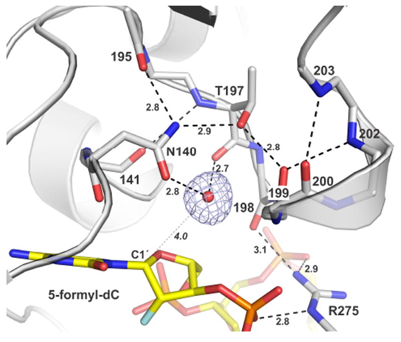
Figure 3.

Binding of the nucleophilic water molecule. TDG is in cartoon and stick format (white), the fdC nucleotide is yellow, and the nucleophile (water) is a red sphere (other waters omitted for clarity). A 2Fo-Fc map, contoured at 1.0 σ, is shown for the nucleophile. Dashed lines are H bonds, some with interatomic distances (Å). The distance from the nucleophile to C1′ of fdCF (4.0 Å) is indicated.
