Abstract
The NS2 (NEP) protein of influenza A virus contains a highly conserved nuclear export signal (NES) motif in its amino-terminal region (12ILMRMSKMQL21, A/WSN/33), which is thought to be required for nuclear export of viral ribonucleoprotein complexes (vRNPs) mediated by a cellular export factor, CRM1. However, simultaneous replacement of three hydrophobic residues in the NES with alanine does not affect NS2 (NEP) binding to CRM1, although the virus with these mutations is not viable. To determine the extent of sequence conservation required by the NS2 (NEP) NES for its export function during viral replication, we randomly introduced mutations by degenerative mutagenesis into the region of NS cDNA encoding the NS2 (NEP) NES and then attempted to generate mutant viruses containing these alterations by reverse genetics. Sequence analysis of the recovered viruses showed that although some of the mutants possessed amino acids other than those conserved in the NES, hydrophobicity within this motif was maintained. Nuclear export of vRNPs representing all of the mutant viruses was completely inhibited in the presence of a CRM1 inhibitor, leptomycin B, as was the transport of wild-type virus, indicating that the CRM1-mediated pathway is responsible for the nuclear export of both wild-type and mutant vRNPs. The vRNPs of some of the mutant viruses were exported in a delayed manner, resulting in limited viral growth in cell culture and in mice. These results suggest that the NES motif may be an attractive target for the introduction of attenuating mutations in the production of live vaccine viruses.
The life cycle of influenza A virus, whose genome harbors eight single-stranded RNA segments, differs from that of many other negative-strand RNA viruses, as it involves a nuclear phase of genomic replication. After their attachment to cell surface receptors, the influenza A virions enter cells by receptor-mediated endocytosis. Within the endosome, after M2 ion channel-mediated acidification of the virion interior, viral ribonucleoprotein (vRNP) complexes of viral genomic RNA, nucleoprotein (NP), and three polymerase subunit proteins (PA, PB1, and PB2) dissociate from the M1 protein, are released into the cytoplasm, and are then transported to the nucleus, where transcription and replication of viral RNAs occur. Subsequently, the newly synthesized vRNPs in the nucleus must be exported to the cytoplasm to be incorporated into progeny virions. Although the mechanism(s) of vRNP nuclear export are only partly understood, some appreciation of this process has begun to emerge. During nuclear export, proteins and vRNPs are ushered through the nuclear pore complexes by specialized receptor proteins.
Export receptors of the importin β-like family bind to the nuclear export signals (NESs) of cargo molecules in the presence of Ran-GTP, the GTP-bound form of the small GTPase Ran. After translocation to the cytoplasm, the intrinsic GTPase activity of Ran is activated by cytoplasmic Ran-GTP-binding proteins and a Ran-GTPase-activating protein, resulting in the release of cargo from the export receptor (12, 20, 21). NESs are generally short stretches of amino acids characterized by multiple hydrophobic residues with typical spacing (e.g., ψXXXψXXψXψ, where ψ indicates important hydrophobic residues such as leucine, isoleucine, methionine, valine, or phenylalanine) that interact with export receptors (5, 18). CRM1, a member of the importin β-like family of transport receptors, is one of the receptors that export cargo molecules carrying NESs (9, 18).
O'Neill et al. (27) demonstrated that the NS2 (NEP) protein of influenza A virus interacts with cellular nucleoporins in a yeast two-hybrid system and that a stretch of sequence in the amino-terminal region of NS2 (NEP) can functionally replace the NES of human immunodeficiency virus type 1 Rev protein. They also proposed that a region containing a stretch of hydrophobic residues in the NS2 (NEP) of WSN virus (i.e., 12ILMRMSKMQL21) serves as an NES for this protein. We then showed that NS2 (NEP) interacts with CRM1 in a mammalian two-hybrid system, suggesting that influenza vRNPs are exported by a CRM1-mediated pathway with NS2 (NEP). In fact, a mutant virus containing alterations in the NS2 (NEP) NES (replacement of three hydrophobic residues with alanine) was not viable (23). Moreover, in the absence of NS2 (NEP), vRNPs remained in the nucleus. In the mammalian two-hybrid system, however, alteration of the NS2 (NEP) NES did not affect the protein's interaction with CRM1. These findings demonstrate that the NS2 (NEP) NES is indeed crucial for vRNP export, but the sequence required for NS2 (NEP) function in vRNP nuclear export in the context of viral replication is not fully understood. Here we generated mutant viruses with altered NS2 (NEP) NES sequences in an effort to provide this information and to further elucidate the molecular basis of vRNP nuclear export in the influenza A virus life cycle.
MATERIALS AND METHODS
Cells.
293T human embryonic kidney cells and Madin-Darby canine kidney (MDCK) cells were maintained in Dulbecco's modified Eagle's medium supplemented with 10% fetal calf serum and in minimal essential medium containing 5% newborn calf serum, respectively. The 293T cell line is a derivative of the 293 line into which the gene for the simian virus 40 T antigen was inserted (7). All cells were maintained at 37°C in 5% CO2.
Degenerative PCR mutagenesis.
To generate plasmids containing random mutations in the NES coding sequence, we performed degenerative PCR mutagenesis. The NES region (amino acid residues 12 to 21) was divided into five parts (NES1N to NES5N; six bases per part), and nucleotides in each part were degenerated to NNNNNN (Table 1). We first constructed a template plasmid for degenerative PCR in which a mutant form of the cDNA of the WSN NS gene (pPolI-WSN-NS) was amplified by inverse PCR (26) by using the back-to-back primers NEStemF and NEStemR (Table 2), which have both restriction enzyme EcoRI and BsmBI sites at the 5′ end. The PCR products were digested with EcoRI, self-ligated, and propagated in Escherichia coli strain DH5α, resulting in the pPolI-NStemplate plasmid. pPolI-NStemplate, which has a 20-base insertion (one EcoRI site, two BsmBI sites, and two additional bases) in the middle of the region encoding NES, was then subjected to inverse PCR with the back-to-back primers NEStemF and NES1N-R, NES2N-R, or NES3N-R and NES4N-F or NES5N-F and NEStemR (Table 2). These primers have a BsmBI site at their 5′ ends. The PCR products were digested with BsmBI and self-ligated, resulting in pPolI-NES1N, pPolI-NES2N, pPolI-NES3N, pPolI-NES4N, and pPolI-NES5N. These ligated plasmids were used directly in the reaction mixtures for the reverse genetics procedure. NS viral RNA (vRNA) derived from pPolI-NStemplate carried over in the reaction mixture would not support viral replication because it contains insertions that would lead to the introduction of 17 unrelated amino acids as well as a frameshift after the insertion.
TABLE 1.
Degenerate NS2 NES sequencesa
| NES region part | Nucleotide sequence encoding amino acidb:
|
|||||||||
|---|---|---|---|---|---|---|---|---|---|---|
| Ile | Leu | Met | Arg | Met | Ser | Lys | Met | Gln | Leu | |
| Whole region | ATA | CTG | ATG | AGG | ATG | TCA | AAA | ATG | CAG | TTG |
| NES1N | NNN | NNN | *** | *** | *** | *** | *** | *** | *** | *** |
| NES2N | *** | *** | NNN | NNN | *** | *** | *** | *** | *** | *** |
| NES3N | *** | *** | *** | *** | NNN | NNN | *** | *** | *** | *** |
| NES4N | *** | *** | *** | *** | *** | *** | NNN | NNN | *** | *** |
| NES5N | *** | *** | *** | *** | *** | *** | *** | *** | NNN | NNN |
Asterisks represent nucleotides identical to those found in the wild-type sequence.
The NES region contains amino acid residues 12 to 21.
TABLE 2.
Primers used for degenerative PCRa
| Resulting plasmid | Forward primer
|
Reverse primer
|
||
|---|---|---|---|---|
| Name | Sequence (5′-3′) | Name | Sequence (5′-3′) | |
| pPolI-NStemplate | NEStemF | CGGAATTCCGTCTCAAAAATGCAGTTGGGGTCCTCAT | NEStemR | CGGAATTCCGTCTCTTGACATCCTCATCAGTATGT |
| pPolI-NES1N | NEStemF | CGGAATTCCGTCTCAAAAATGCAGTTGGGGTCCTCAT | NES1N-R | CGCGCGCGTCTCATTTTTGACATCCTCATNNNNNNGTCCTGGAAGAGA GGGCAGTGGT |
| pPolI-NES2N | NEStemF | CGGAATTCCGTCTCAAAAATGCAGTTGGGGTCCTCAT | NES2N-R | CGCGCGCGTCTCATTTTTGACATNNNNNNCAGTATGTCCTGGAAGAGA |
| pPolI-NES3N | NEStemF | CGGAATTCCGTCTCAAAAATGCAGTTGGGGTCCTCAT | NES3N-R | CGCGCGCGTCTCATTTTNNNNNNCCTCATCAGTATGTCCTGGAA |
| pPolI-NES4N | NES4N-F | CGCGCGCGTCTCTGTCANNNNNNCAGTTGGGGTCCTC ATCGGAGGACTT | NEStemR | CGGAATTCCGTCTCTTGACATCCTCATCAGTATGT |
| pPolI-NES5N | NES5N-F | CGCGCGCGTCTCTGTCAAAAATGNNNNNNGGGTCCTC ATCGGAGGACTT | NEStemR | CGGAATTCCGTCTCTTGACATCCTCATCAGTATGT |
Mutations within the primers are shown in boldface type. EcoRI and BsmBI sites within the primers are identified by double and triple underlining, respectively. Additional bases removed by BsmBI digestion are underlined once.
Plasmid-based reverse genetics.
Influenza A viruses were generated with plasmids possessing the cDNA of A/WSN/33 (WSN; H1N1) viral genes under the control of an RNA polymerase I promoter and terminator (referred to as PolI plasmids) and the eukaryotic protein expression vector pCAGGS/MCS (controlled by the chicken β-actin promoter) (15, 25) as described by Neumann et al. (22). Transfectant viruses were harvested at 48 h posttransfection and plaque purified twice, and stock viruses were made with MDCK cells.
Sequence analysis.
First-strand cDNAs were synthesized from the total RNA of virus-infected cells with a commercial reagent (Isogen; Nippon Gene) and a primer (5′-AGCAAAAGCAGGGTGACAAAGACAT-3′; nucleotides 1 to 25 of the WSN NS gene) together with Moloney murine leukemia virus reverse transcriptase (GIBCO-BRL). PCR was performed with Pfu DNA polymerase (Promega) with the above primer and an additional primer (5′-ACTAGAAACAAGGGTGTGTTTTTATT-3′; nucleotides 890 to 866 of the WSN NS gene). Reverse transcription-PCR products were sequenced by means of a dye-terminator cycle sequencing system (Applied Biosystems).
Immunostaining assay.
Cells were fixed with 4% paraformaldehyde in phosphate-buffered saline and permeabilized with 0.5% Triton X-100. Antigens were detected with an anti-WSN NP monoclonal antibody as the primary antibody and fluorescein isothiocyanate-labeled anti-mouse immunoglobulin G (Cappel Laboratories) as the secondary antibody (Kirkegaard & Perry Laboratories).
Replicative properties of transfectant viruses.
MDCK cells in duplicate wells of 24-well plates were infected with virus at a multiplicity of infection (MOI) of 0.01 PFU/cell, overlaid with minimal essential medium containing 0.3% bovine serum albumin, and incubated at 37°C. At different times, supernatants were measured for infectious virus in plaque assays with MDCK cells.
Experimental infection.
Four-week-old female BALB/c mice, anesthetized with isoflurane, were intranasally infected with 50 μl (105, 104, or 103 PFU) of each virus. Survival and body weights were monitored for 16 days postinfection.
RESULTS
Generation of mutant viruses with an altered NS2 (NEP) NES sequence.
To determine the sequence required by the NES of NS2 (NEP) to function as a nuclear export signal for vRNPs in the context of viral replication, we attempted to generate mutant viruses with random mutations in the NES motif. We produced a pool of degenerative PCR-generated pPolI NS constructs in which every six nucleotides, representing two successive amino acids, within this region were randomly replaced. Reverse genetics with five sets of pPolI NS constructs, covering the entire region of the NS2 (NEP) NES sequence (Table 1), yielded a number of mutant viruses. We analyzed a total of 71 plaques formed by the transfectant viruses from six independent experiments for each set of primers, which yielded 16 mutant viruses, each containing different substitutions. The numbers of mutant viruses obtained for each set may reflect differences in the flexibility of each test region. After plaque-cloning, each virus was analyzed for the sequence in the NS2 (NEP) NES region (Table 3). Five hydrophobic residues (leucine, isoleucine, methionine, valine, and phenylalanine) are known to be important for NES function (5). Reverse genetics with the pPolI-NES1N construct, characterized by the substitution of two hydrophobic residues (12IL13) in the NES sequence, generated a total of eight mutant viruses. Sequencing of their NS genes revealed that one or both of the two residues was still hydrophobic, suggesting that one hydrophobic residue at these two positions is a minimal requirement for a functional NES. Although use of the pPolI-NES2N construct resulted in the generation of only a single mutant (M14Y), it indicated that methionine at position 14 is not essential for the NES sequence. With the pPolI-NES3N construct, three mutant viruses were generated, in which the methionine at position 16 was maintained (S17C) or substituted for hydrophobic isoleucine or leucine, suggesting the functional importance of the hydrophobic residue at this position in the NES sequence. Similarly, all three mutant viruses generated with the pPolI-NES4N construct still possessed a hydrophobic residue at position 19. The pPolI-NES5N construct resulted in the generation of a mutant virus possessing leucine at position 20 instead of position 21. All of the mutant viruses generated in this study have concomitant mutations at amino acid positions 169 to 179 of the NS1 (Table 3). Overall, these results demonstrate the flexibility of the NES sequence in maintaining a functional molecule and suggest that the hydrophobicity of the sequence may exert an important influence on vRNP nuclear export.
TABLE 3.
Amino acid sequences of NES-like regions of mutant virusesa
| Virus type | pPolI plasmid used for reverse genetics (no. of plaques analyzed/ no. of mutants found/no. of transfections performed)b | Name of mutant virus | No. of viruses with identical mutations | NES sequence | Growth competencyc | Associated mutations in NS1 |
|---|---|---|---|---|---|---|
| None (consensus sequence) | ΨXXXΨXXΨXΨd | |||||
| Wild type | ILMRMSKMQL | +++ | HTDEDVKNAVG | |||
| Mutant | pPolI-NES1N (23/15/6) | I12L/L13F | 1 | LF********e | NTf | LVH******** |
| I12L/L13R | 1 | LR******** | NT | P*Y******** | ||
| I12L/L13H | 1 | LH******** | NT | L*Y******** | ||
| I12C/L13V | 6 | CV******** | +++ | LR********* | ||
| I12T/L13V | 1 | TV******** | NT | *CY******** | ||
| I12L | 2 | L********* | +++ | PVN******** | ||
| L13T | 2 | *T******** | NT | *HN******** | ||
| L13H | 1 | *H******** | NT | **Y******** | ||
| pPolI-NES2N (13/1/6) | M14Y | 1 | **Y******* | ++ | **VSH****** | |
| pPolI-NES3N (16/5/6) | M16I/S17R | 2 | ****IR**** | NT | *****SQ**** | |
| M16L | 1 | ****L***** | ++ | ****ALQ**** | ||
| S17C | 2 | *****C**** | +++ | ******Q**** | ||
| pPolI-NES4N (7/3/6) | K18S/M19V | 1 | ******SV** | + | *******RS** | |
| K18R/M19F | 1 | ******RF** | ++ | ******TFS** | ||
| K18C | 1 | ******C*** | NT | ******MH*** | ||
| pPolI-NES5N (12/5/6) | Q20L/L21C | 5 | ********LC | ++ | *********LW |
Reverse transcription-PCR products from mutant viruses were directly sequenced.
The total number of plaques analyzed and the total number of mutants found are listed. The remaining viruses contained the wild-type sequence (e.g., for pPolI-NES1N, among 23 plaques examined, 15 were mutant, and 8 were wild-type viruses).
Growth properties in comparison to the wild-type. +++, grew as well as the wild type; ++, grew at least 1 log lower than the wild type; +, grew over 3 logs lower than the wild type (for details, see Fig. 1).
Ψ indicates hydrophobic residues, including leucine, isoleucine, methionine, valine, and phenylalanine. X indicates any amino acid (see references 5 and 18).
Asterisks represent amino acids identical to those found in the wild type. Hydrophobic residues are shown in boldface type.
NT, not tested.
Growth property of NES mutants in cell culture.
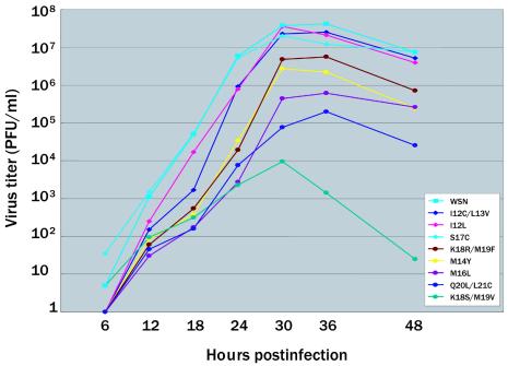
Although the mutations introduced into the NS2 (NEP) NES sequence did not prevent virus replication, we could not rule out an effect on growth. Thus, we compared the growth properties of the mutant viruses with that of wild-type virus in MDCK cells (Fig. 1). Cells were infected with a virus at an MOI of 0.01, and the kinetics of virus yields were monitored. Three growth phenotypes were apparent. First, the I12C/L13V, I12L, and S17C viruses grew as well as the wild-type virus, indicating that none of the amino acid mutations introduced into these viruses had affected viral replication in cell culture and, hence, that one hydrophobic residue at position 12 or 13 is sufficient for optimal NES function. Second, the peak titers of the K18R/M19F, M14Y, M16L, and Q20L/L21C mutants were at least 1 log lower than that of the wild-type virus. Finally, one virus, K18S/M19V, had a peak titer that was more than 3 logs lower than that of the control (wild-type) virus. All of the viruses with slower growth rates had a mutation in one of the hydrophobic residues (M14, M16, M19, or L21) thought to be required for optimal NES function (5). Interestingly, a mutation from one hydrophobic residue to another hydrophobic residue (M16L, M19V, or M19F) affected viral growth. Taken together, these findings indicate that the methionine at positions 14, 16, and 19 and the leucine at position 21 of the sequence contribute importantly to optimal NES function.
FIG. 1.
Growth curves of NES mutant and wild-type WSN viruses. MDCK cells were infected with the indicated virus at an MOI of 0.01. At 6 to 48 h postinfection, virus titers in the supernatant were determined. The values are means of the results from duplicate experiments. The standard deviation is less than 0.59 for each sample.
vRNP nuclear export of the mutant viruses.
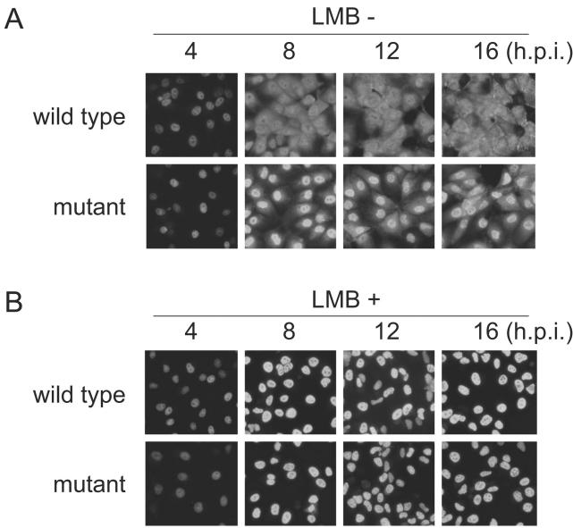
We next assessed whether restricted growth of the mutant viruses resulted from reduced vRNP nuclear export by the mutations introduced into the NS2 (NEP) NES sequence. Immunofluorescence assays with an anti-NP monoclonal antibody revealed a delay in nuclear export of vRNPs for all mutant viruses compared with results for the wild-type virus (Fig. 2A) (Q20L/L21C, depicted as a representative mutant), although there were no appreciable differences in the expression levels among the NS2 mutants by Western blot analysis with an anti-NS2 monospecific antibody (data not shown). Leptomycin B, which is a highly specific and potent inhibitor of CRM1 function (10, 16, 17), completely inhibited nuclear export of the vRNPs of all mutant viruses as well as that of wild-type virus (Fig. 2B), implicating the CRM1 pathway in vRNP export. Thus, the NS2 (NEP) NES mutations appear to have resulted in suboptimal formation of a ternary export complex, NS2 (NEP)/CRM1/Ran-GTP, leading to reduced vRNP nuclear export and restricted growth of the mutant viruses.
FIG. 2.
Intracellular localization of vRNP. MDCK cells were infected with a mutant (Q20L/L21C) or wild-type WSN virus in the absence (−) (A) or presence (+) (B) of a CRM1 inhibitor, leptomycin B (LMB; 5 ng/ml). At 4 to 16 h postinfection (h.p.i.), MDCK cells were fixed with paraformaldehyde and permeabilized with Triton X-100. vRNP localization was assessed by immunofluorescence assay with an anti-NP monoclonal antibody. The vRNP localization for the Q20L/L21C mutant was representative of that for all other mutants.
Virulence of mutant viruses in mice.
Since some of the mutant viruses in this study showed comparable growth to the wild-type virus in cell culture, we next assessed the replicative properties of these mutants in vivo. Mice were intranasally inoculated with three NES mutant viruses, I12L, I12C/L13V, and S17C, or the wild-type virus as a control (105, 104, or 103 PFU/50 μl); another mutant, M14Y, with reduced growth in cell culture was included for comparison. Lethality (Table 4) and body weight changes (data not shown) indicated that two of the mutants, I12L and S17C, were as virulent in mice as the wild-type virus; however, the growth of I12C/L13V was attenuated in mice even though it replicated only slightly more slowly than the wild-type virus during early infection of cell cultures. The M14Y mutant, whose in vitro replication was appreciably slower than that of the wild-type virus, showed highly attenuated growth properties. These results suggest that mutation of certain regions of the NS2 (NEP) NES molecule may generate attenuated influenza A viruses suitable for evaluation as live vaccine candidates.
TABLE 4.
Virulence of mutant viruses in micea
| Virus | Amt of virus inoculated into mice (PFU/mouse) | Cumulative no. of dead mice at post infection day:
|
|||||||||
|---|---|---|---|---|---|---|---|---|---|---|---|
| 5 | 6 | 7 | 8 | 9 | 10 | 11 | 12 | 14 | 16 | ||
| WSN | 105 | 0 | 1 | 3 | 3 | 4 | 4 | 4 | 4 | 4 | 4 |
| 104 | 0 | 1 | 1 | 1 | 1 | 1 | 2 | 3 | |||
| 103 | 0 | ||||||||||
| I12L | 105 | 0 | 1 | 1 | 2 | 3 | 3 | 3 | 3 | ||
| 104 | 0 | 1 | 1 | 1 | 2 | 2 | 2 | ||||
| 103 | 0 | ||||||||||
| S17C | 105 | 0 | 2 | 3 | 3 | 3 | 3 | 3 | 3 | 3 | 3 |
| 104 | 0 | 1 | 1 | 1 | 1 | ||||||
| 103 | 0 | ||||||||||
| I12C/L13V | 105 | 0 | 1 | 1 | 1 | 1 | 1 | ||||
| 104 | 0 | ||||||||||
| 103 | 0 | ||||||||||
| M14Y | 105 | 0 | |||||||||
| 104 | 0 | ||||||||||
| 103 | 0 | ||||||||||
Four-week-old BALB/c mice (n = 4), anesthetized with isoflurane, were intranasally infected with 50 μl (105, 104, or 103 PFU) of each virus. Survival was recorded for 16 days postinfection.
DISCUSSION
Using a series of mutant viruses with alterations in the NS2 (NEP) NES, we show here that the NES sequence does not have to be completely conserved to retain the motif's export signaling function, although some hydrophobic residues appear to play a critical role. This observation accounts for the previous finding (23) that a virus containing three alterations in its NES hydrophobic residues (M16A, M19A, and L21A) was not viable.
Although the most prevalent NES motif found in many cellular and viral proteins consists of a leucine-rich amino acid stretch, which is responsible for their nuclear export through the CRM1-mediated pathway (9), the NS2 (NEP) NES sequence of influenza A virus possesses a methionine-rich amino acid stretch. In fact, none of our mutant viruses had substitutions from methionine to leucine at position 14 or 19 of the NES, and a virus with a Leu substitution for Met at position 16 (M16L) showed appreciably reduced growth compared with that of the wild-type virus. However, all of the mutant viruses used the CRM1-mediated pathway for vRNP nuclear export, as did the wild-type virus. These findings suggest a preference for methionine over leucine at these positions for optimal NS2 (NEP) NES function, even in the CRM1-mediated pathway. It was previously suggested that the NS2 (NEP) protein mediates vRNP nuclear export through an NES-independent interaction with CRM1, unlike most other proteins relying on CRM1 for their export function (11, 28). The results of the present study suggest that methionines in the NS2 (NEP) NES of influenza A virus contribute to this apparently unique mechanism of vRNP nuclear export through the formation of the ternary export complex with Ran-GTP.
The NS2 (NEP) protein of influenza A virus associates with the matrix protein M1 (31, 33), which binds to vRNP (2) and whose nuclear localization is essential for vRNP export (6, 19, 32). Biochemical and two-hybrid assays showed that NS2 (NEP) interacts with CRM1 and with certain nucleoporins (27), supporting a concept whereby vRNPs are exported from the nucleus as a complex, NS2 (NEP)-M1-vRNP. Further support for this notion comes from a recent report on the crystal structure of the C-terminal domain of NS2 (NEP), suggesting a strong interaction of this protein with M1 (1). By contrast, Bui et al. (6) concluded that NS2 (NEP) is not required for vRNP nuclear export, based on the demonstration that vRNPs were retained in the nucleus of virus-infected cells treated with a protein kinase inhibitor, H7, which downregulates expression of M1, and that expression of NS2 (NEP) proteins and vRNA nuclear export could be restored by providing M1 from an expression vector. However, it is possible that an undetectable level of NS2 (NEP) protein may still have been present even in the presence of H7. Elton et al. (8) suggested that vRNP export may be mediated by a direct interaction of NP with CRM1, whereas Huang et al. (13) indicated that the interaction of NP with vRNA and M1 may lead to translocation of vRNPs from the nucleus to the cytoplasm. Collectively, these reports raise the possibility of vRNP nuclear export by mechanisms other than the NS2 (NEP)/CRM1-mediated pathway. Nevertheless, all mutant viruses generated in this study, including those with restricted growth, used the CRM1-mediated pathway, underscoring its importance as a central mechanism of influenza A virus vRNP nuclear export.
We cannot rule out the possibility that mutations in the NS1 protein, introduced together with the NS2 (NEP) NES mutations, may have affected properties of the mutant viruses. Indeed, all of the mutants generated in this study carry mutations in NS1 at amino acid positions 169 to 179 (Table 3). As a multifunctional protein with both N-terminal RNA-binding and C-terminal effector domains, NS1 mutations could produce a variety of ancillary effects. For example, the effector domain has been reported to inhibit the processing and transport of host mRNA or the host innate antiviral response as well as to stabilize the NS1 dimeric structure (30). However, we think it highly unlikely that NS1 mutations affected viral growth properties, since poor growth of the mutants was associated with a delay in their vRNP nuclear export (Fig. 2), a property directly attributable to changes in the NS2 (NEP) NES signaling motif.
The NS2 (NEP) NES mutant viruses we studied were generated by a random mutagenesis protocol with degenerative PCR. This method can be applied to reverse genetics analyses on conserved sequences of any viral protein to determine their biological functions in the virus life cycle. In conventional mutagenesis strategies, individual mutations are introduced into cDNA and then mutant cDNAs are used for viral recovery. Some of the resulting mutants are viable while others are not. Since our degenerative reverse genetics mutagenesis procedure relies on viral replication as an end point, any mutation introduced will yield a live virus, allowing us to identify the minimal sequence in a given motif needed to ensure replicative competency. Hence, this procedure, which eliminates screening steps for viability, should be useful in targeting particular regions of the viral genome to generate attenuated viruses with potential as live vaccine candidates.
Multiple attenuating mutations must be introduced into vaccine strains to prevent their reversion to a virulent phenotype. Since the NS2 (NEP) NES is also present in influenza B and C viruses (29), the restricted growth of our mutant viruses in mice suggests that this site would be a lucrative target for virus attenuation in the development of live attenuated influenza vaccines. A live attenuated vaccine (FluMist) licensed in 2003 in the United States was modeled on cold-adapted type A and B viruses. Five amino acids in the PB1, PB2, and NP segments are responsible for attenuation of the type A virus (14), whereas the basis for attenuation of the type B strain remains unknown. Although these viruses are phenotypically highly stable, including virulence, they confer clinical symptoms (3, 4, 24) that have led to a restriction on the population eligible to receive this vaccine (children and adults 5 to 49 years old). This suggests a need for improved vaccine viruses that show greater attenuation while retaining their immunogenicity. We conclude that further mutation of the NES region of the NS2 (NEP) protein may provide the desired attenuation without comprising vaccine efficacy.
Acknowledgments
We thank John Gilbert for editing the manuscript.
This work was supported by Grants-in-Aid for Scientific Research on Priority Areas from the Ministries of Education, Culture, Sports, Science, and Technology, Japan, by CREST (Japan Science and Technology Corporation), and by Public Health Service research grants from the National Institute of Allergy and Infectious Diseases.
REFERENCES
- 1.Akarsu, H., W. P. Burmeister, C. Petosa, I. Petit, C. W. Müller, R. W. H. Ruigrok, and F. Baudin. 2003. Crystal structure of the M1 protein-binding domain of the influenza virus nuclear export protein (NEP/NS2). EMBO J. 22:4646-4655. [DOI] [PMC free article] [PubMed] [Google Scholar]
- 2.Baudin, F., W. Weissenhorn, and R. W. H. Ruigrok. 2001. In vitro dissection of the membrane binding and RNP binding activities of influenza virus M1 protein. Virology 281:102-108. [DOI] [PubMed] [Google Scholar]
- 3.Belshe, R. B., P. M. Mendelman, J. Treanor, J. King, W. C. Gruber, P. Piedra, D. I. Bernstein, F. G. Hayden, K. Kotloff, K. Zangwill, D. Iacuzio, and M. Wolff. 1998. The efficacy of live attenuated, cold-adapted, trivalent, intranasal influenza virus vaccine in children. N. Engl. J. Med. 338:1405-1412. [DOI] [PubMed] [Google Scholar]
- 4.Belshe, R. B., W. C. Gruber, P. M. Mendelman, I. Cho, K. Reisinger, S. L. Block, J. Wittes, D. Lacuzio, P. Piedra, J. Treanor, J. King, K. Kotloff, D. I. Bernstein, F. G. Hayden, K. Zangwill, L. Yan, and M. Wolff. 2000. Efficacy of vaccination with live attenuated, cold-adapted, trivalent, intranasal influenza virus vaccine against a variant (A/Sydney) not contained in the vaccine. J. Pediatr. 136:168-175. [DOI] [PubMed] [Google Scholar]
- 5.Bogerd, H. P., R. A. Fridell, R. E. Benson, J. Hua, and B. R. Cullen. 1996. Protein sequence requirements for function of the human T-cell leukemia virus type 1 rex nuclear export signal delineated by a novel in vivo randomization-selection assay. Mol. Cell. Biol. 16:4207-4214. [DOI] [PMC free article] [PubMed] [Google Scholar]
- 6.Bui, M., E. G. Wills, A. Helenius, and G. R. Whittaker. 2000. Role of the influenza virus M1 protein in nuclear export of viral ribonucleoproteins. J. Virol. 74:1781-1786. [DOI] [PMC free article] [PubMed] [Google Scholar]
- 7.DuBridge, R. B., P. Tang, H. C. Hsia, P. M. Leong, J. H. Miller, and M. P. Calos. 1987. Analysis of mutation in human cells by using an Epstein-Barr virus shuttle system. Mol. Cell. Biol. 7:379-387. [DOI] [PMC free article] [PubMed] [Google Scholar]
- 8.Elton, D., M. Simpson-Holley, K. Archer, L. Medcalf, R. Hallam, J. McCauley, and P. Digard. 2001. Interaction of the influenza virus nucleoprotein with the cellular CRM1-mediated nuclear export pathway. J. Virol. 75:408-419. [DOI] [PMC free article] [PubMed] [Google Scholar]
- 9.Fornerod, M., M. Ohno, M. Yoshida, and I. W. Mattaj. 1997. CRM1 is an export receptor for leucine-rich nuclear export signals. Cell 90:1051-1060. [DOI] [PubMed] [Google Scholar]
- 10.Fukuda, M., S. Asano, T. Nakamura, M. Adachi, M. Yoshida, M. Yanagida, and E. Nishida. 1997. CRM1 is responsible for intracellular transport mediated by the nuclear export signal. Nature 390:308-311. [DOI] [PubMed] [Google Scholar]
- 11.Fukuda, M., I. Gotoh, Y. Gotoh, and E. Nishida. 1996. Cytoplasmic localization of mitogen-activated protein kinase kinase directed by its NH2-terminal, leucine-rich short amino acid sequence, which acts as a nuclear export signal. J. Biol. Chem. 271:20024-20028. [DOI] [PubMed] [Google Scholar]
- 12.Gorlich, D., and U. Kutay. 1999. Transport between the cell nucleus and the cytoplasm. Annu. Rev. Cell. Dev. Biol. 15:607-660. [DOI] [PubMed] [Google Scholar]
- 13.Huang, X., T. Liu, J. Muller, R. A. Levandowski, and Z. Ye. 2001. Effect of influenza virus matrix protein and viral RNA on ribonucleoprotein formation and nuclear export. Virology 287:405-416. [DOI] [PubMed] [Google Scholar]
- 14.Jin, H., B. Lu, H. Zhou, C. Ma, J. Zhao, C.-F. Yang, G. Kemble, and H. Greenberg. 2003. Multiple amino acid residues confer temperature sensitivity to human influenza virus vaccine strains (FluMist) derived from cold-adapted A/Ann Arbor/6/60. Virology 306:18-24. [DOI] [PubMed] [Google Scholar]
- 15.Kobasa, D., M. E. Rodgeres, K. Wells, and Y. Kawaoka. 1997. Neuraminidase hemadsorption activity, conserved in avian influenza A viruses, does not influence viral replication in ducks. J. Virol. 71:6706-6713. [DOI] [PMC free article] [PubMed] [Google Scholar]
- 16.Kudo, N., N. Matsumori, H. Taoka, D. Fujiwara, E. P. Schreiner, B. Wolff, M. Yoshida, and S. Horinouchi. 1999. Leptomycin B inactivates CRM1/exportin 1 by covalent modification at a cysteine residue in the central conserved region. Proc. Natl. Acad. Sci. USA 96:9112-9117. [DOI] [PMC free article] [PubMed] [Google Scholar]
- 17.Kudo, N., B. Wolff, T. Sekimoto, E. P. Schreiner, Y. Yoneda, M. Yanagida, S. Horinouchi, and M. Yoshida. 1998. Leptomycin B inhibition of signal-mediated nuclear export by direct binding to CRM1. Exp. Cell Res. 242:540-547. [DOI] [PubMed] [Google Scholar]
- 18.Macara, I. G. 2001. Transport into and out of the nucleus. Microbiol. Mol. Biol. Rev. 65:570-594. [DOI] [PMC free article] [PubMed] [Google Scholar]
- 19.Martin, K., and A. Helenius. 1991. Nuclear transport of influenza virus ribonucleoproteins: the viral matrix protein (M1) promotes export and inhibit import. Cell 67:117-130. [DOI] [PubMed] [Google Scholar]
- 20.Mattaj, I. W., and L. Englmeier. 1998. Nucleocytoplasmic transport: the soluble phase. Annu. Rev. Biochem. 67:265-306. [DOI] [PubMed] [Google Scholar]
- 21.Moroianu, J. 1999. Nuclear import and export pathways. J. Cell. Biochem. Suppl. 32-33:76-83. [DOI] [PubMed] [Google Scholar]
- 22.Neumann, G., T. Watanabe, H. Ito, S. Watanabe, H. Goto, P. Gao, M. Hughes, D. R. Perez, R. Donis, E. Hoffmann, G. Hobom, and Y. Kawaoka. 1999. Generation of influenza A viruses entirely from cloned cDNAs. Proc. Natl. Acad. Sci. USA 96:9345-9350. [DOI] [PMC free article] [PubMed] [Google Scholar]
- 23.Neumann, G., M. T. Hughes, and Y. Kawaoka. 2000. Influenza A virus NS2 protein mediates vRNA nuclear export through NES-independent interaction with hCRM1. EMBO J. 19:6751-6758. [DOI] [PMC free article] [PubMed] [Google Scholar]
- 24.Nichol, K. L., P. M. Mendelman, K. P. Mallon, L. A. Jackson, G. J. Gorse, R. B. Belshe, W. P. Glezen, and J. Wittes. 1999. Effectiveness of live, attenuated intranasal influenza virus vaccine in healthy, working adults. JAMA 282:137-144. [DOI] [PubMed] [Google Scholar]
- 25.Niwa, H., K. Yamamura, and J. Miyazaki. 1991. Efficient selection for high-expression transfectants with a novel eukaryotic vector. Gene 108:193-199. [DOI] [PubMed] [Google Scholar]
- 26.Ochman, H., A. S. Gerber, and D. L. Hartl. 1988. Genetics applications of an inverse polymerase chain reaction. Genetics 120:621-623. [DOI] [PMC free article] [PubMed] [Google Scholar]
- 27.O'Neill, R. E., J. Talon, and P. Palese. 1998. The influenza virus NEP (NS2 protein) mediates the nuclear export of viral ribonucleoproteins. EMBO J. 17:288-296. [DOI] [PMC free article] [PubMed] [Google Scholar]
- 28.Ossareh-Nazari, B., and C. Dargemont. 1999. Domains of Crm1 involved in the formation of the Crm1, RanGTP and leucine-rich nuclear export sequences trimeric complex. Exp. Cell Res. 252:236-241. [DOI] [PubMed] [Google Scholar]
- 29.Paragas, J., J. Talon, R. E. O'Neill, D. K. Anderson, A. García-Sastre, and P. Palese. 2001. Influenza B and C virus NEP (NS2) proteins possess nuclear export activities. J. Virol. 75:7375-7383. [DOI] [PMC free article] [PubMed] [Google Scholar]
- 30.Qian, X. Y., F. Alonso-Caplen, and R. M. Krug. 1994. Two functional domains of the influenza virus NS1 protein are required for regulation of nuclear export of mRNA. J. Virol. 68:2433-2441. [DOI] [PMC free article] [PubMed] [Google Scholar]
- 31.Ward, A. C., L. A. Castelli, A. C. Lucantoni, J. F. White, A. A. Azad, and I. G. Macreadie. 1995. Expression and analysis of the NS2 protein of influenza A virus. Arch. Virol. 140:2067-2073. [DOI] [PubMed] [Google Scholar]
- 32.Whittaker, G., M. Bui, and A. Helenius. 1996. Nuclear trafficking of influenza virus ribonucleoproteins in heterokaryons. J. Virol. 70:2743-2756. [DOI] [PMC free article] [PubMed] [Google Scholar]
- 33.Yasuda, J., S. Nakada, A. Kato, T. Toyoda, and A. Ishihama. 1993. Molecular assembly of influenza virus: association of the NEP protein with virion matrix. Virology 196:249-255. [DOI] [PubMed] [Google Scholar]


