Abstract
Infectious bursal disease virus (IBDV) causes a highly immunosuppressive disease in chickens. Currently available, live IBDV vaccines can lead to generation of variant viruses. We have developed an alternative vaccine that will not create variant IBDV. By using the reverse genetics approach, we devised a recombinant Newcastle disease virus (NDV) vector from a commonly used vaccine strain LaSota to express the host-protective immunogen VP2 of a variant IBDV strain GLS-5. The gene encoding the VP2 protein of the IBDV was inserted into the most 3′-proximal locus of a full-length NDV cDNA for high-level expression. We successfully recovered the recombinant virus, rLaSota/VP2. The rLaSota/VP2 was genetically stable, at least up to 12 serial passages in chicken embryos, and was shown to express the VP2 protein. The VP2 protein was not incorporated into the virions of recombinant virus. Recombinant rLaSota/VP2 replicated to a titer similar to that of parental NDV strain LaSota in chicken embryos and cell cultures. To assess protective efficacy of the rLaSota/VP2, 2-day-old specific-pathogen-free chickens were vaccinated with the recombinant virus and challenged with a highly virulent NDV strain Texas GB or IBDV variant strain GLS-5 at 3 weeks postvaccination. Vaccination with rLaSota/VP2 generated antibody responses against both NDV and IBDV and provided 90% protection against NDV and IBDV. Booster immunization induced higher levels of antibody responses against both NDV and IBDV and conferred complete protection against both viruses. These results indicate that the recombinant NDV can be used as a vaccine vector for other avian pathogens.
Infectious bursal disease virus (IBDV) is a pathogen of major economic importance in the poultry industry worldwide. IBDV replicates specifically in developing B-lymphoid cells, resulting in the destruction of the precursors of antibody-producing B cells in the bursa of Fabricius, and consequently, the immunosuppression, which leads to vaccination failures and susceptibility to other infections and diseases (25). Vaccination is the principal method used for the control of infectious bursal disease in chickens. Although IBDV strains of different antigenic types have been incorporated into vaccines, IBDV remains a major problem for the poultry industry. The efficacy of current live IBDV vaccines decreases in the presence of maternal antibodies, which are essential for the protection of young chickens for the critical first few weeks of life. Live IBDV vaccines also cause various degrees of bursal atrophy and may contribute to the emergence of antigenic variant viruses (25). The first antigenic variant strain of IBDV was isolated from vaccinated flocks on the Delmarva Peninsula in 1985 (40). Other variant strains were subsequently isolated in the United States and other countries (17, 35, 41, 42, 46). The antigenic variant strains were serologically different from the so-called classic isolates most typically isolated before 1985 (25, 42, 46). These variant strains lack the epitope(s) defined by neutralizing monoclonal antibodies (MAbs) B69 and R63, which were prepared with classical isolates (40, 41). The variant viruses infect chickens, even those with relatively high levels of maternal antibodies, and cause great economic losses (17, 35, 42, 46). The apparent inability to control IBDV infection through current vaccination warrants a necessity to develop alternate IBDV vaccine strategies that would not result in variant viruses.
IBDV is a member of the genus Avibirnavirus in the family Birnaviridae, and its genome is composed of two segments of double-stranded RNA (25). The smaller segment B encodes VP1, a 90,000-molecular-weight RNA-dependent RNA polymerase. The larger segment A contains two partially overlapping open reading frames (ORFs). The first, smaller ORF encodes a nonstructural protein VP5; whereas the second ORF encodes a precursor polyprotein, which is subsequently cleaved into VP2, VP4, and VP3. VP2 and VP3 are the major capsid proteins of IBDV, constituting 51 and 40% of the viral proteins, respectively (25). The VP2 protein has been identified as the major host-protective immunogen of IBDV and contains major epitopes responsible for eliciting neutralizing antibodies (3, 4, 16). Passive antibodies to VP2 were found to protect chickens (12, 25, 44). Many attempts have been made to express the structural proteins of IBDV as subunit vaccines for the control of this disease. It has been demonstrated that the recombinant VP2 protein expressed in different expression systems provided significant protection against the disease (5, 37, 39, 43, 45). However, those efforts have not been translated for practical use, due to limitations of the delivery systems (11, 21, 34, 48).
The recently developed reverse genetics system to engineer negative-strand RNA viruses (NSV) has provided a new method to express foreign genes (9, 30). Owing to the modular nature of their genomes, it is easy to engineer additional genes into the genomes of NSV. A number of recombinant NSV containing additional foreign genes have been engineered (6, 14, 15, 27, 31, 38, 47). It has also been shown that NSV can serve as highly effective vaccine vectors for protection against other pathogens (7, 13, 29, 36). Earlier studies have shown that recombinant Newcastle disease virus (NDV), an NSV, can be used to express heterologous genes (19, 22, 29).
NDV is a member of the genus Avulavirus in the family Paramyxoviridae (24, 26). The genome of NDV is a nonsegmented, negative-stranded RNA of 15,186 nucleotides (nt) containing six genes in the order of 3′-NP-P-M-F-HN-L-5′ (10, 23). Expression levels of the proteins are attenuated in a sequential manner from the 3′ end to the 5′ end of the viral genome (24, 32). NDV causes an economically important disease in all species of birds worldwide (2). Newcastle disease can vary from clinically inapparent to highly virulent forms depending on the virus strains and host species (2). Currently, naturally occurring avirulent NDV strains are routinely used as live vaccines throughout the world.
Several characteristics of NDV suggest that recombinant NDVs expressing a protective antigen of another avian pathogen would be a very good multivalent vaccine for poultry. Live NDV vaccines are widely used in commercial chickens with proven track records of efficacy and safety. NDV grows to very high titers in many cell lines and embryonated eggs and elicits strong humoral and cellular immune responses in vivo. NDV naturally infects the chicken via the upper respiratory tract and induces strong secretory immunoglobulin A (IgA), which is a critical component of the immunological repertoire to prevent mucosal infections (2, 18). Therefore, we explored the potential of recombinant NDV as a vaccine vector for poultry.
In this paper, we describe a recombinant NDV strain LaSota expressing the host-protective immunogen VP2 of a variant IBDV strain GLS-5. The recombinant rLaSota/VP2 grew to a level comparable to the parental virus. The VP2 gene was stably maintained and expressed even after serial passages in embryonated eggs. Vaccination with the recombinant rLaSota/VP2 induced strong humoral immune responses against both NDV and IBDV and conferred complete protection against both viruses after secondary immunization. These results clearly suggest that it is possible to develop a commercial bivalent recombinant NDV/IBDV vaccine that would provide protection against both of these economically important diseases.
MATERIALS AND METHODS
Cells and viruses.
DF1 (a chicken embryo fibroblast cell line) cells were grown in Dulbecco's modified Eagle medium (DMEM) containing 10% fetal bovine serum. Vero and HEp-2 cells were grown in Eagle's minimum essential medium containing 10% fetal bovine serum. Recombinant and wild-type NDV strains were grown in 9-day-old specific-pathogen-free (SPF) embryonated chicken eggs. The highly virulent NDV strain Texas GB used for challenge studies was originally received from National Veterinary Services Laboratory, Ames, Iowa. The wild-type and cell culture-adapted IBDV variant GLS-5 strains were kindly provided by Vikram Vakharia, University of Maryland Biotechnology Institute. The modified vaccinia strain Ankara expressing the T7 RNA polymerase (a generous gift of Bernard Moss, National Institutes of Health) was grown in primary chicken embryo fibroblast cells.
Plasmid construction and recovery of recombinant virus.
A cDNA fragment containing the VP2 gene (size of VP2 ORF, 1,356 nt; GenBank access number, M97346) of the cell culture-adapted GLS-5 strain was obtained by reverse transcription (RT)-PCR. Briefly, the IBDV strain GLS-5 was grown in Vero cells. Virus was purified from clarified supernatant by centrifugation at 70,000 × g for 2 h on a 26% sucrose cushion. The partially purified virus pellet was resuspended in phosphate-buffered saline (PBS) and treated with sodium dodecyl sulfate (SDS) (0.1%) and proteinase K (100 μg/ml) for 2 h. IBDV genomic RNAs were extracted with TRIzol reagent (Invitrogen, Carlsbad, Calif.) according to the manufacturer's instruction. The VP2 gene was amplified by RT-PCR using IBDV specific primers. The amplified product was sequenced to confirm the identity of the VP2 gene.
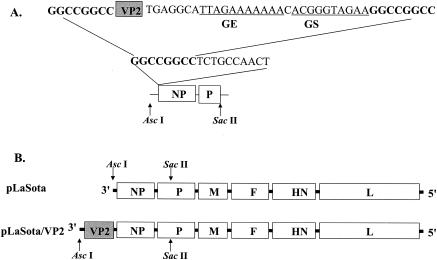
Construction of plasmid pLaSota carrying the full-length cDNA of the NDV vaccine strain LaSota has been described previously (19). To facilitate insertion of the VP2 gene of IBDV into the most 3′-proximal locus of NDV antigenome, the AscI and SacII fragment from pLaSota was subcloned into pGEM-7Z (+) (Promega, Madison, Wis.) using XbaI and HindIII overhang primers. An 18-nt insert (5′-GGCCGGCCTCTGCCAACT-3′) with an FseI site was introduced just before the NP gene ORF as described elsewhere (19). The VP2 gene of IBDV was amplified by PCR with FseI-tagged forward primer 5′-actggccggccATGACAAACCTGCAAGATCAAACCCAACAG-3′ and reverse primer 5′-actggccggccttctacccgtcttttttctaatgcctcaCCTTATGGCCCGGATTATGTCTTTG-3′ (the FseI site is in bold; the NDV gene start and gene end sequences are underlined; sequence specific to the IBDV VP2 gene is in uppercase), digested with FseI, and cloned into the pGEM-7Z (+) subclone. A termination codon (TGA) was placed immediately after the VP2 gene. Four additional nucleotides (GGCA, antigenome sense) were added after the termination codon to maintain the “rule of six” (8, 32, 33). The resulting AscI and SacII fragment in the subclone was excised and used to replace the corresponding part in pLaSota. The AscI and SacII region was sequenced to confirm the presence of the VP2 gene.
The recovery of infectious LaSota virus entirely from cloned cDNA using a mixture of three expression plasmids encoding NP, P, and L proteins of NDV strain LaSota and a fourth plasmid encoding the NDV plus IBDV VP 2 gene was carried out following the procedures described previously (19). Virus recovered in the supernatant was plaque purified prior to amplification and characterization. The recovered recombinant virus was designated rLaSota/VP2.
Identification of recombinant virus by RT-PCR.
Recombinant virus was grown in 10-day-old embryonated SPF chicken eggs. The virus was purified as described previously (20, 22). Genomic RNA was extracted from partially purified virus using TRIzol reagent (Invitrogen). The genomic RNA was reverse transcribed using ThermoScript reverse transcriptase (Invitrogen) and an antigenomic-sense primer 5′-35CGAAGGAGCAATTGAAGTCGCACG58-3′. Two sets of PCR were performed using the above reverse-transcribed cDNA as templates. The first set of PCR was performed with the same antigenomic-sense primer used for RT and a genomic-sense oligonucleotide, 5′-TCTGCTGGTTTACCCTGGGCTGT-3′, which is at position 1959 to 1981 in the NDV genome. The second PCR was performed using the same antigenomic-sense primer and a negative-sense internal IBDV VP2 gene-specific primer, 5′-TAGCCAAACCTGCGTCTGCTAGA-3′, at position 1371 to 1393 in the IBDV genome. Aliquots of the PCR products were analyzed on a 1% agarose gel. The PCR products were purified using the PCR purification kit (QIAGEN, Hilden, Germany) and sequenced to entirety to confirm the presence of the VP2 gene of IBDV in the recovered virus.
Expression of IBDV VP2 protein by recombinant virus.
The expression of VP2 protein was demonstrated in DF1 cells infected with virus stock by immunofluorescence assay. Briefly, confluent DF1 cells on six-well plates were infected with virus at a multiplicity of infection (MOI) of 0.1. After 24 or 48 h, the cells were washed with PBS and fixed with 3% paraformaldehyde in PBS for 20 min at room temperature. The cells were washed three times and permeabilized with PBS containing 0.05% Tween 20 (PBS-T) for 30 min. After further washing with PBS, the cells were incubated with 1:700 dilution of a rabbit anti-IBDV antiserum (a gift from Vikram Vakharia) for 1 h. The cells were rinsed with PBS and incubated with fluorescein isothiocyanate (FITC)-conjugated goat anti-rabbit immunoglobulin G antibody (Kirkegaard & Perry Laboratories [KPL], Gaithersburg, Md.) for 45 min. The cells were washed again with PBS and visualized using a Nikon Eclipse TE (Nikon, Tokyo, Japan) fluorescent microscope.
To further confirm the expression of the VP2 protein by the recombinant virus, Western blot analysis was performed using infected cell lysate and purified recombinant virus. Cell lysate was prepared from DF1 cells infected with the recombinant virus (MOI, 3) at 20 h postinfection. Cells were washed with PBS, scraped, collected by low-speed centrifugation, and lysed with lysis buffer (6.25 mM Tris [pH 6.8], 1% SDS, 10% glycerol, 6 M urea, 0.01% bromophenol blue, 0.01% phenol red) for 30 min on ice. The lysates were clarified by centrifugation at 7,000 × g for 10 min and used for Western blot analysis. The recombinant virus was purified from infective allantoic fluid and subjected to Western blot analysis. To examine whether the VP2 protein is incorporated into the virion, samples of the cell lysate and the purified virions were mixed with Laemmli sample buffer (Bio-Rad, Hercules, Calif.) and boiled for 3 min before electrophoresis. The boiled samples were separated by SDS-polyacrylamide gel electrophoresis on a 12% gel, and the resolved proteins were transferred to polyvinylidene difluoride membranes (Millipore, Bedford, Mass.). The membranes were probed with the rabbit anti-IBDV antiserum, washed with PBS-T, and subsequently incubated with horseradish peroxidase-conjugated goat anti-rabbit IgG antibody (KPL). Proteins were visualized after incubation with 3,3′,5,5′-tetramethyl benzidine (TMB) peroxidase substrate (KPL).
To examine whether the VP2 protein expressed by the recombinant virus retained the conformational antigenic epitopes, antigen capture enzyme-linked immunosorbent assay (AC-ELISA) was performed using a commercial kit (Synbiotics, San Diego, Calif.). The assay utilized a panel of MAbs raised against neutralizing epitopes of IBDV to examine the reactivity profile of the expressed VP2 from both cell lysates and supernatant of virus-infected cells. Briefly, cell lysate or supernatant from virus-infected DF1 cells was diluted (1:50) with antigen dilution buffer (Synbiotics) and added to the wells of MAb-coated plates. The plates were incubated at 4 to 8°C overnight and then washed three times with PBS-T. A positive chicken antiserum against IBDV was added to the plates. After 30 min at room temperature, the plates were washed three times and incubated with peroxidase-labeled goat anti-chicken IgG for 30 min. Following another three washings with PBS-T, the substrate 2,2′-azinobis(3-ethylbenzthiazolinesulfonic acid) (ABTS) was added. Color development was stopped after 15 min with stop solution. Optical density (OD) values were read on an ELISA reader at 405 nm, and the reactivities of each monoclonal were inferred from the OD readings compared to the controls, according to the manufacturer's instructions (Synbiotics).
Virus growth in cell cultures and embryonated SPF chicken eggs.
The growth of the recombinant virus was assessed by a multistep growth assay in DF1 cells. DF1 cells were infected at an MOI of 0.01 and incubated at 37°C in DMEM containing 5% fetal bovine serum and 1 μg of acetyl-trypsin/ml. Supernatants were harvested at 8-h intervals and titrated by plaque assay in DF1 cells. To compare the growth of the recombinant viruses in SPF chicken embryonated eggs, 2 × 10³ PFU of each virus in a volume of 100 μl were inoculated into the allantoic cavity of 10-day-old embryonated chicken eggs. Infective allantoic fluids were harvested 60 h postinoculation for virus titration by plaque assay.
Characterization of recombinant virus in vivo.
The mean death time (MDT) was determined to assess the pathogenicity of the recombinant viruses in embryonated SPF chicken eggs as described previously (1). Briefly, fresh infective allantoic fluid was diluted in PBS to give a 10-fold dilution series. For each dilution, 100 μl was inoculated into the allantoic cavity of each of five 9-day-old embryonated SPF chicken eggs. The eggs were incubated at 37°C and examined three times daily for 7 days. The time that each embryo was first observed dead was recorded. The highest dilution that killed all embryos was considered the minimum lethal dose. The MDT was calculated as the mean time in hours for the minimum lethal dose to kill the embryos.
Evaluation of the immunogenicity and protective efficacy of the recombinant virus in chickens.
We evaluated the immunogenicity and protective efficacy of rLaSota/VP2 in chickens at our U.S. Department of Agriculture-approved biosafety level 3 animal facility. One-day-old SPF white leghorn chickens (SPAFAS, Norwich, Conn.) were randomly assigned to 12 treatment groups of 10 birds each and 2 groups of 5 birds each, as shown in Table 1. Chickens in groups 1 to 7 were used for primary vaccination and challenge, whereas groups 8 to 14 were kept for secondary immunization and challenge later. All chickens were housed in separate poultry isolation chambers with ad libitum access to feed and water. At 2 days of age, chickens were vaccinated via the ocular route with either rLaSota/VP2 (1 × 10⁴ 50% embryo lethal doses [ELD50] per bird) or commercial NDV LaSota vaccine and commercial live IBDV vaccine at the manufacturer's recommended dose. Chickens from each group were bled at 3 weeks postimmunization for assessing NDV or IBDV antibodies using commercially available ELISA kits (Synbiotics). Virus-neutralizing antibody titers in blood samples were also assessed by microtiter virus neutralization (VN) test with the constant-virus diluting-serum technique using homologous viruses. The VN titers were expressed as the reciprocal of the last serum dilution that was capable of neutralizing 100 mean 50% tissue culture infectious doses (TCID50). A geometric mean titer was calculated for each group. Birds in groups 1 to 7 were challenged with virulent NDV strain Texas GB at 1 × 10⁴ ELD50 per bird intramuscularly or virulent IBDV GLS-5 strain 1 × 10³ embryo infectious dose (EID50) per bird ocularly, at 23 days of age, as indicated in Table 1. The birds in groups 8 to 14 were boosted with commercial NDV or IBDV vaccines or recombinant rLaSota/VP2 at 23 days of age. Two weeks after the secondary vaccination, all the chickens were bled for immunological assessment of NDV or IBDV antibodies by ELISA and then challenged as described above.
TABLE 1.
Experimental design
| Exptl. group | No. of chickens | Age (days) at vaccination
|
Vaccinea | Age at challenge (days) | Challenge virusb | |
|---|---|---|---|---|---|---|
| Primary | Secondary | |||||
| 1 | 10 | 2 | — | Commercial NDV vaccine | 23 | Texas GB |
| 2 | 10 | 2 | — | Commercial IBDV vaccine | 23 | GLS-5 |
| 3 | 10 | 2 | — | rLaSota/VP2 | 23 | Texas GB |
| 4 | 10 | 2 | — | rLaSota/VP2 | 23 | GLS-5 |
| 5 | 10 | —c | — | None | 23 | Texas GB |
| 6 | 10 | — | — | None | 23 | GLS-5 |
| 7 | 5 | — | — | None | — | None |
| 8 | 10 | 2 | 23 | Commercial NDV vaccine | 47 | Texas GB |
| 9 | 10 | 2 | 23 | Commercial IBDV vaccine | 47 | GLS-5 |
| 10 | 10 | 2 | 23 | rLaSota/VP2 | 47 | Texas GB |
| 11 | 10 | 2 | 23 | rLaSota/VP2 | 47 | GLS-5 |
| 12 | 10 | — | 23 | None | 47 | Texas GB |
| 13 | 10 | — | 23 | None | 47 | GLS-5 |
| 14 | 5 | — | — | None | — | None |
Vaccination was given by eyedrop instillation with commercial vaccines (manufacturer's recommended dose/chicken) or rLaSota/VP2 (1 × 10⁴ EID50/chicken).
Chickens were challenged with virulent NDV strain Texas GB (1 × 10⁴ EID50/chicken) intramuscularly or virulent IBDV strain GLS-5 (1 × 10³ EID50/chicken) ocularly.
—, no vaccination or no challenge.
The birds challenged with IBDV were examined for clinical signs and mortality for 72 h postchallenge. Three days after challenge, the chickens were euthanized and weighed. Bursa samples from all chickens were weighed for comparison of bursa-to-body weight ratio (BBWR). The gross lesions of the bursa were examined. The obtained bursa was divided into two parts. One part was used for viral antigen detection by AC-ELISA, and the other part was fixed in 10% neutral buffered formalin, embedded in paraffin, sectioned, and stained with hemoxylin and eosin for histopathological examination. Indicators of protection from IBDV challenge were based on the following criteria: (i) lack of morbidity or mortality, (ii) lack of histological lesions in the bursa, (iii) undetectable IBDV antigen in the bursa, and (iv) the individual value of BBWR within 2 standard deviation units of the mean of the corresponding control group, which indicates bursal integrity. To evaluate whether the expression of VP2 protein by recombinant rLaSota/VP2 interferes with protective immunity against NDV, birds challenged with NDV strain Texas GB were observed daily for a period of 10 days for mortality and clinical signs of neurotropic Newcastle disease. Birds were considered protected if they showed no central nervous system signs and survived.
RESULTS
Recovery of recombinant NDV expressing IBDV VP2 protein.
We have previously constructed pLaSota, which encodes a complete 15,816-nt antigenome of NDV strain LaSota, and pLaSota/CAT containing the chloramphenicol acetyltransferase gene as an additional transcriptional unit (19). Here, we used the same approach to construct a plasmid pLaSota/VP2, in which the IBDV VP2 gene was inserted into the NDV antigenome upstream of the NP gene ORF (Fig. 1). IBDV genomic RNA extracted from the purified virus was subjected to RT-PCR to amplify the VP2 gene fragment as described previously (19). A termination codon and four additional nucleotides were inserted after the VP2 gene ORF to maintain the rule of six (8, 32). The resulting plasmid, designated as pLaSota/VP2, contained an additional transcriptional unit and encoded an antigenome of 16,596 nt (Fig. 1). Recombinant rLaSota/VP2 virus was recovered entirely from this cDNA by using our established reverse genetics procedures (19).
FIG. 1.
Construction of the recombinant NDV expressing IBDV VP2 protein. (A) The VP2 gene of IBDV was amplified by RT-PCR with a pair of FseI-tagged primers, digested with FseI, and introduced into the FseI site in the noncoding region of the NP gene. A termination codon (TGA) was placed immediately after the VP2 gene, followed by four additional nucleotides (GGCA) to maintain the rule of six. The VP2 gene was flanked by NDV gene end (GE), intergenic sequence, and gene start (GS) signals. (B) The fragment containing the VP2 gene was excised with AscI and SacII and used to replace the corresponding fragment in pLaSota. The resulting pLaSota/VP2 encodes an antigenomic RNA of 16,596 nt, which is a multiple of six.
RT-PCR analysis of recombinant virus.
To confirm the presence of the IBDV VP2 gene in the genome of the recovered rLaSota/VP2, viral RNA was extracted from purified virus and analyzed by RT-PCR. RT was performed with an antigenomic-sense primer that annealed to the leader sequence at NDV genome positions 35 to 58. Two sets of PCR were performed with the same antigenomic-sense primer along with two other negative-sense primers, as detailed in Materials and Methods. As expected, rLaSota yielded a PCR product of 1.9 kb, which would represent the segment without the foreign gene. In contrast, rLaSota/VP2 produced a single, longer PCR product corresponding to the predicted 3.3-kb fragment containing the inserted VP2 transcriptional unit. No PCR product was present when PCR was performed on extracted viral RNA without RT (data not shown). When using the VP2 gene internal primer for PCR, only rLaSota/VP2 yielded a PCR product of the predicted size of 0.5 kb and no product was observed with rLaSota virus (data not shown). The RT-PCR products were sequenced to confirm the identity of the IBDV VP2 gene.
To determine the stability of the VP2 gene in recombinant rLaSota/VP2 virus, the recovered virus was passaged 12 times in 9-day-old embryonated SPF chicken eggs, and the presence of the VP2 gene in the virus from each passage was examined by RT-PCR. Our results showed that the sequence identity of the VP2 gene was preserved and stably maintained even after 12 passages in developing chicken embryos.
Expression of IBDV VP2 protein by recombinant virus.
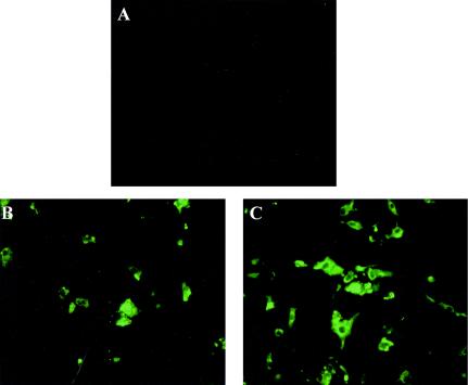
To examine the expression of VP2 protein by the rLaSota/VP2, DF1 cells were infected with rLaSota/VP2 of the 12th passage at an MOI of 0.1. At 24 or 48 h postinfection, the cells were fixed and incubated with a rabbit antiserum directed against IBDV, followed by immunostaining with FITC-conjugated goat anti-rabbit IgG. The results indicated that there was extensive expression of VP2 protein at 24 h postinfection (Fig. 2C), which further increased at 48 h postinfection (data not shown). Compared to rLaSota/VP2, cell culture-adapted IBDV strain GLS-5 displayed low levels of fluorescence at 24 h postinfection (Fig. 2), suggesting a slower replication cycle for IBDV. However, the fluorescence intensities and cellular distribution of VP2 antigen in rLaSota/VP2- and IBDV GLS-5-infected cells were similar at 48 h postinfection (data not shown). Western blot analysis of infected DF1 cell lysates further confirmed the expression of VP2 protein by the rLaSota/VP2 virus (data not shown). We were also able to demonstrate the VP2 protein from the supernatant of rLaSota/VP2-infected cells by ELISA. Western blotting of purified rLaSota/VP2 virus showed no detectable amount of the VP2 protein (data not shown). Taken together, these results suggested that the recombinant rLaSota/VP2 stably expressed the VP2 protein and that the VP2 protein was probably not incorporated into the virions.
FIG. 2.
Immunofluorescence analysis of IBDV VP2 protein expression. Confluent DF1 cells were infected with rLaSota (A), cell culture-adapted GLS-5 (B), or rLaSota/VP2 at 12th passage (C) at an MOI of 0.1. The infected cells were fixed, permeabilized, and probed with rabbit anti-IBDV antiserum, followed by incubation with FITC-conjugated goat anti-rabbit IgG antibody. The cells were visualized under an Eclipse TE (Nikon) fluorescent microscope. Magnification, ×400. Data are shown at 24 h postinfection.
MAb reactivity profile of rLaSota/VP2.
To assess whether the VP2 protein expressed by rLaSota/VP2 virus contains the neutralizing epitopes, both the cell lysate and the supernatant from infected cells were analyzed by AC-ELISA with a panel of well-characterized epitope-specific MAbs. Five virus-neutralizing MAbs (8, 10, 57, R63, and B69) corresponding to epitopes residing on the VP2 protein were used to analyze the antigenic properties of the expressed protein (40, 41). Samples were considered positive if their OD values were equal to or greater than 0.6. MAb 57 corresponds to a unique neutralizing epitope in the VP2 protein of variant GLS-5 strain; whereas MAbs 8 and 10 define neutralizing epitopes present both in the classic and variant GLS-5 strains of IBDV (40, 41, 42). The expressed VP2 protein reacted with both MAbs 57 and 10, as did the variant GLS-5 strain (Table 2). However, the cell lysate and supernatant from rLaSota/VP2-infected cells did not bind to MAb 8, indicating that the presence of this epitope may depend on the presence of other virus-associated proteins. Epitopes defined by MAbs R63 and B69 are present only in classical strains but absent in the GLS-5 strain (41, 42). The recombinant VP2 protein did not bind to R63 and B69 (Table 2). These results suggested that the VP2 protein expressed by rLaSota/VP2 was specific to the GLS-5 strain and retained the neutralizing epitopes. Since these neutralizing epitopes are conformation dependent, the expressed VP2, therefore, should be forming conformationally correct structures for immune recognition. Although direct measurement of the amount of recombinant proteins was not carried out, the results from immunostaining and AC-ELISA suggested that the expression of VP2 protein was at a higher level.
TABLE 2.
Antigenic characterization of the VP2 protein expressed by rLaSota/VP2 with IBDV-neutralizing MAbs by AC-ELISAa
| MAbsb | Antigenic reactivity of IBDVc
|
|||
|---|---|---|---|---|
| Classicd | GLS-5 | rLaSota/VP2 | rLaSota | |
| 8 | + | + | − | − |
| 10 | + | + | + | − |
| 57 | − | + | + | − |
| R63 | + | − | − | − |
| B69 | + | − | − | − |
Cell lysates from DF1 cells infected with recombinant rLaSota/VP2, rLaSota and GLS-5, and homogenized bursa of IBDV classic strain D78, were used for AC-ELISA.
Neutralizing MAbs with known reactivity to epitopes on VP2 protein of IBDV.
Antigenic reactivity determined by AC-ELISA. The presence (+) and the absence (−) of the epitope in viruses are indicated.
IBDV classic strain D78.
Growth characteristics of the recombinant virus.
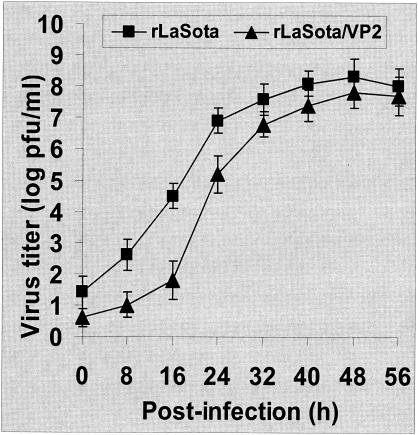
To determine the effect of VP2 protein on growth of recombinant rLaSota/VP2, we analyzed the kinetics and final virus yield under multistep growth conditions on DF1 cells. Triplicate monolayers of DF1 cells were infected with either rLaSota/VP2 or rLaSota virus at an MOI of 0.01, and samples were collected at 8-h intervals to assess the virus titers by plaque assay. The results showed that the kinetics and magnitude of replication for both rLaSota and rLaSota/VP2 were very similar, though the replication of rLaSota/VP2 was delayed slightly (Fig. 3). The final virus yields in DF1 cells and in embryonated SPF chicken eggs 64 h postinfection were comparable for both recombinant viruses. Moreover, there were no significant differences in the plaque size and morphology between the rLaSota/VP2 and the parental rLaSota (data not shown).
FIG. 3.
Multistep growth curve of rLaSota and rLaSota/VP2 in cell culture. Monolayers of DF1 cells in 25-cm2 flasks were infected with viruses at an MOI of 0.01 and incubated at 37°C in DMEM supplemented with 5% fetal calf serum and 1 μg of acetyl-trypsin/ml. Supernatants were harvested at 8-h intervals for virus titration. Values are from two independent experiments, each performed in triplicate. Bars show standard deviations.
MDT in chicken embryos.
The virulence of the parental virus rLaSota and that of the recombinant rLaSota/VP2 were determined by MDT in 10-day-old embryonated SPF chicken eggs. The MDT is one of the internationally accepted methods for assessing the pathogenicity of NDV strains. Strains of NDV are categorized into three groups on the basis of their MDTs: velogenic (less than 60 h), mesogenic (60 to 90 h), and lentogenic (greater than 90 h) (1, 2). The values of MDT for rLaSota and rLaSota/VP2 were 98 and 106 h, respectively. These results indicated that the rLaSota/VP2 virus took slightly longer to kill the embryos and that the pathogenicity of the recombinant virus was not increased after insertion of the IBDV VP2 gene.
Immunogenicity and protective efficacy against virulent IBDV challenge.
To determine the immunogenicity and protective efficacy of recombinant rLaSota/VP2, groups of 2-day-old chickens were vaccinated with a commercial IBDV, or commercial NDV vaccine, or rLaSota/VP2 (Table 1). Three weeks after vaccination, blood samples (prechallenge) were analyzed by ELISA for the presence of antibodies to NDV or IBDV with a commercially available ELISA kit (Synbiotics) and by VN test for neutralizing antibodies. Our results showed that antibody titers from the ELISA and VN test were highly correlated (Table 3). The ELISA titers and VN titers against IBDV were at high levels in chickens vaccinated with rLaSota/VP2 and commercial IBDV vaccine (Table 3). Both the ELISA titers and VN titers against NDV were comparable in birds immunized with rLaSota/VP2 and commercial NDV vaccine. No detectable amounts of NDV or IBDV antibodies were present in the blood samples from the unvaccinated control group (Table 3). The data suggested that the VP2 protein expressed by rLaSota/VP2 induced a very good antibody response in chickens and that the expression of the VP2 protein did not interfere with the induction of NDV antibodies.
TABLE 3.
Immune responses to NDV or IBDV induced by vaccination
| Groups | Vaccinea | Antibody titersb
|
|||||||
|---|---|---|---|---|---|---|---|---|---|
| Against NDV
|
Against IBDV
|
||||||||
| Primary vaccination
|
Secondary vaccination
|
Primary vaccination
|
Secondary vaccination
|
||||||
| ELISAc | VNd | ELISA | VN | ELISA | VN | ELISA | VN | ||
| 1 | Commercial NDV vaccine | 4,720 | 336 | 6,960 | 683 | <10 | ≤2 | <10 | ≤2 |
| 2 | Commercial IBDV vaccine | <10 | ≤2 | <10 | ≤2 | 7,870 | 687 | 8,610 | 1,056 |
| 3 | rLaSota/VP2 | 5,103 | 294 | 7,030 | 629 | 6,580 | 576 | 7,120 | 982 |
| 4 | None | <10 | ≤2 | <10 | ≤2 | <10 | ≤2 | <10 | ≤2 |
Vaccination was given by eyedrop instillation with commercial vaccines (manufacturer's recommended dose/chicken) or rLaSota/VP2 (1 × 10⁴ EID50/chicken).
Immune responses were performed 3 weeks after primary vaccination or 2 weeks after secondary vaccination.
Value was the reciprocal geometric mean titer from ELISA.
VN titer was expressed as geometric mean titer of serum that neutralized 100 50% tissue culture infective doses of the homologous virus.
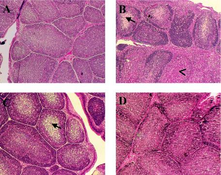
Immunized chickens, along with unvaccinated control birds, were challenged with virulent IBDV strain GLS-5 at 3 weeks postvaccination. Unvaccinated chickens were fully susceptible to the challenge, showing either mortality (10%) or clinical signs (100%). In contrast, no clinical signs or mortality were observed in chickens vaccinated with either rLaSota/VP2 or commercial live IBDV vaccine. Of chickens vaccinated with rLaSota/VP2 or commercial live IBDV vaccine, 20 and 10% had bursal atrophy after challenge, respectively, as indicated by BBWR. The group mean of BBWR was assigned to similar groups by the Kruskal-Wallis test, showing that both commercial IBDV vaccine and rLaSota/VP2 offered significant protection against challenge (Table 4, primary vaccination). Analysis by AC-ELISA showed a protective pattern similar to that of BBWR, with 20 and 10% bursal samples displaying the presence of IBDV antigens (Table 4, primary vaccination). Histopathological examination of representative samples revealed that chickens vaccinated with commercial live IBDV vaccine or rLaSota/VP2 and challenged with GLS-5 virus showed no to only mild bursal lesions, whereas unvaccinated challenged chickens showed severe bursal lesions (Fig. 4). Overall, based on these criteria, primary vaccination with rLaSota/VP2 and commercial IBDV live vaccine conferred 80 and 90% protection, respectively, against challenge with virulent IBDV strain GLS-5 after primary vaccination. Of chickens vaccinated with either commercial live NDV vaccine or rLaSota/VP2, 1 out of 10 in each group died after lethal challenge with virulent NDV strain Texas GB. All other birds were normal and healthy, suggesting 90% protection level for both live NDV vaccine and rLaSota/VP2 after primary vaccination.
TABLE 4.
Protection efficacy for primary and secondary vaccination against IBDV challengea
| Vaccination | Primary vaccination
|
Secondary vaccination
|
||||||
|---|---|---|---|---|---|---|---|---|
| BBWRb
|
AC-ELISAc (no. protected/total) | Overall protection (%) | BBWR
|
AC-ELISA (no. protected/total) | Overall protection (%) | |||
| Mean ± SD | No. protected/total | Mean ± SD | No. protected/total | |||||
| Commercial IBDV vaccine | 5.04 ± 0.77 (A) | 9/10 | 9/10 | 90 | 4.71 ± 0.44 (A) | 10/10 | 10/10 | 100 |
| rLaSota/VP2 | 5.12 ± 0.78 (A) | 8/10 | 8/10 | 80 | 4.91 ± 0.51 (A) | 10/10 | 10/10 | 100 |
| Unvaccinated, challenged | 2.99 ± 0.57 (B) | 0/10 | 0/10 | 0 | 2.89 ± 0.58 (B) | 0/10 | 0/10 | 0/10 |
| Unvaccinated, unchallenged | 5.31 ± 0.79 (A) | 5/5 | 5/5 | 100 | 5.26 ± 0.62 (A) | 5/5 | 5/5 | 100 |
Chickens were vaccinated by eyedrop instillation and challenged ocularly with virulent IBDV strain GLS-5 3 weeks after primary or 2 weeks after secondary vaccination.
BBWR value represents BBWR × 1,000. Data presented in the Mean ± SD column are the group means ± standard deviations followed by a letter (A or B) indicating statistical significance. Group means were assigned to similar groups (A and B) by the Kruskal-Wallis test. Within each column, group means with different letters are statistically different (P < 0.05). Protection was defined as individual value of BBWR within 2 standard deviation units of the mean of the unvaccinated unchallenged control group, with data showing the numbers protected and the total in each group.
Protection was determined by the absence of viral antigen in bursa 3 days postchallenge by AC-ELISA.
FIG. 4.
Histopathology of bursa of Fabricius 3 days after challenge. Chickens were vaccinated and boosted with PBS (A and B), commercial IBDV vaccine (C), or rLaSota/VP2 (D) and challenged with virulent IBDV strain GLS-5 (B, C, D). Panel A shows an unchallenged control. Seventy-two hours after challenge, the bursa was removed and fixed in neutral buffered formalin. Tissues were embedded in paraffin, sectioned, and stained with hemoxylin and eosin for microscopic examination. Magnification, ×40. (A) Bursa from normal chicken: large active follicles with little interfollicular tissue. (B) Bursa from unvaccinated challenged chicken: severe multifocal medullary vacuolation and lymphocytic depletion (arrow), and severe follicular degeneration (arrowhead). (C) Bursa from chicken vaccinated with commercial IBDV vaccine and challenged: some mild multifocal medullary vacuolation and lymphocytic depletion (arrow). (D) Bursa from chicken vaccinated with rLaSota/VP2 and challenged: no histologic lesions observed.
Secondary immunization at 23 days of age with commercial live NDV or IBDV vaccines induced higher levels of antibodies to NDV and IBDV, respectively, than did the primary vaccination (Table 3). Revaccination with rLaSota/VP2 enhanced antibody responses to both NDV and IBDV, as reflected by the higher antibody titers (Table 3). Two weeks after secondary immunization, chickens were challenged with either virulent IBDV strain GLS-5 or NDV strain Texas GB as described above. As indicated by antigen detection, BBWR, and histopathology, both the commercial live IBDV vaccine and recombinant rLaSota/VP2 conferred complete protection against virulent IBDV strain GLS-5 challenge; whereas the unvaccinated control chickens were fully susceptible (Table 4). Chickens vaccinated with rLaSota/VP2 and commercial NDV vaccines were also protected against challenge with virulent strain NDV Texas GB after secondary immunization (Table 5). Overall, the results showed that the levels of protection against both IBDV and NDV challenge were very well correlated with the antibody titers induced by the vaccination and that expression of the IBDV VP2 protein by rLaSota/VP2 would not interfere with immunity against NDV.
TABLE 5.
Protective efficacy of primary and secondary vaccination against NDV challengea
| Vaccine | Primary vaccination
|
Secondary vaccination
|
||
|---|---|---|---|---|
| No. dead/total | Protection (%) | No. dead/total | Protection (%) | |
| Commercial NDV vaccine | 1/10 | 90 | 0/10 | 100 |
| rLaSota/VP2 | 1/10 | 90 | 0/10 | 100 |
| None | 10/10 | 0 | 10/10 | 0 |
Chickens were vaccinated by eyedrop instillation with commercial NDV vaccine or rLaSota/VP2 and challenged intramuscularly with virulent NDV strain Texas GB 3 weeks after primary or 2 weeks after secondary vaccination.
DISCUSSION
The use of biotechnology to create recombinant viral-vectored vaccines holds many promises for the future. In the poultry industry, so far, fowl poxvirus, herpesvirus of turkeys, Marek's disease virus, adenovirus, and members of the retrovirus family have been used most extensively as expression vectors (11, 21, 48). These recombinant viruses have demonstrated protective efficacy against a variety of avian diseases. However, their practical use still needs to be evaluated, particularly with regard to factors such as safety, route of delivery, efficacy, and production cost (11, 21, 48).
NDV provides an efficient vector system for the delivery of protective antigens of other avian pathogens, such as those of the IBDV or infectious bronchitis virus. Live NDV vaccines, such as the LaSota strain, are widely used in poultry industries around the world with proven track records in safety and efficacy. Immunization with LaSota induces not only long-lasting humoral and cellular immunity but also a strong mucosal immunity. NDV grows to very high titers in many cell lines and embryonated eggs, which allows cost-effective and easy manufacture of the vaccine. Unlike other viral vectors, NDV has a simple genome encoding only a few proteins. For generation of specific immune responses, it would be advantageous to have only a limited number of proteins expressed.
IBDV is a pathogen of major economic importance to the poultry industry worldwide. Currently, chickens are routinely vaccinated against IBDV (25). However, infectious bursal disease still has considerable socioeconomic importance at the international level, as the disease is present in almost every country because of the emergence of new antigenic variants or strains of increased virulence (25). Variant and hypervirulent strains probably arise through the accumulation of point mutations due to the error-prone nature of viral RNA polymerase and through reassortment between circulating strains or between vaccines and circulating field strains (25, 42). Another factor that may be contributing to creation of the antigenic variant viruses is the wide use of live attenuated IBDV vaccines (25). It is possible that, over a period of time, these live attenuated IBDV vaccines may revert back to virulence and may change antigenicity (17, 41, 42). Further, live IBDV vaccines cannot break through the high levels of maternal antibodies in young chickens (25). Therefore, there is a need to develop alternative IBDV immunizing strategies, which will deter the development of variant and/or hypervirulent strains but still have the capability to induce local and systemic immunity critical to controlling the disease, in the presence of maternal antibodies.
The VP2 protein is the major host-protective immunogen of IBDV, and most of the variations among isolates appear in this part of the viral genome (4, 12, 16, 40). Since a major neutralizing epitope of the virus is a conformational epitope, delivery of VP2 protein in native conformation is critical for correct antigen processing and presentation (3, 25). Furthermore, in developing a poultry vaccine, a number of factors must be taken into account. Since the price of a single bird is relatively low, one of the most important factors is the cost of the vaccine. The research conducted here leads to the development of a bivalent vaccine candidate for NDV and IBDV and provides an inexpensive approach. This novel vaccine candidate has the following features. The IBDV VP2 protein was stably and correctly expressed and retained the host-protective conformational epitopes. The expressed VP2 protein was not incorporated into the recombinant virus, and the recombinant virus had the similar replicative kinetics as the parental rLaSota virus.
Overall, the recombinant rLaSota/VP2 has several advantages over the existing IBDV vaccines. First, the recombinant NDV/VP2 will be highly economical for the poultry industry, since the cost of current IBDV vaccination will be eliminated for the poultry producers. Second, the recombinant virus can break through and will not be neutralized by maternal immunity against IBDV; therefore, it can be used for vaccination even in the presence of maternal antibody to enhance protection in the first few critical weeks of chicken life. Third, the recombinant rLaSota/VP2 vaccine will not result in new IBDV variants, since no live IBDV will be used for vaccination. Finally, it was reported that the V protein of NDV is associated with viral pathogenesis and functions as an interferon antagonist (20, 28). It was also demonstrated that elimination of the V protein expression in NDV rendered the virus attenuated but still highly immunogenic and that the attenuated NDV vaccine strain would be administered in ovo in uniform dosage by automation to protect chickens with or without maternal antibodies (28). Therefore, the recombinant virus can be tailored with ease for in ovo use. Thus, the recombinant virus described here for the protection of both NDV and IBDV will be highly beneficial to the poultry industry.
Our results demonstrated, for a variant infectious bursal disease, the potential of using NDV recombinant vaccine to protect SPF chickens against a massive challenge dose of homologous virus strains. Successful immunization of commercial chickens against many different field strains is likely to be more complicated. However, we can devise strategies to improve the recombinant vaccines. Since additional, albeit minor, neutralizing epitopes are also present on VP3 subunit (25), it is reasonable to assume that the immunogenicity of the recombinant virus can be enhanced by inserting the gene encoding IBDV polyprotein into the NDV genome. Moreover, correct expression of the polyprotein may result in formation of virus-like particles, which would be more immunogenic and offer better protection (45). Currently, in the poultry industry, classic and variant strains of live IBDV vaccines are simultaneously used to obtain maximal cross-protection. We could make two recombinant viruses, each expressing the VP2 proteins of the classic or variant strains, respectively, and use them as combined products for vaccination. Alternatively, we could express both the VP2 proteins simultaneously from the same NDV genome as two separate transcriptional units. In the case of emergence of a new variant serotype for which current vaccines do not provide enough protection, we could use this system conveniently to update the antigenic specificity of vaccines to best control current circulating field strains. The fact that NDV grows in cell cytoplasm without a DNA stage and has extremely low frequency of recombination (24) would make the use of recombinant NDV expressing the VP2 protein of IBDV more appealing.
The present study demonstrated that recombinant NDV is an excellent vaccine vector for IBDV. The use of an NDV-based vaccine that would reduce the number of circulating IBDV strains may be highly beneficial to the poultry industry. Furthermore, our results suggested that NDV could be used as a vaccine vector for other avian pathogens. Moreover, NDV replicates preferentially in avian cells but is replication deficient in most mammalian cells; therefore, NDV could be exploited as a promising vaccine delivery vector for human pathogens. Armed with basic knowledge, the recombinant NDV could also be used in cancer treatment and gene therapy.
Acknowledgments
We greatly acknowledge Chinta Lamichhane at Synbiotics Corporation for his advice and reagents in immunological assessment. We thank Daniel Rockemann and Peter Savage for their excellent technical assistance.
This work was partially supported by U.S. Department of Agriculture grant 2002-35204-1601.
REFERENCES
- 1.Alexander, D. J. 1989. Newcastle disease, p. 114-120. In H. G. Purchase, L. H. Arp, C. H. Domermuth, and J. E. Pearson (ed.), A laboratory manual for the isolation and identification of avian pathogens, 3rd ed. American Association for Avian Pathologists, Inc., Kennett Square, Pa.
- 2.Alexander, D. J. 1997. Newcastle disease and other avian Paramyxoviridae infection, p. 541-569. In B. W. Calnek (ed.), Diseases of poultry, 10th ed. Iowa State University Press, Ames.
- 3.Azad, A. A., N. M. McKern, I. G. Macreadie, P. Failla, H. G. Heine, A. Chapman, C. W. Ward, and K. J. Fahey. 1991. Physicochemical and immunological characterization of recombinant host-protective antigen (VP2) of infectious bursal disease virus. Vaccine 9:715-722. [DOI] [PubMed] [Google Scholar]
- 4.Becht, H., H. Muller, and H. K. Muller. 1988. Comparative studies on structural and antigenic properties of two serotypes of infectious bursal disease virus. J. Gen. Virol. 69:631-640. [DOI] [PubMed] [Google Scholar]
- 5.Boyle, D. B., and H. G. Heine. 1993. Recombinant fowlpox virus vaccines for poultry. Immunol. Cell Biol. 71:391-397. [DOI] [PMC free article] [PubMed] [Google Scholar]
- 6.Bukreyev, A., E. Camargo, and P. L. Collins. 1996. Recovery of infectious respiratory syncytial virus expressing an additional, foreign gene. J. Virol. 70:6634-6641. [DOI] [PMC free article] [PubMed] [Google Scholar]
- 7.Bukreyev, A., S. S. Whitehead, N. Bukreyev, B.R. Murphy, and P. L. Collins. 1999. Interferon γ expressed by a recombinant respiratory syncytial virus attenuates virus replication in mice without compromising immunogenicity. Proc. Natl. Acad. Sci. USA 96:367-2372. [DOI] [PMC free article] [PubMed] [Google Scholar]
- 8.Calain, P., and L. Roux. 1993. The rule of six, a basic feature for efficient replication of Sendai virus defective interfering RNA. J. Virol. 67:4822-4830. [DOI] [PMC free article] [PubMed] [Google Scholar]
- 9.Conzelmann, K. K. 1996. Genetic manipulation of non-segment negative-strand RNA viruses. J. Virol. 77:381-389. [DOI] [PubMed] [Google Scholar]
- 10.De Leeuw, O., and B. Peeters. 1999. Complete nucleotide sequence of Newcastle disease virus: evidence for the existence of a new genus within the subfamily Paramyxovirinae. J. Gen. Virol. 80:131-136. [DOI] [PubMed] [Google Scholar]
- 11.Dodds, W. J. 1999. More bumps on the vaccine road. Adv. Vet. Med. 41:715-732. [DOI] [PMC free article] [PubMed] [Google Scholar]
- 12.Fahey, K. J., K. Erny, and J. Crooks. 1989. A conformational immunogen on VP-2 of infectious bursal disease virus that induces virus-neutralizing antibodies that passively protect chickens. J. Gen. Virol. 70:1473-1481. [DOI] [PubMed] [Google Scholar]
- 13.Haglund, K., J. Forman, H. G. Krausslich, and J. K. Rose. 2000. Expression of human immunodeficiency virus type 1 Gag protein precursor and envelope proteins from vesicular stomatitis virus recombinant: high-level production of virus-like particles containing HIV envelope. Virology 268:112-121. [DOI] [PubMed] [Google Scholar]
- 14.Hasan, M. K., A. Kato, T. Shioda, Y. Sakai, D. Yu, and Y. Nagai. 1997. Creation of an infectious recombinant Sendai virus expressing the firefly luciferase gene from the 3′ proximal first locus. J. Gen. Virol. 78:2813-2820. [DOI] [PubMed] [Google Scholar]
- 15.He, B., R. G. Paterson, C. D. Ward, and R. A. Lamb. 1997. Recovery of infectious SV5 from cloned DNA and expression of a foreign gene. Virology 237:249-260. [DOI] [PubMed] [Google Scholar]
- 16.Heine, H. G., and D. B. Boyle. 1993. Infectious bursal disease virus structural protein VP2 expressed by a fowlpox virus recombinant confers protection against disease in chickens. Arch. Virol. 131:277-292. [DOI] [PubMed] [Google Scholar]
- 17.Heine, H. G., M. Haritou, P. Failla, K. Fahey, and A. Azad. 1991. Sequence analysis and expression of the host-protective immunogen VP2 of a variant strain of infectious bursal disease virus which can circumvent vaccination with standard type I strains. J. Gen. Virol. 72:1835-1843. [DOI] [PubMed] [Google Scholar]
- 18.Huang, Z., S. Elankumaran, A. Panda, and S. K. Samal. 2003. Recombinant Newcastle disease as a vaccine vector. Poult. Sci. 82:899-906. [DOI] [PubMed] [Google Scholar]
- 19.Huang, Z., S. Krishnamurthy, A. Panda, and S. K. Samal. 2001. High-level expression of a foreign gene from the most 3′-proximal locus of a recombinant Newcastle disease virus. J. Gen. Virol. 82:1729-1736. [DOI] [PubMed] [Google Scholar]
- 20.Huang, Z., S. Krishnamurthy, A. Panda, and S. K. Samal. 2003. Newcastle disease virus V protein is associated with viral pathogenesis and functions as an alpha interferon antagonist. J. Virol. 77:8676-8685. [DOI] [PMC free article] [PubMed] [Google Scholar]
- 21.Jackwood, M. W. 1999. Current and future recombinant viral vaccines for poultry. Adv. Vet. Med. 41:517-522. [DOI] [PubMed] [Google Scholar]
- 22.Krishnamurthy, S., Z. Huang, and S. K. Samal. 2000. Recovery of a virulent strain of Newcastle disease virus from cloned cDNA: expression of a foreign gene results in growth retardation and attenuation. Virology 278:168-182. [DOI] [PubMed] [Google Scholar]
- 23.Krishnamurthy, S., and S. K. Samal. 1998. Nucleotide sequences of the trailer, nucleocapsid protein gene and intergenic regions of Newcastle disease virus strain Beaudette C and completion of the entire genome sequence. J. Gen. Virol. 79:2419-2424. [DOI] [PubMed] [Google Scholar]
- 24.Lamb, R. A., and D. Kolakofsky. 2001. Paramyxoviridae: the viruses and their replication, p. 1305-1340. In D. M. Knipe and P. M. Howley (ed.), Fields virology. Lippincott Williams & Wilkins, Philadelphia, Pa.
- 25.Lukert, P. D., and Y. M. Saif. 1991. Infectious bursal disease, p. 648-663. In B. W. Calnek, H. J. Barnes, C. W. Beard, W. M. Reid, and H. W. Yoder, Jr. (ed.), Diseases of poultry. Iowa State University Press, Ames.
- 26.Mayo, M. A. 2002. A summary of taxonomic changes recently approved by ICTV. Arch. Virol. 147:1655-1656. [DOI] [PubMed] [Google Scholar]
- 27.Mebatsion, T., M. J. Schnell, J. H. Cox, S. Finke, and K. K. Conzelmann. 1996. Highly stable expression of a foreign gene from rabies virus vectors. Proc. Natl. Acad. Sci. USA 93:7310-7314. [DOI] [PMC free article] [PubMed] [Google Scholar]
- 28.Mebatsion, T., S. Verstegen, L. T. C. de Vaan, A. Rómer-Oberdórfer, and C. C. Schrier. 2001. A recombinant Newcastle disease virus with low-level V protein expression is immunogenic and lacks pathogenicity for chicken embryos. J. Virol. 75:420-428. [DOI] [PMC free article] [PubMed] [Google Scholar]
- 29.Nakaya, T., J. Cros, M. S. Park, Y. Nakaya, H. Zheng, A. Sagrera, E. Villar, A. García-Sastre, and P. Palese. 2001. Recombinant Newcastle disease virus as a vaccine vector. J. Virol. 75:11868-11873. [DOI] [PMC free article] [PubMed] [Google Scholar]
- 30.Palese, P., H. Zheng, O. G. Engelhardt, S. Pleschka, and A. García-Sastre. 1996. Negative-strand RNA viruses: genetic engineering and applications. Proc. Natl. Acad. Sci. USA 93:11354-11358. [DOI] [PMC free article] [PubMed] [Google Scholar]
- 31.Park, K., T. Huang, F. F. Correia, and M. Krystal. 1991. Rescue of a foreign gene by Sendai virus. Proc. Natl. Acad. Sci. USA 88:5537-5541. [DOI] [PMC free article] [PubMed] [Google Scholar]
- 32.Peeters, B. P. H., Y. K. Cruthuisen, O. S. de Leeuw, and A. L. Gielkens. 2000. Genome replication of Newcastle disease virus: involvement of the rule-of-six. Arch. Virol. 145:1829-1845. [DOI] [PubMed] [Google Scholar]
- 33.Peeters, B. P. H., O. S. de Leeuw, G. Koch, and A. L. Gielkens. 1999. Rescue of Newcastle disease virus from cloned cDNA: evidence that cleavability of the fusion protein is a major determinant for virulence. J. Virol. 73:5001-5009. [DOI] [PMC free article] [PubMed] [Google Scholar]
- 34.Phenix, K. V., K. Wark, C. J. Luke, M. A. Skinner, J. A. Smyth, K. A. Mawhinney, and D. Todd. 2001. Recombinant Semliki Forest virus vector exhibits potential for avian virus vaccine development. Vaccine 19:3116-3123. [DOI] [PubMed] [Google Scholar]
- 35.Reddy, S. K., and A. Silim. 1991. Comparison of neutralizing antigens of recent isolates of infections bursal disease virus. Arch. Virol. 117:287-296. [DOI] [PubMed] [Google Scholar]
- 36.Roberts, A., E. Kretzschmar, A. S. Perkins, J. Forman, R. Price, L. Buonocore, Y. Kawaoka, and J. K. Rose. 1998. Vaccination with a recombinant vesicular stomatitis virus expressing an influenza virus hemagglutinin provides complete protection from influenza virus challenge. J. Virol. 72:4704-4711. [DOI] [PMC free article] [PubMed] [Google Scholar]
- 37.Sakaguchi, M., H. Nakamura, K. Sonoda, H. Okamura, K. Yokogawa, K. Matsuo, and K. Hira. 1998. Protection of chickens with or without maternal antibodies against both Marek's and Newcastle disease by one-time vaccination with recombinant vaccine of Marek's disease virus type 1. Vaccine 16:472-479. [DOI] [PubMed] [Google Scholar]
- 38.Sakai, Y., K. Kiyotani, M. Fukumura, M. Asakawa, A. Kato, T. Shioda, T. Yoshida, A. Tanaka, M. Hasegawa, and Y. Nagai. 1999. Accommodation of foreign genes into the Sendai virus genome: sizes of inserted genes and viral replication. FEBS Lett. 456:221-226. [DOI] [PubMed] [Google Scholar]
- 39.Sheppard, M., W. Werner, E. Tsatas, R. McCoy, S. Prowse, and M. Johnson. 1998. Fowl adenovirus recombinant expressing VP2 of infectious bursal disease virus induces protective immunity against bursal disease. Arch. Virol. 143:915-930. [DOI] [PMC free article] [PubMed] [Google Scholar]
- 40.Snyder, D. B., D. P. Lana, B. R. Cho, and W. W. Marquardt. 1988. Group and strain-specific neutralizing sites of infectious bursal disease virus defined with monoclonal antibodies. Avian Dis. 32:527-534. [PubMed] [Google Scholar]
- 41.Snyder, D. B., D. P. Lana, P. K. Savage, F. S. Yancey, S. A. Mengel, and W. W. Marquardt. 1988. Differentiation of infectious bursal disease viruses directly from infected tissues with neutralizing antibodies: evidence of a major antigenic shift in recent field isolates. Avian Dis. 32:535-539. [PubMed] [Google Scholar]
- 42.Snyder, D. B., V. N. Vakharia, and P. K. Savage. 1992. Naturally occurring-neutralizing monoclonal antibody escape variants define the epidemiology of infectious bursal disease viruses in the United States. Arch. Virol. 127:89-101. [DOI] [PubMed] [Google Scholar]
- 43.Tsukamoto, K., S. Saito, S. Saeki, T. Sato, N. Tanimura, T. Isobe, M. Mase, T. Imada, N. Yuasa, and S. Samaguchi. 2002. Complete, long-lasting protection against lethal infectious bursal disease virus challenge by a single vaccination with an avian herpesvirus vector expressing VP2 antigens. J. Virol. 76:5637-5645. [DOI] [PMC free article] [PubMed] [Google Scholar]
- 44.Vakharia, V. N., D. B. Snyder, J. He, G. H. Edwards, P. K. Savage, and S. A. Mengel-Whereat. 1993. Infectious bursal disease virus structural proteins expressed in a baculovirus recombinant confer protection in chickens. J. Gen. Virol. 74:1201-1206. [DOI] [PubMed] [Google Scholar]
- 45.Vakharia, V. N., D. B. Snyder, D. Lütticken, S. A. Mengel-Whereat, P. K. Savage, G. H. Edwards, and M. A. Goodwin. 1994. Active and passive protection against variant and classic infectious bursal disease virus strains induced by baculovirus-expressed structural proteins. Vaccine 12:452-456. [DOI] [PubMed] [Google Scholar]
- 46.Van den Berg, T. P., M. Gonze, and G. Meulemans. 1991. Acute infectious bursal disease in poultry: isolation and characterization of a highly virulent strain. Avian Pathol. 20:133-143. [DOI] [PubMed] [Google Scholar]
- 47.Walsh, E. P., M. D. Baron, J. Anderson, and T. Barrett. 2000. Development of a genetically marked recombinant rinderpest vaccine expressing green fluorescent protein. J. Gen. Virol. 81:709-718. [DOI] [PubMed] [Google Scholar]
- 48.Yokoyama, N., K. Maeda, and T. Mikami. 1997. Recombinant viral vector vaccines for the veterinary use. J. Vet. Med. Sci. 59:311-322. [DOI] [PubMed] [Google Scholar]


