Abstract
This article describes the method to make a do it yourself smartphone-based fundus camera which can image the central retina as well as the peripheral retina up to the pars plana. It is a cost-effective alternative to the fundus camera.
Key words: Fundus camera, fundus photography, mobile phone fundus camera, retinal imaging, smartphone
Fundus photography plays a key role in monitoring and follow-up of patients. Recently, smartphones have been used for fundus documentation.[1] Here, we describe a cost-effective method of a do it yourself (DIY) smartphone fundus camera (DIYretCAM) using commonly available materials [Table 1, Fig. 1].
Table 1.
Materials required
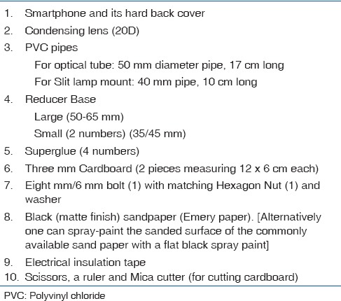
Figure 1.
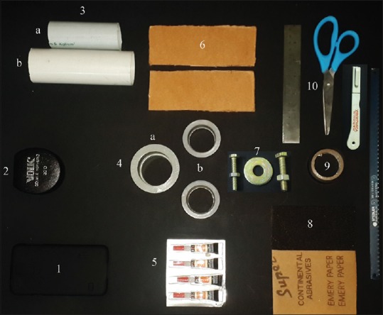
Materials required for the do it yourself smartphone fundus camera
Assembling the Do it Yourself Smartphone Fundus Camera
Align and center the narrow end of the large reducer base on the camera hole of the smartphone cover. The area of contact of the reducer and cover is then carefully glued [Fig. 2]. The 50 mm pipe is used as the optical tube. A piece of sandpaper 17 cm × 14.8 cm, is rolled and inserted into the tube and glued, the sanded surface facing inward to prevent dazzle. Another 15.5 cm × 2 cm sandpaper is glued inside the reducer base leaving 1.0 cm bare area toward the wider end [Fig. 3].
Figure 2.
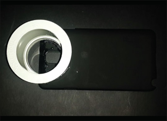
The reducer glued to the back cover
Figure 3.
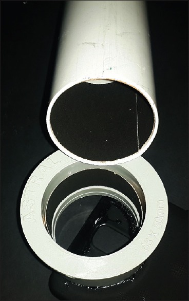
Sandpapers used to reduce the dazzle
Insulation tape is used to camouflage the optical tube and reducer base. At one end, for the condensing lens, apply 8–12 rounds of tape for snug fitting of the lens [Fig. 4a and b]. The phone is positioned in the cover, the optical tube inserted into the reducer, the 20 D fixed and the handheld device is ready [Fig. 4c]. For phones where the centers of the camera and flash are separated beyond 1 cm, a longer optical tube will be required.
Figure 4.
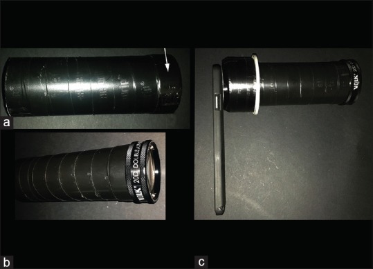
(a) The optical tube camouflaged with insulation tape. To fit the 20 D, few more rounds are applied to increase the thickness (arrow). (b) The condensing lens fixed on the optical tube. (c) The handheld device is ready to use
Assembling the Slit Lamp Mount
On each cardboard, make a hole to fit the bolt (8 mm for Haag-Strait model/Carl Zeiss Slit lamps or 6 mm for Topcon Model Slit lamps), using a pointed knife/Mica cutter and scissors, centered at 2.5 cm from one of its edges. The nut is tightened on the bolt, and the bolt passed through the holes in the cardboard pieces and tightened. The washer glued, centering the inner ring on the bolt at the under surface of the cardboards. Two cardboards are then glued together forming the platform for the slit lamp mount. Glue is applied around the nut also [Fig. 5a]. The small reducer is glued on the platform with its wider side facing up at the opposite edge [Fig. 5b]. A 40 mm pipe, 10 cm long, is fixed to the reducer [Fig. 5c]. For the optical tube holder, a 50 mm pipe is cut transversely, at approximately two-third of its diameter, to a length of 10 cm, and the smaller piece is discarded. The larger piece is then glued with its concave surface facing up, to the second small reducer, on to its narrower end as in Fig. 5d. The mount is then assembled as shown in the Fig. 5e. The shorter part of the optical tube holder faces the patient's eye. The mount is also camouflaged with the insulation tape. Alternatively, using spray painting can enhance the appearance (optional) [Video 1].
Figure 5.
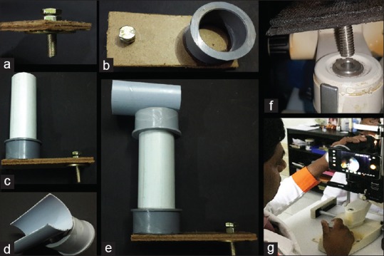
Slit lamp mount. (a) The nut, bolt, and washer in place. (b) Reducer is fixed. (c) 40 mm pipe fixed. (d) Optical tube holder. (e) Assembled mount. (f) Bolt placed in the slot. (g) The do it yourself smartphone fundus camera on the slit lamp
Using the Do it Yourself Smartphone Fundus Camera
To use the DIYretCAM on the slit lamp, move the observation illumination columns to one side, and fix the mount by placing the bolt in the slot for the focusing rod [Fig. 5f]. Different slit lamps have different diameters for the slot. For slit lamps with larger diameter slots, a few rounds of insulation tape on the bolt will improve stability. The optical tube is placed on the holder. The DIYretCAM can now be used like a fundus camera using the joystick [Fig. 5g], with the camera in the continuous flash on mode.
As in indirect ophthalmoscopy, the images are laterally reversed and vertically inverted, and therefore the movements to align the field of view, will be in opposite direction to the images seen. In the handheld method, one can hold the optical tube or the camera or the 20 D lens [Fig. 6a and b], and use the camera in the photo or video mode. In video mode, imaging of the peripheral retina till pars plana [Fig. 7] is possible with simultaneous scleral depression [Fig. 6c].
Figure 6.
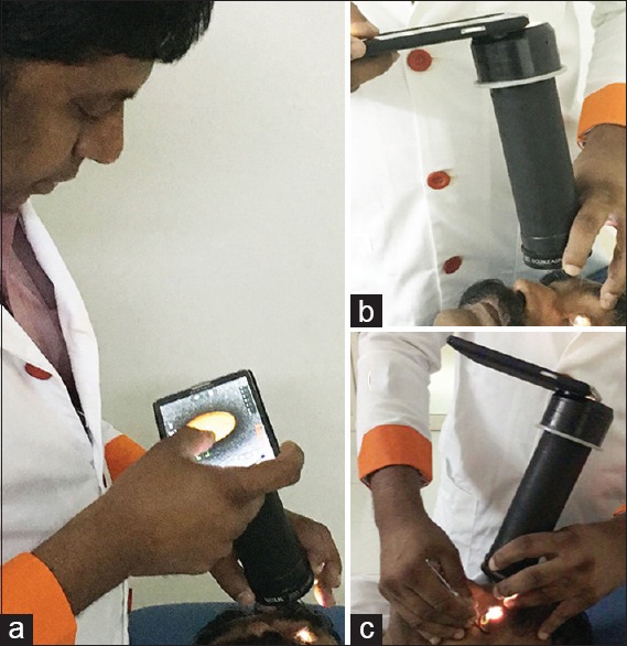
(a) The do it yourself smartphone fundus camera used as a hand held the device. (b) The do it yourself smartphone fundus camera can be held at the condensing lens and supported with the other hand on the camera. (c) Like in indirect ophthalmoscopy, scleral depression is done after stabilizing the do it yourself smartphone fundus camera
Figure 7.
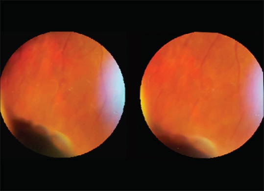
Stereo pair showing pars plana cysts
The camera app which is preloaded in the smartphone is good enough for basic fundus photography. We have used Camera FV-5 and Cinema FV-5 (FGAE Studios, Germany, http://www.cinemafv5.com/index.php) for Android and Camera Plus (Global Delight Technologies, Udupi, India, www.globaldelight.com/iphone/cameraplus) for iPhone and these apps give more control on the image capture with various options such as focus lock and exposure lock.
Image editing can be done on the smartphone itself using Adobe Photoshop Express (Adobe Systems Incorporated, www.photoshop.com/products/photoshopexpress). Editing involves capturing the screenshot of the desired frame from the video and opening the screenshot or image in Photoshop [Fig. 8a]. The image is then cropped, rotated to proper orientation [Fig. 8b] and corrected for brightness and contrast [Fig. 8c] as required. The central reflections may be removed using the blemish removal tool [Fig. 8d] and e]. Finally, the image mask (circle black) is applied [Fig. 8f] and the image is saved.
Figure 8.
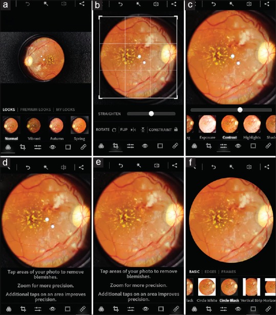
Editing the images. (a) The images opened in Photo Express. (b) The image cropped, rotated to correct orientation. (c) Improve the contrast, brightness, etc., (d) The blemish removal tool is used to remove the reflections (e) After removing the reflections. (f) The circle black mask is applied
DIYretCAM captures cost-effective [Table 2] quality fundus images. It is also capable of imaging up to the pars plana with scleral depression. Stereo fundus photography of the central [Fig. 9] and peripheral retina [Fig. 7] are possible with the DIYretCAM. The DIYretCAM is a cost-effective option for documenting the fundus changes in retinopathy of prematurity [Fig. 10] and in bedridden patients. Although useful in such settings, this device is not intended to replace the fundus camera especially in macular imaging, where, the fundus camera with its reflex free, high-quality imaging gives a better resolution of the macular details.
Table 2.
Cost of DIYretCAM
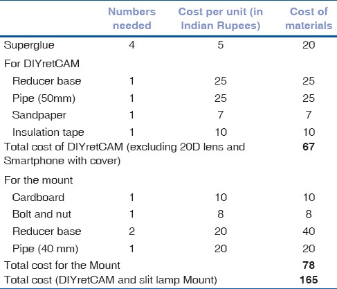
Figure 9.
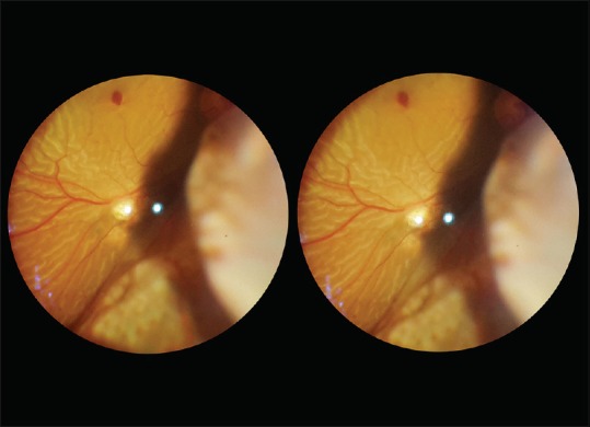
Stereo pair taken with the do it yourself smartphone fundus camera shows a recurrent retinal detachment with macular hole. Note the bullous detachment inferonasally
Figure 10.
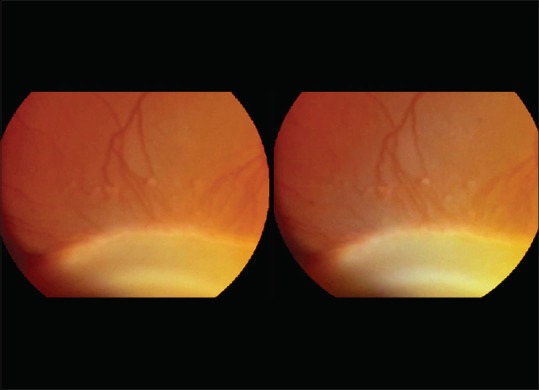
Stereo pair of a Zone II Stage 3 retinopathy of prematurity showing popcorn lesions. The mount of the scleral depressor is clearly made out in stereo
Holding the phone with one hand and the 20 D lens with the other hand helps to stabilize the device during the learning curve. Once familiar with this technique, the device can be held with the hand holding the 20 D lens, with the optical tube resting within the web of the thumb and the index finger. We made this device with commonly available materials on the “Do-It-Yourself” concept. With professional help, the mount can be made from sturdier materials like medium density fibreboard (MDF) or Polyvinyl chloride (PVC) pipes, with custom made anchoring bolt, giving a better appearance, and stability to the device. Fluorescein angiography is also possible [Fig. 11] with this device using matched filters as described by Suto et al.[2] This device is also capable of capturing good quality anterior segment photographs as well [Fig. 12a and b].
Figure 11.
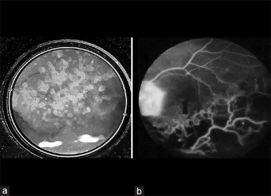
Do it yourself smartphone fundus camera fluorescein angiography. (a) Preinjection photo with only the barrier filter, showing dense asteroid hyalosis. (b) The mid-phase fundus fluorescein angiography showing capillary nonperfusion, macular ischemia and neovascularization on the disc suggestive of neovascular branch retinal vein occlusion with macular ischemia
Figure 12.
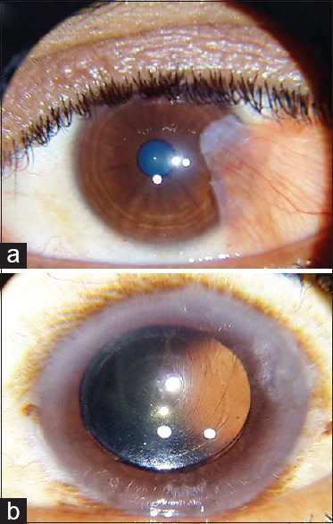
Anterior segment photographs taken with the do it yourself smartphone fundus camera. (a) Vascular pterygium (b) wrinkling of the posterior capsule in a pseudophakic eye
Financial support and sponsorship
Nil.
Conflicts of interest
There are no conflicts of interest.
Video available on www.ijo.in
References
- 1.Raju B, Raju NS. Regarding fundus imaging with a mobile phone: A review of techniques. Indian J Ophthalmol. 2015;63:170–1. doi: 10.4103/0301-4738.154407. [DOI] [PMC free article] [PubMed] [Google Scholar]
- 2.Suto S, Hiraoka T, Oshika T. Fluorescein fundus angiography with smartphone. Retina. 2014;34:203–5. doi: 10.1097/IAE.0000000000000041. [DOI] [PubMed] [Google Scholar]
Associated Data
This section collects any data citations, data availability statements, or supplementary materials included in this article.


