

Abstract
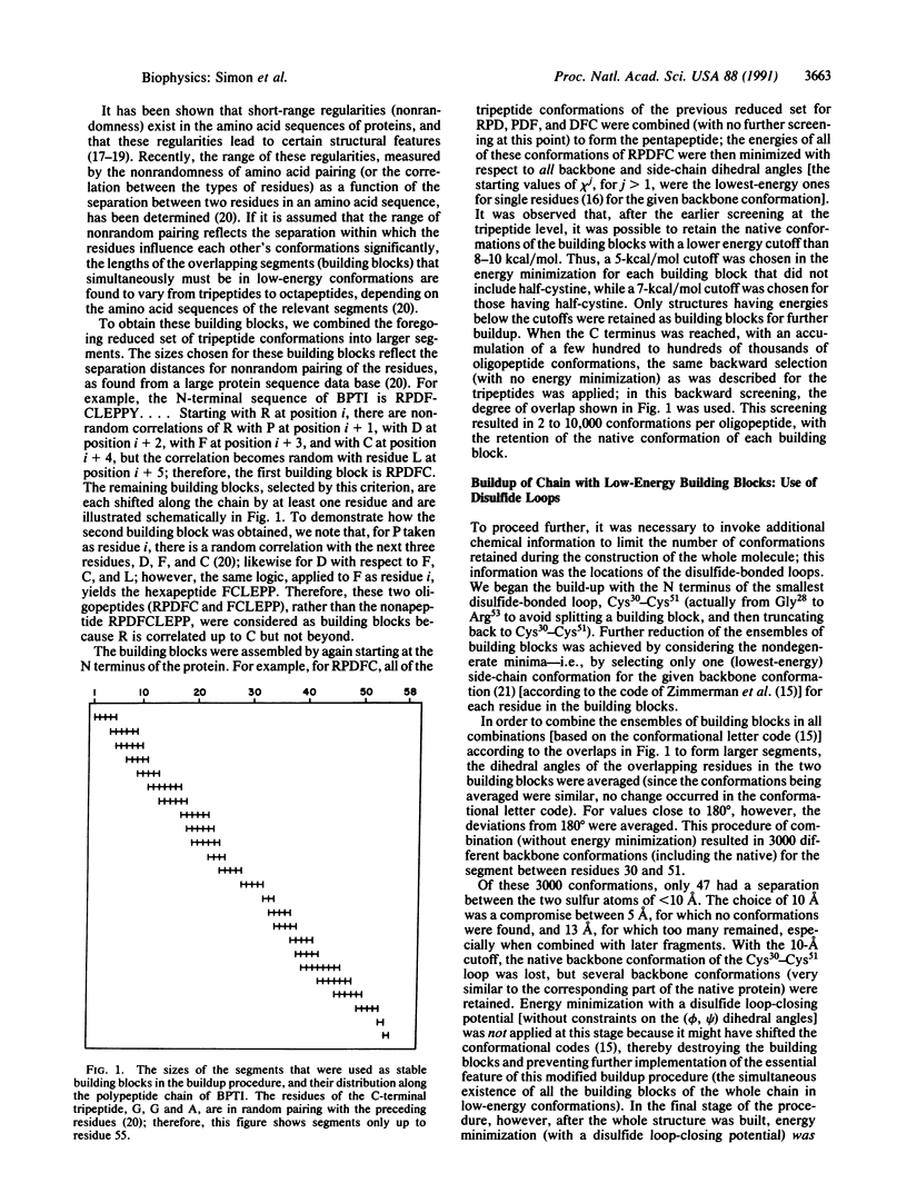
Conformations of bovine pancreatic trypsin inhibitor were calculated by assuming that the final structure as well as properly chosen overlapping segments thereof are simultaneously in low-energy (not necessarily the lowest-energy) conformational states. Therefore, the whole chain can be built up from building blocks whose conformations are determined primarily by short-range interactions. Our earlier buildup procedure was modified by taking account of a statistical analysis of known amino acid sequences that indicates that there is nonrandom pairing of amino acid residues in short segments along the chain, and by carrying out energy minimization on only these segments and on the whole chain [without minimizing the energies of intermediate-size segments (20-30 residues long)]. Results of this statistical analysis were used to determine the variable sizes of the overlapping oligopeptide building blocks used in the calculations; these varied from tripeptides to octapeptides, depending on the amino acid sequence. Successive stages of approximations were used to combine the low-energy conformations of these building blocks in order to keep the number of variables in the computations to a manageable size. The calculations led to a limited number of conformations of the protein (only two different groups, with very similar structure within each group), most residues of which were in the same conformational state as in the native structure.
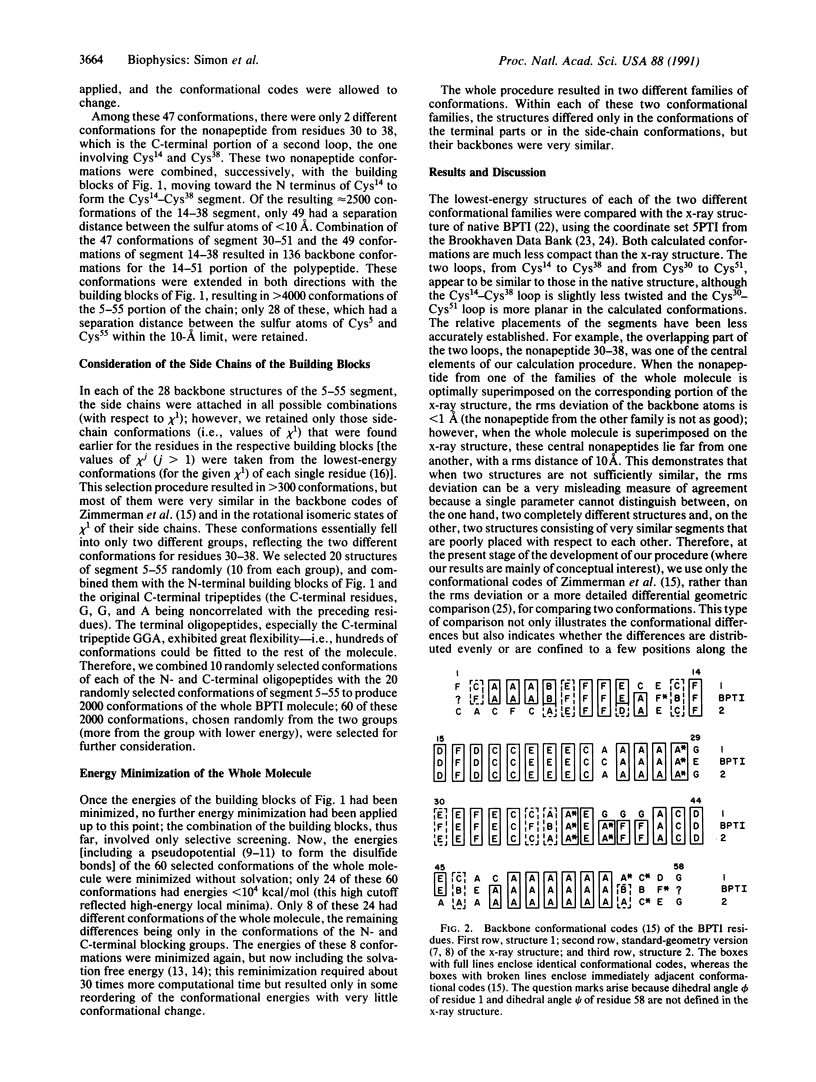
Full text
PDF

Selected References
These references are in PubMed. This may not be the complete list of references from this article.
- Bernstein F. C., Koetzle T. F., Williams G. J., Meyer E. F., Jr, Brice M. D., Rodgers J. R., Kennard O., Shimanouchi T., Tasumi M. The Protein Data Bank: a computer-based archival file for macromolecular structures. J Mol Biol. 1977 May 25;112(3):535–542. doi: 10.1016/s0022-2836(77)80200-3. [DOI] [PubMed] [Google Scholar]
- Burgess A. W., Scheraga H. A. Assessment of some problems associated with prediction of the three-dimensional structure of a protein from its amino-acid sequence. Proc Natl Acad Sci U S A. 1975 Apr;72(4):1221–1225. doi: 10.1073/pnas.72.4.1221. [DOI] [PMC free article] [PubMed] [Google Scholar]
- Chan H. S., Dill K. A. Polymer principles in protein structure and stability. Annu Rev Biophys Biophys Chem. 1991;20:447–490. doi: 10.1146/annurev.bb.20.060191.002311. [DOI] [PubMed] [Google Scholar]
- Chothia C. Hydrophobic bonding and accessible surface area in proteins. Nature. 1974 Mar 22;248(446):338–339. doi: 10.1038/248338a0. [DOI] [PubMed] [Google Scholar]
- Cserzö M., Simon I. Regularities in the primary structure of proteins. Int J Pept Protein Res. 1989 Sep;34(3):184–195. doi: 10.1111/j.1399-3011.1989.tb00229.x. [DOI] [PubMed] [Google Scholar]
- Pincus M. R., Klausner R. D., Scheraga H. A. Calculation of the three-dimensional structure of the membrane-bound portion of melittin from its amino acid sequence. Proc Natl Acad Sci U S A. 1982 Aug;79(16):5107–5110. doi: 10.1073/pnas.79.16.5107. [DOI] [PMC free article] [PubMed] [Google Scholar]
- Scheraga H. A. Recent progress in the theoretical treatment of protein folding. Biopolymers. 1983 Jan;22(1):1–14. doi: 10.1002/bip.360220104. [DOI] [PubMed] [Google Scholar]
- Simon I. Investigation of protein refolding: a special feature of native structure responsible for refolding ability. J Theor Biol. 1985 Apr 21;113(4):703–710. doi: 10.1016/s0022-5193(85)80187-9. [DOI] [PubMed] [Google Scholar]
- Tüdös E., Cserzö M., Simon I. Predicting isomorphic residue replacements for protein design. Int J Pept Protein Res. 1990 Sep;36(3):236–239. doi: 10.1111/j.1399-3011.1990.tb00973.x. [DOI] [PubMed] [Google Scholar]
- Vonderviszt F., Mátrai G., Simon I. Characteristic sequential residue environment of amino acids in proteins. Int J Pept Protein Res. 1986 May;27(5):483–492. doi: 10.1111/j.1399-3011.1986.tb01046.x. [DOI] [PubMed] [Google Scholar]
- Vonderviszt F., Simon I. A possible way for prediction of domain boundaries in globular proteins from amino acid sequence. Biochem Biophys Res Commun. 1986 Aug 29;139(1):11–17. doi: 10.1016/s0006-291x(86)80072-9. [DOI] [PubMed] [Google Scholar]
- Vásquez M., Scheraga H. A. Calculation of protein conformation by the build-up procedure. Application to bovine pancreatic trypsin inhibitor using limited simulated nuclear magnetic resonance data. J Biomol Struct Dyn. 1988 Feb;5(4):705–755. doi: 10.1080/07391102.1988.10506425. [DOI] [PubMed] [Google Scholar]
- Vásquez M., Scheraga H. A. Variable-target-function and build-up procedures for the calculation of protein conformation. Application to bovine pancreatic trypsin inhibitor using limited simulated nuclear magnetic resonance data. J Biomol Struct Dyn. 1988 Feb;5(4):757–784. doi: 10.1080/07391102.1988.10506426. [DOI] [PubMed] [Google Scholar]
- Wlodawer A., Deisenhofer J., Huber R. Comparison of two highly refined structures of bovine pancreatic trypsin inhibitor. J Mol Biol. 1987 Jan 5;193(1):145–156. doi: 10.1016/0022-2836(87)90633-4. [DOI] [PubMed] [Google Scholar]
- Zimmerman S. S., Pottle M. S., Némethy G., Scheraga H. A. Conformational analysis of the 20 naturally occurring amino acid residues using ECEPP. Macromolecules. 1977 Jan-Feb;10(1):1–9. doi: 10.1021/ma60055a001. [DOI] [PubMed] [Google Scholar]