Abstract
Purpose
Retinal vascular occlusions in sickle cell anemia patients cause tissue ischemia and the release of angiogenic mediators that promote the development of retinal neovascularization, initiating proliferative sickle retinopathy (PSR). Laser photocoagulation (LPC) has emerged as the most common treatment for PSR. Nonetheless, only two randomized controlled clinical trials have evaluated the use of LPC for PSR, and both failed to definitively demonstrate efficacy of this approach. This may be due to a lack of knowledge regarding the appropriate location for placement of laser coagulations in PSR eyes. To help address this question, we examined the expression of hypoxia-inducible factor (HIF)-1α and vascular endothelial growth factor (VEGF) in PSR eyes.
Methods
The expression pattern of HIF-1α and VEGF in PSR (n = 5) and control (n = 3) eyes was examined by immunohistochemistry in different retinal regions defined by the presence or absence of retinal vessels.
Results
Hypoxia-inducible factor 1α and VEGF were expressed in the inner retina of 5/5 untreated PSR eyes adjacent to retinal neovascularization; expression of HIF-1α was not detected (and VEGF only lightly detected) in normal retinal and choroidal vasculature of 3/3 control eyes. Hypoxia-inducible factor 1α and VEGF were strongly expressed in retinal cells within avascular (nonperfused) retina, anterior to the boundary between perfused and nonperfused retina, as well as in posterior ischemic retina in the presence or absence of neovascular sea fans.
Conclusions
If the goal of LPC in PSR is to quench the expression of HIF-1–driven angiogenic mediators, our results support broad application of peripheral laser for its treatment.
Keywords: sickle cell retinopathy, neovascularization, hypoxia-inducible factor, vascular endothelial growth factor
Sickle cell anemia is the most common hemoglobinopathy in the African American population in the United States, with approximately 1 in 500 African American children affected by this disease.1 Approximately 8% of African Americans are carriers of the sickle cell hemoglobin (Hb) S allele (i.e., they are heterozygous for the Hb S mutation).2 Sickle cell patients homozygous for Hb S (SS disease) or heterozygous for Hb S and either Hb C (SC disease) or β-thalassemia (S–β thalassemia), are all at increased risk for vascular occlusions.3,4
In the retina, vaso-occlusions in sickle cell patients most often occur in the small vessels in the peripheral retina and cause tissue ischemia.5,6 The classic severity classification of sickle cell retinopathy includes five stages, ranging from nonproliferative to proliferative changes.7 Arteriolar occlusion and capillary loss at the retinal periphery (stage 1) is the most common feature of sickle cell retinopathy and occurs even in very young children.6 As patients age, the occlusion and ischemia can progress, and extensive remodeling of the peripheral vasculature occurs, leading to the development of abnormal arteriovenous communications at the boundary between vascularized and nonvascularized retina (stage 2).6,8 From these abnormal vessels, retinal neovascularization (or NV; e.g., classic sea fan lesions) may occur (stage 3), indicating the development of proliferative sickle retinopathy (PSR), the leading cause of vision loss in sickle cell patients. Peripheral vaso-occlusion can progress posteriorly, reaching or even extending centrally to the equator. Vitreous hemorrhage (stage 4) or retinal detachment (stage 5) ultimately can lead to profound loss of vision in PSR patients.9
Laser photocoagulation (LPC) has emerged as the most common intervention for PSR, and is often initiated when retinal NV develops (stage 3), with the long-term goal of preventing progression of PSR to stages 4 or 5.10 Laser photocoagulation has been successfully used for the treatment of other ischemic retinopathies, most notably proliferative diabetic retinopathy (PDR),11 but also for retinopathy of prematurity (ROP) and retinal vein occlusions. However, clinical trials have failed to demonstrate a clear benefit of LPC for patients with PSR,10 raising questions as to whether current treatment approaches result in better outcomes than observation alone.
The lack of substantial efficacy of LPC therapy for PSR patients may be due in part to an absence of consensus regarding the appropriate location for placement of laser coagulations. Designing a clinical trial that could help address this unresolved question has been challenging. Indeed, a recent review published in the Cochrane Database of Systematic Reviews reports that there are only two randomized controlled clinical trials that compare LPC to no treatment in patients with PSR,12,13 and both have failed to clearly demonstrate its efficacy for PSR patients.10
Here, we took an alternative approach: we examined the expression of the transcription factor hypoxia-inducible factor (HIF)-1α, the master regulator of ischemia-driven pathologic angiogenesis,14 and the HIF-1–regulated angiogenic mediator, vascular endothelial growth factor (VEGF), in different regions of retinas from sickle cell patients with the goal of identifying a rational location for laser treatment in PSR patients.
Materials and Methods
Autopsy Eyes
All studies were in accordance with the Declaration of Helsinki. Institutional Review Board approval from the Johns Hopkins University School of Medicine was obtained for all autopsy eyes used in this study. Clinical characteristics of previously untreated PSR and control eyes are described in Supplementary Table S1. Human eyes were provided by the Medical Eye Bank of Maryland, Baltimore, and the National Disease Research Interchange, Philadelphia, and processed as previously described.15 Briefly, the eye cups were fixed at room temperature for 60 minutes in 2% paraformaldehyde in 0.1 mol/1 phosphate buffer (pH 7.4) with 5% sucrose. The tissue was washed three times in 0.1 mol/1 phosphate with 5% sucrose at room temperature for 10 min/wash. All washes were performed on a Pelco Infiltron (Ted Pella, Inc., Tustin, CA, USA). The tissue then was washed (30 min/wash) at room temperature in phosphate buffer with increasing concentrations of sucrose and held overnight at 4°C in 20% sucrose in 0.1 mol/1 phosphate on the tissue infiltron. The choroid, retina, and vitreous were removed from the eye cup, cut into quadrants, and placed in an embedding solution consisting of a 2:1 mixture of 20% sucrose in 0.1 mol/1 phosphate:OCT compound (Tissue-Tek; Baxter Scientific, Columbia, MD, USA) and infiltrated for 30 minutes at room temperature. The quadrants then were placed in flat embedding molds with fresh embedding solution and frozen in isopentane cooled with dry ice. Tangential or cross-sections 8-μm thick were cut on a Reichert-Jung (Deerfield, IL, USA) Frigocut N cryostat at −25°C and mounted on gelatin-coated slides. After the slides were air dried, they were stored desiccated at −70°C until used.
Immunohistochemistry and Immunofluorescence
Immunohistochemical detection of HIF-1α (Abcam, Cambridge, MA, USA), VEGF (Santa Cruz, Dallas, TX, USA), and CD34 (Covance, Princeton, NJ, USA) was performed in cryopreserved human tissue sections by using a nitroblue tetrazolium development system as previously described.16–19 Immunohistochemical detection of glial fibrillary acidic protein (GFAP; Sigma-Aldrich Corp., St. Louis, MO, USA) and CD34 (Covance) was performed in paraffin-embedded tissue sections by using an ABC system as previously described.20
Immunofluorescence detection of CD34, GFAP, HIF-1α, using Alexa Fluor 555, Alexa Fluor 488, Alexa Fluor 647 fluorochromes used in combination with DAPI (Invitrogen, Carlsbad, CA, USA) was performed on cryopreserved human tissue sections as previously described.16–18 Images were captured with the confocal microscope 710 LSM (Carl Zeiss, Inc., Oberkochen, Germany).
Results
Expression of HIF-1α and VEGF in Neovascular Sea Fans From PSR Eyes
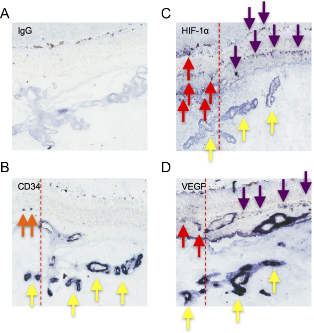
We examined five autopsy eyes from patients with known PSR, which have been previously characertized,6,8,21–24 as well as three eyes from control patients (Supplementary Table S1). We observed that VEGF is expressed by vascular endothelial cells lining neovascular sea fans (highlighted by expression of the endothelial cell marker CD34) in PSR patients (Fig. 1), as has previously been reported.21 Conversely, we observed only modest expression of VEGF in normal inner retinal and choroidal vessels of control eyes (Supplementary Fig. S1). We further observed expression of HIF-1α, a transcription factor that regulates the expression of VEGF in ischemic retinal disease, in neovascular sea fans (Fig. 1), consistent with an autocrine mechanism for the promotion of retinal NV in PSR.21 Examination of the retina adjacent to the NV in PSR eyes also demonstrated expression of both HIF-1α and VEGF within the inner retina in the region between perfused and nonperfused retina (termed the “marginal zone”) where retinal NV most typically develops (Fig. 1), consistent also with paracrine-driven retinal NV in PSR. Expression of HIF-1α was not detected in the inner retina of control eyes, including an eye from a patient with sickle cell trait (Supplementary Fig. S1).
Figure 1.
Expression of HIF-1α and VEGF in neovascular sea fans from PSR eyes without a prior history of LPC. (A) Immunohistochemistry for IgG (negative control). (B) Expression of CD34 in vascular endothelial cells lining neovascular sea fans (yellow arrows) and in the few remaining vessels in the inner retina (orange arrows) at the margin between perfused and nonperfused retina (red dashed line) in an eye from a sickle cell patient with PSR. (C, D) Expression of HIF-1α (C) and VEGF (D) in vascular endothelial cells lining neovascular sea fans (yellow arrows) and in the inner retina posterior (red arrows) and anterior (purple arrows) to the margin between perfused and nonperfused retina (red dashed line) in an eye from a sickle cell patient with PSR. Similar results were obtained in 5/5 eyes with PSR.
Glial Fibrillary Acidic Protein Expression in Hypoxic Retinal Müller Glial Cells in the Ischemic Retina of PSR Eyes
It has been reported that “activated” retinal Müller glial cells play an important role in the expression of HIF-1–regulated angiogenic factors in the ischemic retina in diabetic eye disease.17,18 We therefore determined if retinal Müller cells in the ischemic peripheral retina were similarly activated in PSR eyes where they might therefore contribute to the expression of HIF-1–regulated angiogenic factors in sickle cell retinopathy. To this end, we examined expression of GFAP, an astrocyte marker that is also expressed by injured or activated Müller cells, in the peripheral retina of patients with known sickle cell retinopathy. We observed the presence of GFAP-expressing Müller cells in a broad area that extended peripherally and centrally to the “marginal zone” (Fig. 2).
Figure 2.
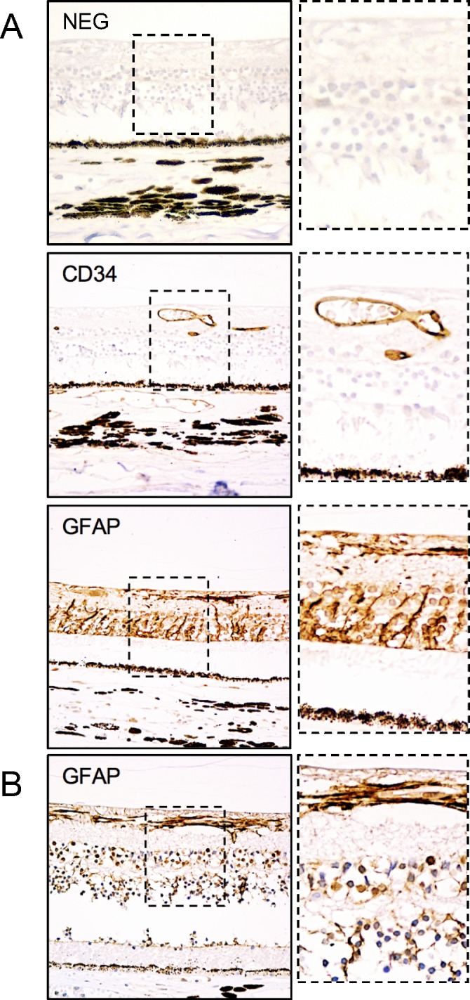
Broad expression of GFAP in retinal Müller cells in the ischemic peripheral retina of PSR eyes. (A) Expression of GFAP in activated (injured) retinal Müller glial cells in ischemic peripheral retina extends anteriorly and posteriorly from the border between perfused (CD34+) and nonperfused (CD34−) retina. (B) Glial fibrillary acidic protein expression was observed in astrocytes but not retinal Müller cells in posterior (perfused) retina. Immunoglobulin G was used as a negative control. Similar results were obtained in 5/5 eyes with PSR.
Accumulation of HIF-1α Expression in Inner Retinal and RPE Cells in the Peripheral Retina of PSR Eyes
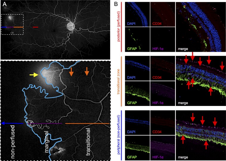
Ultra-widefield fluorescein angiography (FA) from patients with PSR demonstrated areas of peripheral nonperfusion characterized by a well-defined boundary where the retinal vasculature terminates (an example is shown in Fig. 3A). Careful examination of this region revealed three zones (Fig. 3A, inset): (1) a peripheral nonperfused retina in which there are no perfused retinal vessels on FA; (2) a posterior region of largely perfused retina with focal areas of capillary drop out (termed the “transitional zone”); and (3) a region between the nonperfused retina and the transitional zone, which we term the “marginal zone” (corresponding to the histopathologic description described above). While the nonperfused retina is well defined on FA and varies in size, the “marginal zone” can be semiarbitrarily defined as the region extending 1 mm anteriorly and 1 mm posteriorly to the border between perfused and nonperfused retina (approximately 2 mm in total width). The transitional zone is less well defined and can extend 2 mm (or more) centrally to the “marginal zone.” Interestingly, when we examined these three zones (as defined by the distance anterior and posterior to the border between the absence and presence of CD34-expressing vascular endothelial cells), we observed strong expression of HIF-1α within GFAP+ Müller cells in both the nonperfused and transitional zones—similar to the expression of HIF-1α in the marginal zone—in 5/5 PSR eyes examined (Fig. 3B), similar to what has been reported for diabetic eye disease.17,18 Within these three zones, we further observed expression of HIF-1α in other inner retinal cells (e.g., microglia) as well as in the retinal pigment epithelium (RPE; Fig. 3B). We did not observe significant HIF-1α expression in the inner retina or RPE in posterior (perfused) retina. Collectively, these results suggest that inner retinal cells and RPE (within a broad area of retina peripheral and central to neovascular sea fans) may also participate in the paracrine production of HIF-1–regulated angiogenic mediators in PSR eyes.
Figure 3.
Expression of HIF-1α and VEGF in the different regions of peripheral retina of PSR eyes. (A) Ultra-widefield FA image from a patient with PSR with inset highlighting the different zones of retinal perfusion in the peripheral retina anterior and posterior to a retinal neovascular sea fan (yellow arrow) at the margin (blue line) between perfused and nonperfused retina (“marginal zone”; purple). The “transitional zone” (orange) between the marginal zone and the posterior perfused retina (red) has areas of capillary drop out (orange arrows) within areas of perfusion. (B) Expression of the endothelial cell marker CD34 (red), GFAP (green), and HIF-1α (purple) in perfused retina, “transitional zone,” and avascular retina. Expression of HIF-1α was detected in activated (GFAP-expressing) retinal Müller cells, microglial cells, and RPE cells in the transitional zone as well as in avascular retina (red arrows), but not in posterior (perfused) retina. Similar results were obtained in 5/5 PSR eyes.
Expression of HIF-1α and VEGF in the Peripheral Retina Distant From Retinal Neovascular Sea Fans
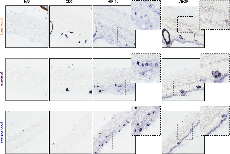
To assess the possible contribution of ischemic retina distant from (i.e., not adjacent to) retinal neovascular sea fans to the promotion of retinal NV in PSR eyes, we next examined the expression of HIF-1α and VEGF proximal and distal to the margin between perfused and nonperfused retina in areas without neovascular sea fans. We again observed expression of HIF-1α and VEGF in the “marginal zone,” “transitional zone,” and in nonperfused (avascular) retina in 5/5 sickle cell patients with PSR (Fig. 4).
Figure 4.
Expression of HIF-1α and VEGF in the peripheral retina of PSR eyes. Expression of HIF-1α and VEGF in avascular (nonperfused), marginal, and transitional zone distant from retinal NV. CD34 expression highlights the presence or absence of vascular endothelial cells and defines these zones.
Discussion
For over 4 decades, LPC has remained the most common intervention for patients with PSR.25 There have been three principal approaches for the use of LPC in PSR treatment: (1) specifically targeting feeder vessels of neovascular sea fans (now, largely abandoned); (2) sectoral laser to treat the (presumably) ischemic retina immediately surrounding the neovascular sea fan; or (3) circumferential laser to treat all of the ischemic retina, whether affected or not affected by sea fans (largely reserved for patients who are at risk for not returning for follow-up care).10 The use of sectoral or circumferential scatter LPC in PSR patients may be supported by extrapolation from extensive studies demonstrating the successful use of panretinal laser photocoagulation (PRP) for the treatment of PDR in diabetic patients; clinical trials have demonstrated the clear benefit of PRP for high-risk PDR.11 However, an equivalent benefit of scatter LPC has not yet been unequivocally demonstrated for patients with PSR.10
The poor efficacy of LPC for the treatment of NV in some patients with PSR, in comparison with PDR, may be a consequence of the lack of agreement regarding the optimal location for photocoagulation placement in PSR patients. The use of local laser coagulation of feeder vessels was initially supported by a prior report that VEGF expression is increased specifically in endothelial cells lining neovascular sea fans.21 Based on this observation, it was speculated that the angiogenic drive promoting the growth and survival of neovascular sea fans may be dependent on the expression of VEGF by the vascular endothelial cells within the neovascular tissue. LPC of the feeder vessels results in regression of neovascular sea fans and would therefore quench this autocrine angiogenic drive. We demonstrated that vascular endothelial cells lining sea fans express both HIF-1α and VEGF. However, we further observed that Müller glial cells, microglial cells, and RPE adjacent to retinal NV also express both HIF-1α and VEGF, suggesting that the ischemic retina surrounding neovascular sea fans can support their growth and survival by secreting VEGF through a paracrine mechanism. These findings suggest that sectoral scatter laser coagulation may be more effective than LPC of feeder vessels as a therapeutic intervention in the treatment of PSR. Indeed, owing to poor efficacy and frequent complications (e.g., vitreous hemorrhage), coagulation of feeder vessels for the treatment of retinal NV has largely been abandoned in favor of sectoral scatter laser.
In a clinical trial comparing sectoral scatter LPC to no treatment for PSR, sectoral scatter laser is surprisingly shown to lead to complete regression of only one-third of preexisting retinal sea fans.12 We provided evidence here that may shed light on the poor efficacy of sectoral laser therapy in some PSR patients. Examination of the inner retina in regions not immediately adjacent to neovascular sea fans also demonstrated expression of HIF-1α and VEGF. Although not sufficient to drive the local formation of retinal NV, it is tempting to speculate that the expression of VEGF in ischemic retina—distant from neovascular sea fans—may still be sufficient to promote the survival of preexisting remote sea fans.
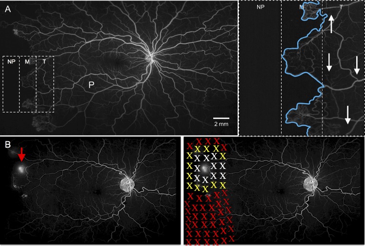
Current principles for the treatment of ischemic retinal disease support the application of scatter LPC to ischemic peripheral retina (i.e., within the “marginal zone” between perfused and nonperfused retina; Fig. 5). Importantly, we demonstrated here that retinal cells both central and peripheral to this “marginal zone”—within a “transitional zone” posteriorly and within avascular (“nonperfused”) retina anteriorly (Fig. 5)—also express HIF-1α and VEGF. The expression of HIF-1α and VEGF within retinal tissue in the transitional zone adjacent to focal areas of capillary drop out is not surprising. However, the observation that avascular retina peripheral to the marginal zone is viable and able to express angiogenic factors was unexpected. This finding suggests that the choroidal vasculature is sufficient to support the survival of some avascular peripheral retina in PSR eyes, the surviving inner (and outer) retina remains ischemic. Accumulation of HIF-1α within the ischemic avascular inner retina and the underlying RPE and their expression of VEGF may be sufficient to prevent regression of neovascular sea fans despite scatter laser treatment that is limited only to the marginal (and transitional) zones. These findings help provide important anatomic landmarks for optimal placement of laser coagulation and provide a foundation for designing a clinical trial comparing the efficacy of traditional sectoral scatter laser with more broad application of LPC, including circumferential scatter laser, for the treatment of patients with PSR (Fig. 5).
Figure 5.
Peripheral vascular zones in patients with sickle cell retinopathy. (A) Ultra-widefield FA image from a patient with sickle cell retinopathy with inset highlighting the three zones of retinal perfusion in the peripheral retina anterior and posterior to the border (blue line) between perfused (P) and nonperfused retina (NP). The transitional zone (T) between the marginal zone (M) and the posterior perfused retina has areas of capillary drop out (white arrows) within areas of perfusion. (B) When a sickle cell patient presents with retinal NV (red arrow), treatment with LPC (Xs) is most commonly limited to the area immediately surrounding the retinal NV (white Xs). However, treatment of all three zones (marginal, nonperfused, and transitional; yellow Xs) may be required to quench the production of HIF-1–regulated angiogenic mediators in patients with sickle cell retinopathy. It remains to be determined whether additional treatment distal from the area of NV (red Xs) is also necessary to prevent progression despite local treatment. M, marginal zone: margin (2 mm) between nonperfused and transitional zone where retinal NV is most likely to occur; NP, nonperfused retina: complete loss of inner retinal vasculature; P, perfused retina: no capillary drop out; T, transitional zone: perfused retina posterior to marginal zone (2 mm) but with focal areas of capillary drop out (arrows).
Our observations may have wider implications. The anterior boundary of circumferential (360°) scatter laser treatment of infants with ROP is often limited to the marginal zone; it has been assumed that the avascular retina peripheral to this zone is not viable. However, our results in PSR patients suggest that this assumption may not be correct. Similar to PSR eyes, the ischemic avascular peripheral retina in ROP eyes may also express HIF-1α and VEGF, and promote the survival (and progression) of retinal NV in some ROP infants despite laser coagulation that spares this region. It is therefore possible that the reason why some infants with ROP fail peripheral scatter laser therapy may be due to inadequate treatment of the avascular—but viable—retina anterior to the marginal zone.
A limitation of this study was the reliance on the detection of proteins in autopsy eyes. Epitope detection in autopsy samples has been reported to inversely correlate with postmortem times. Accordingly, detection of total HIF-1α and VEGF in autopsy eyes may be reduced in specimens with longer postmortem times. Conversely, expression of HIF-1α is unique in that its accumulation could theoretically increase immediately after tissue hypoxia associated with circulatory arrest. In a recent study looking at basal HIF-1α and VEGF levels in normal human retina, a modest negative correlation has been found between the expression of HIF-1α and postmortem times,26 suggesting that HIF-1α behaves similarly to other proteins in human eyes. For our studies, we used autopsy specimens that were acquired (enucleated) within 5 hours of death. Following enucleation, the specimens were immediately placed on ice before processing, which occurred within 31 hours of enucleation. Keeping the eyes on ice during the interval between enucleation and fixation preserves epitopes and would theoretically reduce degradation of HIF-1α and VEGF. Moreover, we performed immunohistochemical analyses comparing different regions of the same PSR eye, thereby providing intraspecimen controls. This latter approach minimizes many of the limitations of immunohistochemical analyses on autopsy specimens.
Of note, the recent publication of the landmark Diabetic Retinopathy Clinical Research Network (DRCRnet) Protocol S has demonstrated that anti-VEGF therapy may be as effective as PRP for the treatment of patients with PDR. Accordingly, Protocol S has had an immediate impact on the treatment of PDR and may have broad implications for the treatment of retinal NV in other ischemic retinopathies currently treated with scatter laser. However, PSR does not behave like other ischemic retinopathies (as exemplified by the fact that unlike retinal NV in patients with PDR, ROP, and ischemic retinal vein occlusions, scatter laser treatment has not been shown to be effective for retinal NV in patients with PSR). Currently, the only clinical studies available examining the efficacy of anti-VEGF therapy for PSR include a handful of case reports; it therefore remains uncertain whether Protocol S can be extrapolated to PSR. Moreover, frequent hospitalizations for sickle cell crises, combined with development of PSR at a young age, may make PSR patients poor candidates for a therapy that may require long-term treatment with frequent (e.g., monthly) intravitreal injections. Our findings instead support the use of broad sectoral or circumferential scatter laser applied peripherally and centrally to the boundary between perfused and nonperfused retina to quench HIF-1–driven expression of angiogenic mediators and promote regression of retinal NV in patients with PSR.
Supplementary Material
Acknowledgments
Supported by the National Eye Institute (NEI), National Institutes of Health Grant K08-EY021189 (AS), the Microscopy and Imaging Core Module of the NEI-sponsored Core Grant (EYEY001765), and an Unrestricted Grant from Research to Prevent Blindness (AS). AS gratefully acknowledges the support he receives as a Special Scholar Award recipient from Research to Prevent Blindness, Inc., and from the Owens Research Foundation.
Disclosure: M. Rodrigues, None; F. Kashiwabuchi, None; M. Deshpande, None; K. Jee, None; M.F. Goldberg, None; G. Lutty, None; G.L. Semenza, None; S. Montaner, None; A. Sodhi, None
References
- 1. Yawn BP,, Buchanan GR,, Afenyi-Annan AN,, et al. Management of sickle cell disease: summary of the 2014 evidence-based report by expert panel members. JAMA. 2014; 312: 1033–1048. [DOI] [PubMed] [Google Scholar]
- 2. Lerner NB,, Platania BL,, LaBella S. Newborn sickle cell screening in a region of Western New York State. J Pediatr. 2009; 154: 121–125. [DOI] [PubMed] [Google Scholar]
- 3. Manwani D,, Frenette PS. Vaso-occlusion in sickle cell disease: pathophysiology and novel targeted therapies. Blood. 2013; 122: 3892–3898. [DOI] [PMC free article] [PubMed] [Google Scholar]
- 4. Dean J,, Schechter AN. Sickle-cell anemia: molecular and cellular bases of therapeutic approaches (third of three parts). N Engl J Med. 1978; 299: 863–870. [DOI] [PubMed] [Google Scholar]
- 5. Goldberg MF. Retinal vaso-occlusion in sickling hemoglobinopathies. Birth Defects Orig Artic Ser. 1976; 12: 475–515. [PubMed] [Google Scholar]
- 6. McLeod DS,, Goldberg MF,, Lutty GA. Dual-perspective analysis of vascular formations in sickle cell retinopathy. Arch Ophthalmol. 1993; 111: 1234–1245. [DOI] [PubMed] [Google Scholar]
- 7. Goldberg MF. Classification and pathogenesis of proliferative sickle retinopathy. Am J Ophthalmol. 1971; 71: 649–665. [DOI] [PubMed] [Google Scholar]
- 8. McLeod DS,, Merges C,, Fukushima A,, Goldberg MF,, Lutty GA. Histopathologic features of neovascularization in sickle cell retinopathy. Am J Ophthalmol. 1997; 124: 455–472. [DOI] [PubMed] [Google Scholar]
- 9. Moriarty BJ,, Acheson RW,, Condon PI,, Serjeant GR. Patterns of visual loss in untreated sickle cell retinopathy. Eye (Lond). 1988; 2 (pt 3): 330–335. [DOI] [PubMed] [Google Scholar]
- 10. Myint KT,, Sahoo S,, Thein AW,, Moe S,, Ni H. Laser therapy for retinopathy in sickle cell disease. Cochrane Database Syst Rev. 2015; 10: CD010790. [DOI] [PMC free article] [PubMed] [Google Scholar]
- 11. Evans JR,, Michelessi M,, Virgili G. Laser photocoagulation for proliferative diabetic retinopathy. Cochrane Database Syst Rev. 2014; 11: CD011234. [DOI] [PMC free article] [PubMed] [Google Scholar]
- 12. Farber MD,, Jampol LM,, Fox P,, et al. A randomized clinical trial of scatter photocoagulation of proliferative sickle cell retinopathy. Arch Ophthalmol. 1991; 109: 363–367. [DOI] [PubMed] [Google Scholar]
- 13. Jampol LM,, Farber M,, Rabb MF,, Serjeant G. An update on techniques of photocoagulation treatment of proliferative sickle cell retinopathy. Eye (Lond). 1991; 5 (pt 2): 260–263. [DOI] [PubMed] [Google Scholar]
- 14. Semenza GL. Oxygen sensing, hypoxia-inducible factors, and disease pathophysiology. Annu Rev Pathol. 2014; 9: 47–71. [DOI] [PubMed] [Google Scholar]
- 15. Bhutto IA,, McLeod DS,, Hasegawa T,, et al. Pigment epithelium-derived factor (PEDF) and vascular endothelial growth factor (VEGF) in aged human choroid and eyes with age-related macular degeneration. Exp Eye Res. 2006; 82: 99–110. [DOI] [PMC free article] [PubMed] [Google Scholar]
- 16. Babapoor-Farrokhran S,, Jee K,, Puchner B,, et al. Angiopoietin-like 4 is a potent angiogenic factor and a novel therapeutic target for patients with proliferative diabetic retinopathy. Proc Natl Acad Sci U S A. 2015; 112: E3030–E3039. [DOI] [PMC free article] [PubMed] [Google Scholar]
- 17. Rodrigues M,, Xin X,, Jee K,, et al. VEGF secreted by hypoxic Muller cells induces MMP-2 expression and activity in endothelial cells to promote retinal neovascularization in proliferative diabetic retinopathy. Diabetes. 2013; 62: 3863–3873. [DOI] [PMC free article] [PubMed] [Google Scholar]
- 18. Xin X,, Rodrigues M,, Umapathi M,, et al. Hypoxic retinal Muller cells promote vascular permeability by HIF-1-dependent up-regulation of angiopoietin-like 4. Proc Natl Acad Sci U S A. 2013; 110: E3425–E3434. [DOI] [PMC free article] [PubMed] [Google Scholar]
- 19. Bhutto IA,, Kim SY,, McLeod DS,, et al. Localization of collagen XVIII and the endostatin portion of collagen XVIII in aged human control eyes and eyes with age-related macular degeneration. Invest Ophthalmol Vis Sci. 2004; 45: 1544–1552. [DOI] [PubMed] [Google Scholar]
- 20. Montaner S,, Sodhi A,, Molinolo A,, et al. Endothelial infection with KSHV genes in vivo reveals that vGPCR initiates Kaposi's sarcomagenesis and can promote the tumorigenic potential of viral latent genes. Cancer Cell. 2003; 3: 23–36. [DOI] [PubMed] [Google Scholar]
- 21. Cao J,, Mathews MK,, McLeod DS,, Merges C,, Hjelmeland LM,, Lutty GA. Angiogenic factors in human proliferative sickle cell retinopathy. Br J Ophthalmol. 1999; 83: 838–846. [DOI] [PMC free article] [PubMed] [Google Scholar]
- 22. Kim SY,, Mocanu C,, McLeod DS,, et al. Expression of pigment epithelium-derived factor (PEDF) and vascular endothelial growth factor (VEGF) in sickle cell retina and choroid. Exp Eye Res. 2003; 77: 433–445. [DOI] [PubMed] [Google Scholar]
- 23. Kunz Mathews M,, McLeod DS,, Merges C,, Cao J,, Lutty GA. Neutrophils and leucocyte adhesion molecules in sickle cell retinopathy. Br J Ophthalmol. 2002; 86: 684–690. [DOI] [PMC free article] [PubMed] [Google Scholar]
- 24. Lutty GA,, Merges C,, Crone S,, McLeod DS. Immunohistochemical insights into sickle cell retinopathy. Curr Eye Res. 1994; 13: 125–138. [DOI] [PubMed] [Google Scholar]
- 25. Mohamed Q,, Gillies MC,, Wong TY. Management of diabetic retinopathy: a systematic review. JAMA. 2007; 298: 902–916. [DOI] [PubMed] [Google Scholar]
- 26. Hughes JM,, Groot AJ,, van der Groep P,, et al. Active HIF-1 in the normal human retina. J Histochem Cytochem. 2010; 58: 247–254. [DOI] [PMC free article] [PubMed] [Google Scholar]
Associated Data
This section collects any data citations, data availability statements, or supplementary materials included in this article.


