Abstract
1 T phase incorporation into 2H-MoS2 via an optimal electron irradiation leads to induce a weak ferromagnetic state at room temperature, together with the improved transport property. In addition to the 1T-like defects, the electron irradiation on the cleaved MoS2 surface forms the concentric circle-type defects that are caused by the 2 H/1 T phase transition and the vacancies of the nearby S atoms of the Mo atoms. The electron irradiation-reduced bandgap is promising in vanishing the Schottky barrier to attaining spintronics device. The simple method to control and improve the magnetic and electrical properties on the MoS2 surface provides suitable ways for the low-dimensional device applications.
Recently, based on the reduced dimensionality of transition metal dichalcogenides (TMDs), the successful realization of field-effect transistors1 and the thickness-dependent, indirect-direct bandgap transition2,3 have boosted the development of two-dimensional (2D) materials for high-performance flexible electronic and optoelectronic devices4,5. Additionally, similar to graphene6, developing approaches to effectively induce the ferromagnetism into the diamagnetic 2H-MoS2 have attracted a great interest for possible spintronics and quantum information devices, but it is still challenging7,8,9,10,11,12,13,14,15,16,17,18,19,20,21,22. Extensive experimental and theoretical studies have effectively modified magnetic properties of MoS2 with the various forms of thin films7, nanoribbons8, nanosheets9, and even in bulk limit10,11. The used methods are by the formation of vacancies11,12,13, adsorbing non-metal atoms14,15 substitutional doping TM atoms16,17 and application of an external electric field18,19 or elastic strain20,21, and so on. Notably, together with high-Curie-temperature, relatively large ferromagnetism has been only realized in the significant presence of zigzag edges7,22. However, the possible degradation of transport properties due to the scattered morphology needs to be resolved for nanoscale device applications23.
The local introduction of the metallic 1 T phase into the 2 H matrix via Li intercalation24,25 or electron irradiation26 has been shown to improve the transistor performance27. Especially, novel fabrication methods using electron beam-based techniques have produced semiconducting MoS2 nanoribbons28 and metallic MoS2 nanowires29. Accordingly, for a magnetic MoS2, 1 T phase incorporation has been also explored by using the chemical exfoliation30,31,32. As a separate method, irradiation with low-energy electrons has been also used to modify the magnetic and transport properties13. However, although the achieved magnetic moments are remarkable13,32, it is still obscure whether the magnetism originates intrinsically from the existence of 1 T phase or edge effect driven by the various defects20,33. Thus, with a full understanding of 1 T phase incorporation, it is desirable to find more effective ways to control and improve the magnetic and electrical properties of MoS2. In this paper, we propose a simple method to improve transport property and induce room-temperature ferromagnetism through the optimal electron irradiation on the MoS2 surface. The magnetic moments are found to be attributed to the unpaired spins of Mo4+ ions induced by exotic defects, which form a specific shape of concentric circles on the surface region along the 2 H/1 T phase transition.
Results and Discussion
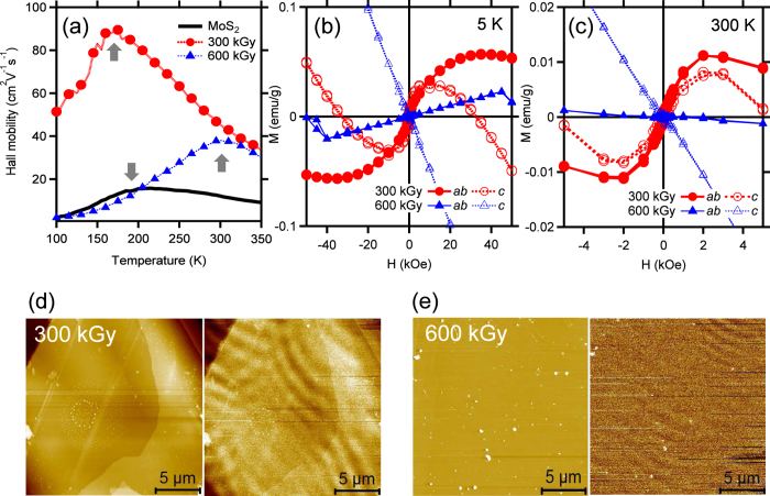
We found a certain condition to increase and induce the Hall mobility and the diamagnetic to ferromagnetic phase transition, after electron irradiation on the cleaved MoS2 surfaces by changing the electron dose and the acceleration energy, respectively: the electron dose of 300 kGy (6.70 × 10¹⁴ electrons/cm2) and the acceleration energy of 0.7 MeV34,35. As shown in the temperature dependence of the Hall mobility (Fig. 1a), the electron irradiation of this condition improves the Hall mobility of the pristine MoS2, but slightly reduces a crossover temperature (TC, as indicated by arrow) of the pristine MoS2 (200 K) to 175 K. Above and below TC, the mobility is mainly subject to the phonon and impurity scatterings, respectively36,37. On the other hand, the higher electron dose of 600 kGy (1.39 × 10¹⁵ electrons/cm2) increases the TC over room temperature. Such shift of TC implies that the mobility is limited dominantly by charged impurities, while the phonon scattering plays a minor role.
Figure 1.
Comparison of single-crystalline MoS2 and electron-irradiated samples for (a) Hall mobility as a function of temperature, magnetic hysteresis loops of (b) 5 K and (c) 300 K, and (d,e) images of AFM (left) and MFM (right) with scan areas of 20 × 20 μm2. The magnetic field (H) is applied parallel (ab) and perpendicular (c) to the basal plane of samples.
Figures 1b shows the magnetizations as a function of the magnetic field strength (H) up to ±50 kOe at low (5 K) temperature. In comparison with the diamagnetic susceptibility11 of the pristine MoS2, the electron dose of 300 kGy induces the diamagnetic to a ferromagnetic phase transition. Interestingly, along the out-of-plane (the c-axis) direction, the diamagnetic behavior still remains for higher magnetic fields than ±10 kOe. The saturated magnetizations along the in-plane (the ab-plane) and out-of-plane directions are 0.057 emu/g (1.634 × 10⁻³ μB/Mo ion) and 0.030 emu/g (8.60 × 10⁻⁴ μB/Mo ion) at the H = 35 kOe and 1 kOe, respectively. These weak ferromagnetic states persist up to room temperature, but the saturated magnetizations of 5 K (Fig. 1b) are significantly reduced to 0.011 emu/g (0.315 × 10⁻³ μB/Mo ion) and 0.008 emu/g (0.229 × 10⁻³ μB/Mo ion) at the H = 2 kOe along the in-plane and out-of-plane directions, respectively (Fig. 1c). The coercivities (0.2 kOe) of both directions at 5 K are also reduced to 0.1 kOe at room temperature.
On the other hand, the higher electron dose of 600 kGy induces the diamagnetic to a paramagnetic phase transition along the in-plane direction while the out-of-plane direction still remains diamagnetic (Fig. 1b). Especially along the in-plane direction, the diamagnetic state also retains over the magnetic field of ±40 kOe, similarly to the case of the out-of-plane direction for the sample irradiated at 300 kGy. At room temperature, however, the temperature-dependent paramagnetic state disappears, while the relatively temperature-insensitive diamagnetic state remains (Fig. 1c)10,11. It is evident from the time-of-flight secondary ion mass spectroscopy measurements (not shown) that the electron irradiation of the current condition34,35 has influences on a few top layers of the cleaved MoS2 single crystals. Furthermore, the different magnetic states due to the different electron doses are elucidated in Fig. 1(d,e) of the atomic and magnetic force microscopy (AFM and MFM) images taken at room temperature. Similarly to shown in the previous study11, undulating magnetic domains representing the ferromagnetic state are clearly observed in the MFM image of 300 kGy (Fig. 1d), whereas the magnetic domains get much weakened in that of 600 kGy (Fig. 1e). This confirms that the electron dose of 300 kGy efficiently induces the ferromagnetic state on the MoS2 surface.
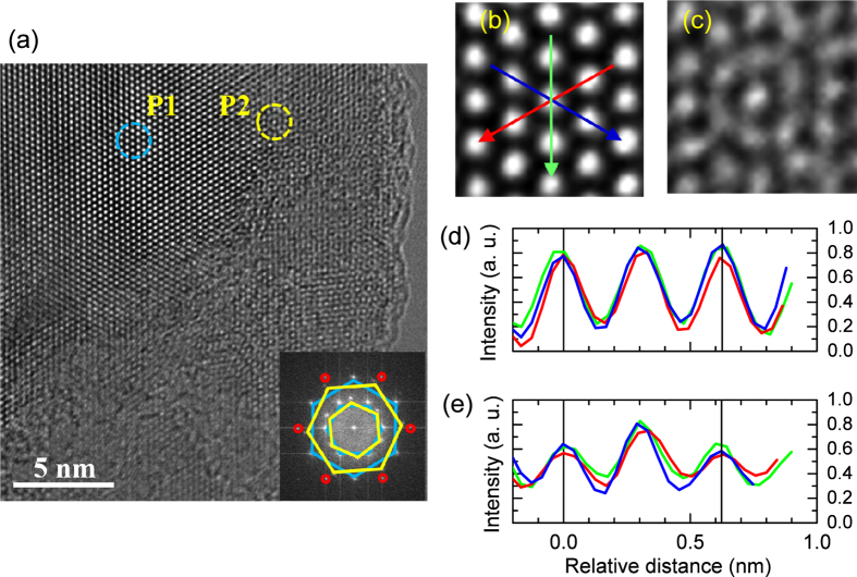
To elucidate the magnetic domains in more detail, atomic structures on the electron-irradiated surface of 300 kGy were investigated by high-resolution transmission electron microscopy (HRTEM) after the sample was exfoliated using ultrasonic. The fringes of the HRTEM image (Fig. 2a) indicate that the thickness of the MoS2 layers is about mono- or bi-layers38. With the lack of honeycomb lattices, two types of defects are dominantly found; 1T-phase-like defect24,25,26 (P1) and concentric circle-type vacancies (P2). The inset of Fig. 2a shows the fast Fourier transform (FFT) image, where the inner and outer hexagons correspond to the (100) and (110) planes39,40. The two defects lead to having two (yellow and cyan) hexagons with a twist angle 24° at each plane, respectively. The electron irradiation-induced 1T-phase-like defect (Fig. 2b) is in good agreement with a previous study26 and supported by the negligible intensities between the main peaks of the line profiles24 (Fig. 2d). Additionally, the structural difference of the 1T-phase-like defect is confirmed by comparing of the TEM image with the 1 H phase (Fig. 3a), which is half of the unit cell of bulk 2H-MoS2. It is notable that the total energy of 1T-MoS2 is much higher than that of 1H-MoS2 by 0.8 eV41. However, the 1 H to 1 T phase transition can be driven by lowering the energy barrier via the charge injection of electron irradiation42.
Figure 2.
(a) HRTEM image of the electron-irradiated sample of 300 kGy. Two types of defects (P1 and P2) are magnified in (b) and (c), respectively. (d,e) Corresponding line profiles are obtained as the colored arrows in (b) and provide the estimated lattice constants of (b) a = 3.15 Å and (c) 3.11 Å, respectively. In the inset of (a), FFT image displays twisted (yellow and cyan) hexagons at the (100) and (110) planes. The six red circles indicate the (200) plane.
Figure 3.
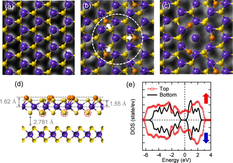
(a) Calculated 1 H phase of 2 × 2 supercells is overlapped in the TEM image of Fig. 2b. TEM image of Fig. 2c is compared to the calculated (b) 1T-like and (c) 1T-3VS phases of 6 × 6 supercells. (d) Side view of 2L-MoS2. 1T-3VS defect is formed by gliding the topmost (orange) S atoms and removing the bottom (dotted) S atoms at the top layer of 6×6 supercells of 2 H phase. Large (blue) and small (yellow and orange) spheres correspond to the Mo and S atoms, respectively. (e) Corresponding calculated total spin polarized DOSs of upper and bottom layers. Up and down arrows indicate positive and negative spin states. DOSs consist of the distribution of Mo 4d and S 3p electrons at the upper and bottom layers with a closed and ~0.64 eV bandgaps, respectively. The Fermi level is at zero energy.
Now, we focus on the concentric-circle-type defect of Fig. 2c. Compared to the line profiles of Fig. 2d, the profiles of Fig. 2e indicate that the central and nearby atoms within the circle (Fig. 2c) correspond to the Mo and S atoms, respectively. In the (dotted) circle of Fig. 3b, the upper three (orange) S atoms of the 1 H phase glide as indicated by the arrows and form partly the 1 T phase. Then such the 1 T phase (Fig. 3b) is turned further into the concentric-circle-type phase (Fig. 3c) after pushing away the inner three S atoms denoted as dotted circles in Fig. 3d. Thus, we will refer to the latter phase as a 1T-3VS defect from the vacancies of the inner three S atoms. The estimated lattice constant of the 1T-phase-like defect (Fig. 2d, a = 3.15 Å) is slightly reduced in the 1T-3VS defect (Fig. 2e, a = 3.11 Å). The details are compared to the calculated results as described in the methods. However, the Raman signals of the characteristic 1 T phase43 observed in the chemically exfoliated MoS2 are not found in the electron-irradiated sample because of the finite thickness of the defect depth. Figure 4a shows two strong Raman peaks at 383 and 408 cm−1, which correspond to the E12g and A1g modes, respectively. The Raman spectrum of the electron-irradiated sample is nearly identical to that of the pristine MoS2.
Figure 4.
Comparison of (a) Raman and (b–d) XPS spectra between the single-crystalline MoS2 and the electron-irradiated sample (300 kGy), where Mo 3d and S 2p core-level spectra include the curve-fitting results of two components, C1 and C2, relating to the contribution of 2 H phase and electron irradiation-induced defects, respectively. BG indicates the background.
On the other hand, the calculations reveal that the 1T-3VS defect doped bilayer MoS2 in a ferromagnetic state is more stable by energy difference of 0.420 eV per formula unit (fu) than a nonmagnetic one and has a magnetic moment of 0.084 μB/Mo. Notably, the magnetic moment of the 1T-3VS defect doped monolayer is 0.168 μB/Mo. The thickness-dependent magnetic moment manifests that, compared to the calculated magnetic moments, the significantly reduced magnetic moments of the electron-irradiated samples (Fig. 1b,c) are attributed to the diamagnetic states of the subsurface layers remaining in the non-defective status. Furthermore, compared to the 1T-3VS doped monolayer, the 1T-phase doped monolayer MoS2 (Fig. 3b) is more favored by the difference of 0.328 eV per fu and has a larger magnetic moment of 0.175 μB/Mo (0.0875 μB/Mo for bilayer MoS2). However, it is notable that 1T-3VS defects were only obtained at the specific condition of 300 kGy, while the higher electron dose of 600 kGy mainly produced the 1T-phase-like defects (not shown) and induced the diamagnetic to the paramagnetic phase transition instead of the ferromagnetic phase. It is contrary to a simple consideration that the higher electron dose may produce more 1T-3VS defects than 1T-phase-like defects. Thus, the 1T-phase-like defects are considered to be closely related to the VS2-like defects33, where each Mo atom lacks six nearby S atoms (3VS2). In other words, the remained S atoms of 1T-3VS defects in Fig. 3c may be pushed away or moved further into the vdW gap at the higher electron dose. The first-principles calculations of 3VS2 (not shown) indicate that, similar to the VS2-doped monolayer MoS2, the 1 × 1 bilayer MoS2 is nonmagnetic (after removing the topmost S layer)20,21,22, and the 2 × 2 bilayer MoS2 is more likely to be antiferromagnetic than ferromagnetic (after removing the S layers at the top layer). Therefore, the undulating magnetic domains of the MFM image due to the ferromagnetic state (Fig. 1d) are considerably related to the 1T-3VS defects. More interestingly, these 1T-3VS defects can be obtained on the sliding surfaces44 and the large-area CVD trilayer-MoS2 film with the plasma treated substrate45. However, there are no 1T-phase-like or VS2-like defects on both samples. In the former case, 1T-3VS defects are simulated by rotation of the two single hexagonal lattices by a misfit angle of 30°. The calculated density of states (DOSs) of the 1T-3VS defect doped bilayer MoS2 show that the bandgap is closed at the top layer while it is open at the bottom layer (Fig. 3e). In order to investigate the bandgap change due to the electron irradiation, surface-sensitive measurements were performed. Figure 4b–d show that the x-ray photoelectron spectroscopy (XPS) spectra of the electron-irradiated sample shift toward the low binding energy side compared to those of the pristine MoS2. However, the stoichiometry of the electron-irradiated sample estimated from the respective integrated peak area of the Mo 3d and S 2p XPS core levels (Fig. 4b,c) retains the ratio (1:2) of the pristine MoS2. Deconvolution fits46 (Fig. 4b,c) elucidate that both Mo 3d (d5/2, 229.77 eV) and S 2p peaks (p3/2, 162.58 eV) of the pristine MoS2 (component C1 of the 2 H phase) are found to consist of two components after electron irradiation. The intensity ratio of C1 to C2 is estimated to be 0.5. The electron irradiation-induced peaks (component C2 of the 1T-3VS phase) are located at lower binding energies of 229.59 eV (Mo 3d5/2) and 162.29 eV (S 2p3/2), respectively. It is similar to the 1T-phase doped monolayer47. The valence-band maximum (VBM) also moves toward the Fermi energy (EF) from 0.99 eV to 0.77 eV as indicated by arrows (Fig. 4d). Notably, the influence of the oxygen, which is inevitable during the electron irradiation, is considered to be negligible from the lack of the change at the weak peak of 236.20 eV (Fig. 4b), corresponding to Mo6+ oxidation state of Mo.
In addition to the shift of XPS spectra toward EF, more complementary measurements such as the spectroscopic ellipsometry and optical absorption were measured. Figure 5a,b show the refractive index n and extinction coefficient k of the spectroscopic ellipsometry, respectively. The sharp feature of Fig. 5b, denoted by E0, corresponds to the direct-gap transition at the K point with following by the E0 + Δ0 peak, which corresponds to the spin-orbit splitting of the valence band at the same K point3. These two features of the direct gap, designated as A and B excitons by PL measurements2,3, are not responsive to the electron irradiation, while the indirect bandgap shows the oscillating features below 1.5 eV after electron irradiation. The optical absorption results (Fig. 5c) confirm the reduction of the bandgap energy (Eg) by using the relation: α = A/hv(E − Eg)n, where A is the constant, hν is the incident photon energy, and the exponent n depends on the kind of optical transition48. The electron irradiation leads to decrease the indirect Eg of the pristine MoS2 by approximately 0.12 eV (Fig. 5d), while the change of direct Eg is insensitive to electron irradiation as revealed in the spectroscopic ellipsometry. The bandgap reduction is promising in vanishing the Schottky barrier to attaining spintronics device49.
Figure 5.
Comparison of (a) refractive index n and (b) extinction coefficient k between the single-crystalline MoS2 and the electron-sample (300 kGy). (c) Optical absorption coefficient shows the dependence of (ahv)1/2 on the photon energy (hv) for two samples. (d) Extrapolating the linear part of each curve toward energy axis gives the corresponding indirect bandgap energy (Eg).
Conclusions
The electron irradiation with the electron dose of 300 kGy (6.70 × 10¹⁴ electrons/cm2) and the acceleration energy of 0.7 MeV creates the 1T-phase-like (VS2) and 1T-3VS defects on the MoS2 surface. These defects reduce the bandgap and improve the transport property. The undulating magnetic domains of the MFM image due to weak ferromagnetic state are considerably related to the 1T-3VS defects. This optimal electron irradiation to improve the magnetic and transport properties at the atomic-layer scale is a key step for the successful integration of 2D TMDs into possible device applications.
Methods
Sample preparation
The natural-single crystalline MoS2 samples (SPI) were snipped from a large piece and, after a several exfoliation to take the clean surface, irradiated with different exposure times at the electron acceleration energy (ELV-8 linear accelerators) of 0.7 MeV and 2.0 MeV, respectively, in ambient conditions at room temperature. The area of the electron irradiation at the specific point of 400 ± 50 mm was of width 600 ± 20 × length 20 ± 5 mm2 with beam diameter of 25~35 mm. The stability of the beam energy and dose was less than ± 5%. The electron dose was checked by the dosimeter films.
Characterization
The dc magnetic and hysteresis loop measurements (ca. area of 3 × 3 mm2 and thickness of ~100 μm) were performed from 2 to 300 K using a SQUID magnetometer (MPMS XL-7). The MFM measurements were performed with non-contact mode AFM (Bruker-Nano N8 Neo) at room temperature. For the MFM measurements, conductive Pt tips with a radius of ~25 nm were used after Co coating. The MFM images were obtained with a distance of 80 nm between the tip and the sample surface. The electrical conductivity, carrier density, and the Hall mobility were measured as a function of temperature from 100 K to 350 K with a fixed magnetic field of 0.5 T using a Hall measurement system with Au contacts (HMS 5000). HRTEM (JEM-2100F) images for the exfoliated samples by sonication in methanol were taken at an energy of electron beam (200 keV)40. The electron irradiation-induced defects are supposed to be hardly influenced by the TEM measurements. From the depth profiles obtained by time-of-flight secondary ion mass spectroscopy, the possible (magnetic) impurities such as O, C, H, and Fe, were found to mostly exist at the electron-irradiated surfaces. There was a negligible reduction of S intensity compared to the Mo intensity on the sample of 300 kGy. Micro-Raman spectroscopy was operated with an Ar ion laser at 514.5 nm. The excitation laser beam of an average power less than 2.5 mW was focused onto samples of interest. The XPS measurements were performed with an Al Kα X-ray source in the vacuum of 1 × 10⁻¹⁰ torr (ESCALAB 250XI). The energy calibrations were referenced to adventitious carbon at 284.50 eV with eliminating the charging of the sample during analysis. In fitting of Mo 3d and S 2p core-level spectra46, the Gaussian width was fixed at the instrumental resolution of 0.65 eV and 0.60 eV, respectively. The values of the spin-orbit splitting and the branching ratios [I (3d5/2)/I(3d3/2) and I(2p3/2)/I(2p1/2)] were 3.17 eV and 0.67 for Mo 3d peaks and 1.18 eV and 0.5 for S 2p peaks, respectively. The refractive index n, extinction coefficient k, and optical absorption spectra were measured by using spectroscopic ellipsometer (UVISEL) and UV-Vis-NIR spectrophotometer (Cary 5000) in the 300–1600 nm wavelength range at room temperature. Optical absorption coefficient were obtained from the transmission mode at room temperature. The thickness of the pristine MoS2 and electron-irradiated sample is ca. 20 and 50 μm, respectively.
Theoretical calculations
First-principles calculations based on density functional theory (DFT) were performed using the Vienna ab initio simulation package (VASP)50. For the exchange-correlation potential, the generalized gradient approximation (GGA) was adopted51. Wave functions were expanded by a plane-wave basis set with a cut-off energy of 400 eV. The k mesh in the Brillouin zones sampling is 3 × 3 × 1. We account for bilayer MoS2, and a large spacing of between two-dimensional unit cells (15 Å) was employed to avoid interlayer interactions (Fig. 3d). To simulate the 1T-3VS doped bilayer MoS2 (Fig. 3c,d), we adopted a 6 × 6 supercell. The in-plane lattice parameter of the bilayer MoS2 was used to be the experimental bulk value46 of 3.160 Å and the atomic positions were fully relaxed. After gliding three S atoms of the topmost layer along the 2 H to 1 T pathway as indicated by the arrows of the (dotted) circle in Fig. 3b, the concentric-circle-type pattern was constructed by removing the inner three S atoms as shown in Fig. 3c. The bond length and the projected distance (dMo-S = 2.41 Å and 1.82 Å) of the 2 H phase in Fig. 3a increases to 3.72 Å and 3.64 Å in Fig. 3b and to 3.60 Å and 2.74 Å in Fig. 3c, respectively. In Fig. 3d, the bond length (2.41 Å) and bond angle (θS-Mo-S = 80.70°) decrease to 2.38 Å and 79.18° with increasing the dMo-S of the 1T-3VS defect to 2.43 Å and 80.9°.
Additional Information
How to cite this article: Han, S. W. et al. Electron beam-formed ferromagnetic defects on MoS2 surface along 1T phase transition. Sci. Rep. 6, 38730; doi: 10.1038/srep38730 (2016).
Publisher's note: Springer Nature remains neutral with regard to jurisdictional claims in published maps and institutional affiliations.
Acknowledgments
This work was supported by the Basic Science Research Program and Priority Research Centers Program through the National Research Foundation of Korea (NRF) funded by the Ministry of Education (Grant Nos 2009-0093818, 2015R1A2A2A01003621, 2015R1D1A1A01058332, and 2014R1A1A2003970).
Footnotes
Author Contributions S.W.H. designed and prepared all electron-irradiated samples, participated in the magnetic measurements. Y.S.P., W.G.L., and G.D.L. participated in TEM measurements and analyzed the data. Y.H.H. performed the Hall-effect measurements and participated in XPS measurement. M.I.K. performed the ellipsometry measurements. W.Y. measured the MFM. S.J. and S.C.H. performed the first-principles calculations. S.C.H. supported the project. S.W.H. wrote the manuscript. All authors contributed to analyzing and interpreting the data and to writing the manuscript.
References
- Radisavljevic B. et al. A. Single-layer MoS2 transistors. Nature Nanotech. 6, 147 (2011). [DOI] [PubMed] [Google Scholar]
- Splendiani A. et al. Emerging photoluminescence in monolayer MoS2. Nano Lett. 10, 1271 (2010). [DOI] [PubMed] [Google Scholar]
- Mak K. F. et al. Atomically thin MoS2: A new direct-gap semiconductor. Phys. Rev. Lett. 105, 136805 (2010). [DOI] [PubMed] [Google Scholar]
- Wang Q. H. et al. M. S. Electronics and optoelectronics of two-dimensional transition metal dichalcogenides. Nature Nanotech. 7, 699 (2012). [DOI] [PubMed] [Google Scholar]
- Xu M., Liang T., Shi M. & Chen H. Graphene-like two-dimensional materials. Chem. Rev. 113, 3766 (2013). [DOI] [PubMed] [Google Scholar]
- Han W., Kawakami R. K., Gmitra M. & Fabian J. Graphene spintronics. Nature Nanotech. 9, 794 (2014). [DOI] [PubMed] [Google Scholar]
- Zhang J. et al. Magnetic molybdenum disulfide nanosheet films. Nano Lett. 7, 2370 (2007). [DOI] [PubMed] [Google Scholar]
- Li Y., Zhou Z., Zhang S. & Chen Z. MoS2 nanoribbons: high stability and unusual electronic and magnetic properties. J. Am. Chem. Soc. 130, 16739 (2008). [DOI] [PubMed] [Google Scholar]
- Gao D. et al. Ferromagnetism in freestanding MoS2 nanosheets. Nanoscale Res. Lett. 8, 129 (2013). [DOI] [PMC free article] [PubMed] [Google Scholar]
- Tongay S. et al. Magnetic properties of MoS2: Existence of ferromagnetism. Appl. Phys. Lett. 101, 123105 (2012). [Google Scholar]
- Han S. W. et al. Controlling ferromagnetic easy axis in a layered MoS2 single crystal. Phys. Rev. Lett. 110, 247201 (2013). [DOI] [PubMed] [Google Scholar]
- Mathew S. et al. Magnetism in MoS2 induced by proton irradiation. Appl. Phys. Lett. 101, 102103 (2012). [Google Scholar]
- Karmakar D. et al. Optimal electron irradiation as a tool for functionalization of MoS2: Theoretical and experimental investigation. J. Appl. Phys. 117, 135701 (2015). [Google Scholar]
- He J., Wu K., Sa R., Li Q. & Wei Y. Magnetic properties of nonmetal atoms absorbed MoS2 monolayer. Appl. Phys. Lett. 96, 082504 (2010). [Google Scholar]
- Yue Q., Shao Z., Chang S. & Li J. Adsorption of gas molecules on monolayer MoS2 and effect of applied electric field. Nanoscale Res. Lett. 8, 425 (2013). [DOI] [PMC free article] [PubMed] [Google Scholar]
- Dolui K., Rungger I., Pemmaraju C. D. & Sanvito S. Possible doping strategies for MoS2 monolayers: An ab initio study. Phys. Rev. B 88, 075420 (2013). [Google Scholar]
- Cheng Y. C. et al. Prediction of two-dimensional diluted magnetic semiconductors: Doped monolayer MoS2 systems. Phys. Rev. B 87, 100401(R) (2013). [Google Scholar]
- Dolui K., Pemmaraju C. D. & Sanvito S. Electric field effects on armchair MoS2 nanoribbons. ACS Nano 6, 4823 (2012). [DOI] [PubMed] [Google Scholar]
- Kou L. et al. Tuning magnetism and electronic phase transitions by strain and electric field in zigzag MoS2 nanoribbons. J. Phys. Chem. Lett. 3, 2934 (2012). [DOI] [PubMed] [Google Scholar]
- Zhou Y., Su Q., Wang Z., Deng H. & Zu X. Controlling magnetism of MoS2 sheet by embedding transition-metal atoms and applying strain. Phys. Chem. Chem. Phys. 15, 18464 (2013). [DOI] [PubMed] [Google Scholar]
- Zheng H. et al. Tuning magnetism of monolayer MoS2 by doping vacancy and applying strain. Appl. Phys. Lett. 104, 132403 (2014). [Google Scholar]
- Yang Z. et al. Realization of high Curie temperature ferromagnetism in atomically thin MoS2 and WS2 nanosheets with uniform and flower-like morphology. Nanoscale 7, 650 (2015). [DOI] [PubMed] [Google Scholar]
- Ghorbani-Asl M. et al. Defect-induced conductivity anisotropy in MoS2 monolayers. Phys. Rev. B 88, 245440 (2013). [Google Scholar]
- Eda G. et al. M. Coherent atomic and electronic heterostructures of single-layer MoS2. ACS Nano 6, 7311 (2012). [DOI] [PubMed] [Google Scholar]
- Wang L., Xu Z., Wang W. & Bai X. Atomic mechanism of dynamic electrochemical lithiation processes of MoS2 nanosheets. J. Am. Chem. Soc. 136, 6693 (2014). [DOI] [PubMed] [Google Scholar]
- Lin Y.-C., Dumcenco D. O., Huang Y.-S. & Suenaga K. Atomic mechanism of the semiconducting-to-metallic phase transition in single-layered MoS2. Nature Nanotech. 9, 391 (2014). [DOI] [PubMed] [Google Scholar]
- Kappera R. et al. Phase-engineered low-resistance contacts for ultrathin MoS2 transistors. Nature Mater. 13, 1128 (2014). [DOI] [PubMed] [Google Scholar]
- Liu X. et al. Top–down fabrication of sub-nanometre semiconducting nanoribbons derived from molybdenum disulfide sheets. Nature Commun. 4, 1776 (2013). [DOI] [PMC free article] [PubMed] [Google Scholar]
- Lin J. et al. Flexible metallic nanowires with self-adaptive contacts to semiconducting transition-metal dichalcogenide monolayers. Nature Nanotech. 9, 436 (2014). [DOI] [PubMed] [Google Scholar]
- Yan S. et al. Enhancement of magnetism by structural phase transition in MoS2. Appl. Phys. Lett. 106, 012408 (2015). [Google Scholar]
- Luxa J. et al. Origin of exotic ferromagnetic behavior in exfoliated layered transition metal dichalcogenides MoS2 and WS2. Nanoscale 8, 1960 (2016). [DOI] [PubMed] [Google Scholar]
- Cai L. et al. Vacancy-induced ferromagnetism of MoS2 nanosheets. J. Am. Chem. Soc. 137, 2622 (2015). [DOI] [PubMed] [Google Scholar]
- Zhou W. et al. Intrinsic structural defects in monolayer molybdenum disulfide. Nano Lett. 13, 2615 (2013). [DOI] [PubMed] [Google Scholar]
- Komsa H.-P. et al. Two-dimensional transition metal dichalcogenides under electron irradiation: Defect production and doping. Phys. Rev. Lett. 109, 035503 (2012). [DOI] [PubMed] [Google Scholar]
- Fivaz R. & Mooser E. Mobility of charge carriers in semiconducting layer structures. Phys. Rev. 163, 743755 (1967). [Google Scholar]
- Kaasbjerg K., Thygesen K. S. & Jacobsen K. W. Phonon-limited mobility in n-type single-layer MoS2 from first principles. Phys. Rev. B 85, 115317 (2012). [Google Scholar]
- The current condition of acceleration energies is higher than the displacement energy of Mo atoms (0.56 MeV) [S atoms (0.09 MeV)].
- Zong X. et al. Enhancement of photocatalytic H2 evolution on CdS by loading MoS2 as cocatalyst under visible light irradiation. J. AM. Chem. Soc. 130, 7176 (2008). [DOI] [PubMed] [Google Scholar]
- Brivio J., Alexander D. T. L. & Kis A. Ripples and layers in ultrathin MoS2 membranes. Nano Lett. 11, 5148 (2011). [DOI] [PubMed] [Google Scholar]
- Han S. W. et al. Hydrogenation-induced atomic stripes on the 2H–MoS2 surface. Phys. Rev. B 92, 241303(R) (2015). [Google Scholar]
- Calandra M. Chemically exfoliated single-layer MoS2: Stability, lattice dynamics, and catalytic adsorption from first principles. Phys. Rev. B 88, 245428 (2013). [Google Scholar]
- Gao G. et al. Charge mediated semiconducting-to-metallic phase transition in molybdenum disulfide monolayer and hydrogen evolution reaction in new 1T′ phase. J. Phys. Chem. C 119, 13124 (2015). [Google Scholar]
- Fan X. et al. Fast and efficient preparation of exfoliated 2H MoS2 nanosheets by sonication-assisted lithium intercalation and infrared laser-induced 1T to 2H phase reversion. Nano Lett. 15, 5965 (2015). [DOI] [PubMed] [Google Scholar]
- Martin J. M., Donnet C. & Mogne Th. Le. Superlubricity of molybdenum disulphide. Phys. Rev. B 48, 14 (1993). [DOI] [PubMed] [Google Scholar]
- Jeon J. et al. Layer-controlled CVD growth of large-area two-dimensional MoS2 films. Nanoscale 7, 1688 (2015). [DOI] [PubMed] [Google Scholar]
- Han S. W. et al. Band-gap transition induced by interlayer van der Waals interaction in MoS2. Phys. Rev. B 84, 045409 (2011). [Google Scholar]
- Eda G. et al. Photoluminescence from chemically exfoliated MoS2. Nano Lett. 11, 5111 (2011). [DOI] [PubMed] [Google Scholar]
- Pankove J. I. Optical Processes in Semiconductors Ch. 3. Absorption 34–46 (Dover Publications, 1971). [Google Scholar]
- Chen J.-R. et al. Control of Schottky barriers in single layer MoS2 transistors with ferromagnetic contacts. Nano Lett. 13, 3106 (2013). [DOI] [PubMed] [Google Scholar]
- Kresse G. & Furthmüller J. Efficient interative schemes for ab initio total-energy calculations using a plane-wave basis set. Phys. Rev. B 54, 11169 (1996). [DOI] [PubMed] [Google Scholar]
- Perdew J. P., Burke K. & Ernzerhof M. Generalized gradient approximation made simple. Phys. Rev. Lett. 77, 3865 (1996). [DOI] [PubMed] [Google Scholar]


