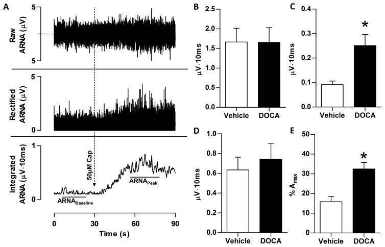
Figure 3.
Following DOCA-salt treatment, resting afferent renal nerve activity (ARNA) was measured in anesthetized rats. Panel A: A sample tracing of raw (top), rectified (middle), and integrated (bottom) nerve activity from a Vehicle-Sham animal is depicted, in which ARNA was recorded before and during the response to intrapelvic administration of 50μM capsaicin to establish the peak level of afferent renal nerve discharge. ARNA was quantified from the integrated (∫ARNA) signal. Panel B: Raw integrated signal of resting total renal nerve activity is no difference between Vehicle and DOCA rats. Panel C: Following proximal sectioning of the renal nerve, signal recorded represents only ∫ARNA. The resting ∫ARNA was elevated in DOCA versus Vehicle (0.24±0.04 versus 0.10±0.01 μV·sec). Panel D: Peak afferent nerve response to intrapelvic perfusion of 50μM capsaicin was no different between Vehicle and DOCA. Panel E: Basal ARNA, expressed as a percentage of peak ARNA response to intrapelvic capsaicin (%Amax). After normalization, resting activity remained increased in DOCA-salt rats compared Vehicle (32.0±5.7 versus 13.8±2.7%Amax). All data presented as mean±SEM (n=10/group). *p<.05 versus Vehicle-Sham.

