ABSTRACT
Cardiac sarcoidosis (CS) can cause sudden death, which is the leading cause of mortality in patients with sarcoidosis in Japan. However, it is difficult to diagnose CS because of the lack of a sensitive diagnostic method for the condition. Late gadolinium-enhanced cardiac magnetic resonance (MR) imaging demonstrates improved sensitivity for diagnosing CS. Therefore, it is important to know the late gadolinium-enhancement (LGE) characteristics of CS on cardiac MR images in order to diagnose CS accurately. In this study, we investigated the most common sites of LGE on cardiac MR images in CS. Late gadolinium-enhanced MR images of 9 consecutive patients with CS (obtained between August 2009 and July 2015) were reviewed by two radiologists. The distribution of LGE was evaluated using the American Heart Association 17-segment model of the left ventricle. The LGE in each segment was also classified into 4 patterns according to the myocardial layer in which it occurred (the subepicardial, subendocardial, intramural, and transmural layer patterns). All 9 patients exhibited LGE in their left ventricle, and 70 of 153 (46%) myocardial segments were enhanced. All of the patients displayed LGE in the basal septal wall. The patients' LGE layer patterns were as follows: subepicardial: 40% (28/70), intramural: 30% (21/70), subendocardial: 16% (11/70), and transmural: 14% (10/70). The basal septum wall and subepicardial layer often exhibit LGE on cardiac MR images in CS patients. LGE can be observed in other segments and layers in some cases.
Key Words: cardiac sarcoidosis, magnetic resonance imaging, late gadolinium enhancement, SPIR
INTRODUCTION
Sarcoidosis is a multisystem disease that is histologically characterized by the formation of non-caseating granulomas in various organs. Cardiac sarcoidosis (CS) can be almost asymptomatic; however, it is a life-threatening disorder that can cause fatal ventricular tachyarrhythmia, conduction disturbances, left ventricular dysfunction, and sudden death.1) Although cardiac involvement is only clinically diagnosed in about 5% of patients with systemic sarcoidosis,2) the incidence of autopsy-proven sarcoidosis involving the myocardium ranges from 13–27%1) and is especially high in Japan.3) CS-related death is the leading cause of mortality in patients with sarcoidosis in Japan.3, 4)
Myocardial biopsy, which is considered to be the gold standard method for detecting CS, is highly specific, but it is invasive and exhibits poor sensitivity (the diagnostic yield can be as low as 19%) because of the patchy nature of the disease.5) Non-invasive imaging techniques, such as echocardiography and nuclear medicine scans, such as thallium-201 (201Tl) and gallium-67 (67Ga) scintigraphy, have been used to detect CS, but with limited success. Recent studies have suggested that fluorine-18 fluorodeoxyglucose positron emission tomography (18F-FDG PET) might be useful for diagnosing CS.6-8) However, this method is expensive, only available at a limited number of facilities, and involves a lengthy pretreatment process (e.g., it requires prolonged fasting). Thus, the antemortem diagnosis of CS is difficult because of the lack of a sensitive diagnostic method.
The use of late gadolinium enhancement (LGE) during cardiac magnetic resonance (MR) imaging is a relatively new technique that allows the visualization of minute amounts of myocardial scarring and fibrotic tissue.9) Diagnostic methods based on detecting LGE on cardiac MR images have demonstrated improved sensitivity for diagnosing CS.10-12) The presence of LGE on cardiac MR imaging was added as a minor criterion to the 2006 revision of the Japanese Ministry of Health and Welfare (JMHW) guidelines for sarcoidosis,13, 14) which are used as consensus criteria for the diagnosis of CS. It has been reported that in CS LGE is most commonly seen on the right ventricular side of the basal septal wall;12) however, the site of LGE can vary from case to case, which makes it difficult to distinguish CS from other diseases, such as ischemic heart disease, myocarditis, and dilated cardiomyopathy. In CS, early diagnosis and the use of corticosteroids can improve atrioventricular block15) and survival;16) therefore, it is important to diagnose CS accurately. Knowledge of the characteristic LGE features of CS on cardiac MR imaging would aid this.
In the present study, we investigate the sites at which LGE was seen on cardiac MR images of the CS patients treated at our hospital.
MATERIAL AND METHODS
Our institutional review board approved this retrospective study and waived the requirement to obtain informed consent from the patients.
Patient selection
From February 2009 to July 2015, 21 consecutive patients underwent cardiac MR imaging under a suspicion of CS at our hospital. Their clinical data were obtained from the medical records at our hospital. Nine of the 21 patients fulfilled the diagnostic criteria outlined in the guidelines for CS developed by the JMHW, including the minor criterion regarding cardiac MR imaging (histological diagnosis group n=2, clinical diagnosis group n=7). The other patients, who were excluded from this study, were diagnosed with dilated cardiomyopathy (n=4), hypertrophic cardiomyopathy (n=1), arrhythmia of unknown cause (n=3), or heart failure of unknown cause (n=2) or did not fulfill the diagnostic criteria for CS (n=2). We studied the late gadolinium-enhanced MR images of the 9 patients. All 9 patients also underwent coronary angiography. One patient (patient No. 5) exhibited right coronary artery occlusion, but the others had normal coronary arteries.
MR protocol
Cardiac MR imaging was performed with a 1.5-T MR imaging scanner (MAGNETOM Avanto or MAGMETOM Aera, Siemens, Erlangen, Germany). An 8-channel phased-array body coil was used in all examinations. To evaluate the anatomy of the left ventricle, cine images and black blood T2-weighted images (short-axis slices from the base to the apex of the left ventricle and a cross-sectional long-axis view scan) were obtained. Ten minutes after the administration of 0.1 mmol/kg of the gadolinium-based contrast medium, MR images (contiguous 8-mm short-axis slices from the base to the apex of the left ventricle) were obtained using a breath-holding electrocardiogram-gated two-dimensional phase-sensitive inversion recovery (PSIR) sequence (repetition time: 700–1188.8 ms, echo time: 1.2–3.4 ms, inversion time: 300–700 ms, flip angle: 25°–45°, matrix: 127–156/256, field of view: 233–276/340 mm).17) An additional cross-sectional long-axis view scan was also acquired.
Image assessment
Two cardiovascular radiologists (with 4 and 11 years' experience, respectively), who were blinded to the patients' clinical findings assessed the late gadolinium-enhanced cardiac MR images by consensus. Short-axis slices and a cross-sectional long-axis view scan were available for 8 cases, whereas only short-axis slices were available in one case (patient No. 2). LGE was considered to be present when the signal intensity of the target myocardial segment was greater than 6 standard deviations (SD) stronger than the remote normal myocardial signal.18)
The American Heart Association developed a 17-segment model of the left ventricle,19) and the extent of LGE was evaluated in each of the 9 patients' 153 cardiac segments. In addition, the LGE in each segment was visually classified into 4 patterns depending on the myocardial layer in which it occurred (the subepicardial, subendocardial, intramural, and transmural layer patterns).
RESULTS
Population
Table 1 summarizes the patients' clinical characteristics. The patients were predominantly elderly (60.8±9.8 y.o.) and female (n=9, 100%). The other organs that exhibited involvement included the lymph nodes (n=8, 89%), lungs (n=6, 67%), eyes (n=5, 56%), and skin (n=3, 33%). The symptoms exhibited by the patients were as follows: no symptoms (n=4), dyspnea (n=3), slow pulse (n=1), and palpitations (n=1).
Table 1.
Patient characteristics
| Patient No. | Age/sex | Symptoms | Other involved organs | Diagnostic criteria for cardiac sarcoidosis | Sarcoidosis positive biopsy site | Myocardial biopsy | Cardiac catheter test | LVEF on echocardiogram (%) | ACE (IU/L) |
|---|---|---|---|---|---|---|---|---|---|
| 1 | 59/F | Dyspnea | LN Lungs | Clinical diagnosis | N.A. | N.A. | Normal | 32 | 5.2 |
| 2 | 57/F | No | LN, Lungs, Eye | Clinical diagnosis | N.A. | Negative | Normal | 44 | 18.0 |
| 3 | 62/F | Dyspnea | LN, Lungs, Eye | Clinical diagnosis | N.A. | Negative | Normal | 26 | 43.6 |
| 4 | 45/F | Dyspnea | LN, Lungs | Histological diagnosis | Heart | Positive | Normal | 35 | 25.1 |
| 5 | 65/F | No | Lungs, Eye, Skin | Clinical diagnosis | Skin | N.A. | RCA occlusion | 55 | 31.2 |
| 6 | 65/F | Slow pulse | LN, Eye, Skin | Clinical diagnosis | Skin | Negative | Normal | 69 | 24.6 |
| 7 | 73/F | No | LN, Eye | Clinical diagnosis | LN | Negative | Normal | 61 | 22.3 |
| 8 | 73/F | Palpitations | LN | Histological diagnosis | Heart | Positive | Normal | 37 | 11.9 |
| 9 | 48/F | No | LN, Lungs, Skin | Clinical diagnosis | Skin | Negative | Normal | 45 | 30.9 |
Notes: LVEF: left ventricular ejection fraction, ACE: angiotensin-converting enzyme, LN: lymph nodes, N.A.: not available, RCA: right coronary artery
Sites of LGE
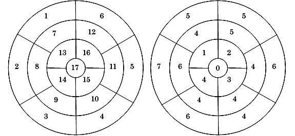
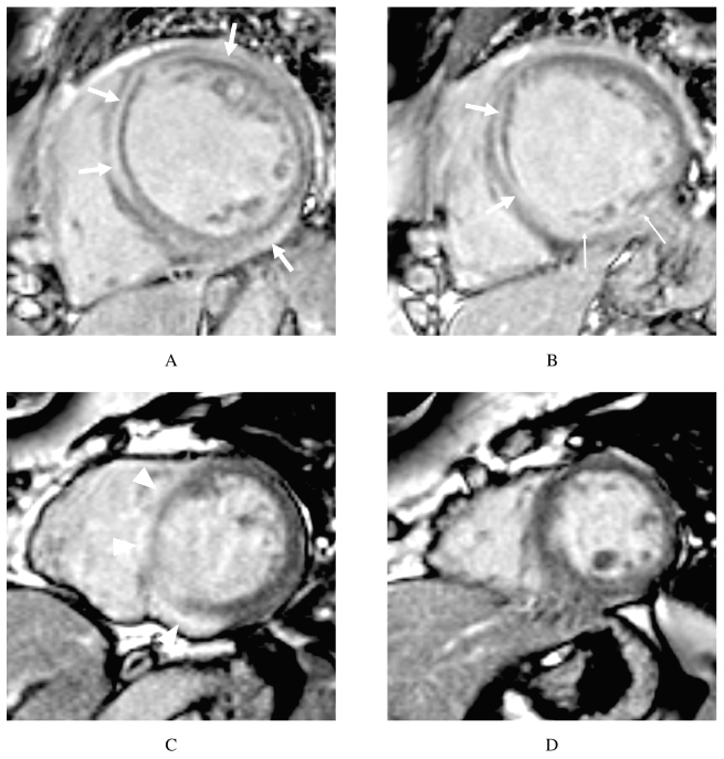
Table 2 summarizes the segments and myocardial layers that exhibited LGE on cardiac MR images in each patient. LGE was found in each case regardless of the perfusion area of the coronary arteries (Fig. 1). Among the 9 patients' 153 myocardial segments, LGE was observed in 70 (46%) segments (Fig. 2). All of the patients exhibited LGE in the basal septal wall (segment #2 and/or #3). The layer patterns of the 70 enhanced myocardial segments were classified as follows: subepicardial: 40% (28/70), intramural: 30% (21/70), subendocardial: 16% (11/70), and transmural: 14% (10/70).
Table 2.
Segments and layers in which late gadolinium enhancement was detected
| Patient No. | Segment | #1 | #2 | #3 | #4 | #5 | #6 | #7 | #8 | #9 | #10 | #11 | #12 | #13 | #14 | #15 | #16 | #17 |
|---|---|---|---|---|---|---|---|---|---|---|---|---|---|---|---|---|---|---|
| 1 | C | C | C | C | C | C | B | B | ||||||||||
| 2 | A | A | A | A | A | A | A | A | A | A | A | |||||||
| 3 | C | D | D | A | A | B | C | |||||||||||
| 4 | C | C | A | B | B | B | A | A | A | C | C | A | A | |||||
| 5 | C | B | ||||||||||||||||
| 6 | A | A | A | C | ||||||||||||||
| 7 | B | B | C | |||||||||||||||
| 8 | C | C | A | D | B | C | D | D | D | D | A | A | A | |||||
| 9 | C | B | C | D | C | D | A | A | D | |||||||||
Notes: A: subepicardial layer pattern, B: subendocardial layer pattern, C: intramural layer pattern, D: transmural layer pattern
The colors indicate the territories of the left anterior descending artery (LAD), right coronary artery (RCA), and left circumflex coronary artery (LCX).
Fig. 1.
Late gadolinium-enhanced cardiac MR images of a 59-year-old woman (patient No. 1) (A and B)
The late gadolinium-enhanced cardiac MR images show LGE in the intramural layers of the septal, anterior, and inferior walls (broad arrows, A) in the basal ventricle, and the intramural layer of the septal wall (broad arrows, B) and the subendocardial layer of the inferior wall (narrow arrows, B) in the mid-ventricle.
Late gadolinium-enhanced cardiac MR images of a 65-year-old woman (patient No. 6) (C and D)
The late gadolinium-enhanced cardiac MR images show LGE in the subepicardial layers of the septal and inferior walls (arrowheads, C) of the basal ventricle.
Fig. 2.

Bullseye plots demonstrating left ventricular segmentation (left) and the numbers of segments that exhibited LGE in 9 patients (right)
Among the 9 patients' 153 segments, LGE was observed in 70 (46%) segments.
DISCUSSION
In this study of consecutive patients who underwent cardiac MR imaging and fulfilled the diagnostic criteria outlined in the guidelines for CS developed by the JMHW, all of the patients exhibited LGE. LGE was detected at various sites, but was seen in the basal septal wall (segment #2 and/or #3) in all cases. Subepicardial layer patterns of LGE were more common than subendocardial layer patterns.
It has been reported that cardiac MR images display LGE in patients with CS.10, 12, 20-22) Smedema et al. detected LGE on cardiac MR images in 19 of 58 patients with sarcoidosis, mainly in the basal and lateral segments.12) Tadamura et al. reported that LGE occurred in the basal septal wall, especially in the right ventricle.10) In our study, we detected LGE in various segments and layers; however, all of the patients demonstrated LGE in the basal septal wall. Thus, the basal septal wall seems to be the most common site of sarcoid lesions, which corresponds with the findings of a postmortem study of CS patients.4) With respect to the layer in which LGE was observed, ischemic heart disease causes LGE in the subendocardial and transmural layers due to the flow of blood, and non-ischemic heart disease, including CS, tends to produce LGE in the subepicardial and intramural layers.22, 23) In our study, the subepicardial and intramural layers patterns were the most common, but subendocardial or transmural LGE was observed in some segments. Interestingly, the sites at which LGE was detected did not correspond to the distribution of the coronary arteries. This information might be useful for distinguishing CS from ischemic heart disease. CS sometimes presents with diffuse dilation of the left ventricle, e.g., in dilated cardiomyopathy (DCM), in which LGE is commonly seen in the intramural layer of interventricular septum. So, the site of LGE is a key difference between CS and DCM.23) It is important to know the LGE characteristics of CS in order to be able to accurately diagnose the condition.
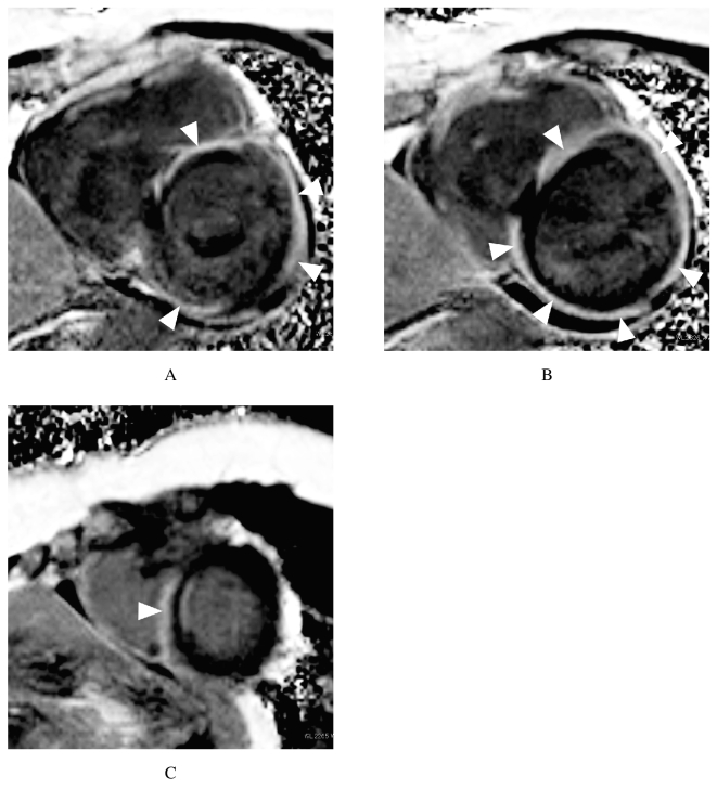
In our study, we used a PSIR sequence to obtain the late gadolinium-enhanced cardiac MR images. The most widely used cardiac MR imaging method is the inversion recovery sequence with magnitude reconstruction.10, 12, 20-22, 24, 25) The inversion recovery sequence typically requires the selection of an appropriate inversion recovery time (TI) in order to null the normal myocardial signal and maximize the contrast ratio between the normal and damaged myocardium. The performance of late gadolinium-enhanced cardiac MR imaging with the inversion recovery sequence depends on the selected TI. An error during TI selection leads to a reduction in contrast, which can reduce the size of the late gadolinium-enhanced area. Some cases (patient Nos. 2, 4, and 8) of CS exhibit broad contrast-enhanced regions (Fig. 3). In such cases, it can be difficult to select an appropriate TI for the inversion recovery sequence because it is not possible to distinguish the normal myocardium from sarcoid lesions beforehand. The TI-insensitive PSIR sequence is affected less by changes in the optimal TI for the normal myocardium with time.26) Thus, using the PSIR sequence can reduce the need for optimal null point setting, which might aid the diagnosis of CS. Further studies are needed to determine whether the PSIR or inversion recovery sequence is better for detecting CS.
Fig. 3.
Late gadolinium-enhanced cardiac MR images (A-C) of a 57-year-old woman (patient No. 2)
The late gadolinium-enhanced cardiac MR images show LGE in the subepicardial layers of various ventricular walls (arrowheads: A, B, and C). It was difficult to distinguish the normal myocardium from the lesional myocardium before the injection of contrast material.
Smedema et al. reported that the sensitivity and specificity of gadolinium-enhanced cardiac MR imaging for sarcoidosis were 100% and 78%, respectively.12) Patel et al. found that sarcoidosis patients that displayed LGE on cardiac MR imaging had a 9-fold higher rate of adverse events and an 11.5-fold higher rate of cardiac death than patients who did not display LGE.20) Nadel et al. reported that patients that exhibited CS on cardiac MR suffered a high rate of adverse cardiovascular events, but found that the use of implantable cardioverter–defibrillators reduced the risk of sudden death.27 Late gadolinium-enhanced cardiac MR imaging for sarcoidosis plays a key role in both diagnostic and prognostic assessments. Therefore, it is important to correctly interpret LGE in patients with sarcoidosis. Information about the sites at which LGE occurs might aid biopsy site selection because LGE is correlated with the locations of fibrous and scar tissue, which are found in sarcoid lesions.
This study has several limitations. First, it involved a relatively small number of patients. Second, although we pathologically confirmed the diagnosis of CS in two cases, we could not in the other cases. In this study, myocardial biopsy was performed in 7 cases, and 2 patients demonstrated positive findings. Myocardial biopsy is the gold standard method for diagnosing CS, but it is a high risk procedure and exhibits low sensitivity for CS. Therefore, other examinations including cardiac MR imaging are important for diagnosing the condition. Third, coronary angiography was performed in all cases, and one patient (patient No. 5) was found to be suffering from right coronary obstruction. Although the latter patient had abundant collateral vessels from the left coronary artery, it is possible that ischemic changes were responsible for the LGE she exhibited. Fourth, we did not employ the inversion recovery sequence in this study; therefore, we could not compare the utility of the PSIR sequence for detecting CS with that of the inversion recovery sequence. Fifth, the minor clinical criteria outlined in the guidelines for CS developed by the JMHW include the presence of LGE on cardiac MR imaging.
CONCLUSIONS
In CS, LGE occurs at various sites in the left ventricle, which vary on a case by case basis, and the most common site of LGE is the basal septal wall. LGE most commonly occurs in the subepicardial and intramural layers in CS patients. However, it can sometimes develop in the subendocardial and transmural layers, and such LGE needs to be distinguished from ischemic heart disease. Therefore, it might be useful to consider the sites at which LGE occurs relative to the distribution of the coronary arteries.
CONFLICT OF INTEREST
The authors have no conflict of interest.
REFERENCES
- 1).Silverman KJ, Hutchins GM, Bulkley BH. Cardiac sarcoid: a clinicopathologic study of 84 unselected patients with systemic sarcoidosis. Circulation, 1978; 58: 1204–1211. [DOI] [PubMed]
- 2).Sharma OP, Maheshwari A, Thaker K. Myocardial sarcoidosis. Chest, 1993; 103: 253–258. [DOI] [PubMed]
- 3).Iwai K, Tachibana T, Takemura T, Matsui Y, Kitaichi M, Kawabata Y. Pathological studies on sarcoidosis autopsy. I. Epidemiological features of 320 cases in Japan. Acta Pathol Jpn, 1993; 43: 372–376. [DOI] [PubMed]
- 4).Matsui Y, Iwai K, Tachibana T, Fruie T, Shigematsu N, Izumi T, et al. Clinicopathological study of fatal myocardial sarcoidosis. Ann N Y Acad Sci, 1976; 278: 455–469. [DOI] [PubMed]
- 5).Uemura A, Morimoto S, Hiramitsu S, Kato Y, Ito T, Hishida H. Histologic diagnostic rate of cardiac sarcoidosis: evaluation of endomyocardial biopsies. Am Heart J, 1999; 138: 299–302. [DOI] [PubMed]
- 6).Ishimaru S, Tsujino I, Takei T, Tsukamoto E, Sakaue S, Kamigaki M, et al. Focal uptake on 18F-fluoro-2-deoxyglucose positron emission tomography images indicates cardiac involvement of sarcoidosis. Eur Heart J, 2005; 26: 1538–1543. [DOI] [PubMed]
- 7).Koiwa H, Tsujino I, Ohira H, Yoshinaga K, Otsuka N, Nishimura M. Images in cardiovascular medicine: Imaging of cardiac sarcoid lesions using fasting cardiac 18F-fluorodeoxyglucose positron emission tomography: an autopsy case. Circulation, 2010; 122: 535–536. [DOI] [PubMed]
- 8).Youssef G, Leung E, Mylonas I, Nery P, Williams K, Wisenberg G, et al. The use of 18F-FDG PET in the diagnosis of cardiac sarcoidosis: a systematic review and metaanalysis including the Ontario experience. J Nucl Med, 2012; 53: 241–248. [DOI] [PubMed]
- 9).Kim RJ, Fieno DS, Parrish TB, Harris K, Chen EL, Simonetti O, et al. Relationship of MRI delayed contrast enhancement to irreversible injury, infarct age, and contractile function. Circulation, 1999; 100: 1992–2002. [DOI] [PubMed]
- 10).Tadamura E, Yamamuro M, Kubo S, Kanao S, Saga T, Harada M, et al. Effectiveness of delayed enhanced MRI for identification of cardiac sarcoidosis: Comparison with radionuclide imaging. AJR Am J Roentgenol, 2005; 185: 110–115. [DOI] [PubMed]
- 11).Serra JJ, Monte GU, Mello ES, Coral GP, Avila LF, Parga JR, et al. Images in cardiovascular medicine. Cardiac sarcoidosis evaluated by delayed-enhanced magnetic resonance imaging. Circulation, 2003; 107: e188–189. [DOI] [PubMed]
- 12).Smedema JP, Snoep G, van Kroonenburgh MP, van Geuns RJ, Dassen WR, Gorgels AP, et al. Evaluation of the accuracy of gadolinium-enhanced cardiovascular magnetic resonance in the diagnosis of cardiac sarcoidosis. J Am Coll Cardiol, 2005; 45: 1683–1690. [DOI] [PubMed]
- 13).Houston BA, Mukherjee M. Cardiac sarcoidosis: clinical manifestations, imaging characteristics, and therapeutic approach. Clin Med Insights Cardiol, 2014; 8: 31–37. [DOI] [PMC free article] [PubMed]
- 14).Diagnostic standard and guidelines for sarcoidosis. Jpn J Sarcoidosis, 2007; 27: 89–102.
- 15).Yodogawa K, Seino Y, Shiomura R, Takahashi K, Tsuboi I, Uetake S, et al. Recovery of atrioventricular block following steroid therapy in patients with cardiac sarcoidosis. J Cardiol, 2013; 62: 320–325. [DOI] [PubMed]
- 16).Yazaki Y, Isobe M, Hiroe M, Morimoto S, Hiramitsu S, Nakano T, et al. Prognostic determinants of long-term survival in Japanese patients with cardiac sarcoidosis treated with prednisone. Am J Cardiol, 2001; 88: 1006–1010. [DOI] [PubMed]
- 17).Kellman P, Arai AE, McVeigh ER, Aletras AH. Phase-sensitive inversion recovery for detecting myocardial infarction using gadolinium-delayed hyperenhancement. Magn Reson Med, 2002; 47: 372–383. [DOI] [PMC free article] [PubMed]
- 18).Nazarian S, Bluemke DA, Lardo AC, Zviman MM, Watkins SP, Dickfeld TL, et al. Magnetic resonance assessment of the substrate for inducible ventricular tachycardia in nonischemic cardiomyopathy. Circulation, 2005; 112: 2821–2825. [DOI] [PMC free article] [PubMed]
- 19).Cerqueira MD, Weissman NJ, Dilsizian V, Jacobs AK, Kaul S, Laskey WK, et al. Standardized myocardial segmentation and nomenclature for tomographic imaging of the heart. A statement for healthcare professionals from the Cardiac Imaging Committee of the Council on Clinical Cardiology of the American Heart Association. Circulation, 2002; 105: 539–542. [DOI] [PubMed]
- 20).Patel MR, Cawley PJ, Heitner JF, Klem I, Parker MA, Jaroudi WA, et al. Detection of myocardial damage in patients with sarcoidosis. Circulation, 2009; 120: 1969–1977. [DOI] [PMC free article] [PubMed]
- 21).Matoh F, Satoh H, Shiraki K, Odagiri K, Saitoh T, Urushida T, et al. The usefulness of delayed enhancement magnetic resonance imaging for diagnosis and evaluation of cardiac function in patients with cardiac sarcoidosis. Journal of Cardiology, 2008; 51: 179–188. [DOI] [PubMed]
- 22).Ichinose A, Otani H, Oikawa M, Takase K, Saito H, Shimokawa H, et al. MRI of cardiac sarcoidosis: basal and subepicardial localization of myocardial lesions and their effect on left ventricular function. AJR Am J Roentgenol, 2008; 191: 862–869. [DOI] [PubMed]
- 23).Satoh H, Sano M, Suwa K, Saitoh T, Nobuhara M, Saotome M, et al. Distribution of late gadolinium enhancement in various types of cardiomyopathies: Significance in differential diagnosis, clinical features and prognosis. World J Cardiol, 2014; 6: 585–601. [DOI] [PMC free article] [PubMed]
- 24).Smedema JP, Snoep G, van Kroonenburgh MP, van Geuns RJ, Cheriex EC, Gorgels AP, et al. The additional value of gadolinium-enhanced MRI to standard assessment for cardiac involvement in patients with pulmonary sarcoidosis. Chest, 2005; 128: 1629–1637. [DOI] [PubMed]
- 25).Patel AR, Klein MR, Chandra S, Spencer KT, Decara JM, Lang RM, et al. Myocardial damage in patients with sarcoidosis and preserved left ventricular systolic function: an observational study. Eur J Heart Fail, 2011; 13: 1231–1237. [DOI] [PMC free article] [PubMed]
- 26).Elgeti T, Abdel-Aty H, Wagner M, Busjahn A, Schulz-Menger J, Kivelitz D, et al. Assessment of late gadolinium enhancement in nonischemic cardiomyopathy: comparison of a fast Phase-Sensitive Inversion Recovery Sequence (PSIR) and a conventional segmented 2D gradient echo recall (GRE) sequence--preliminary findings. Invest Radiol, 2007; 42: 671–675. [DOI] [PubMed]
- 27).Nadel J, Lancefield T, Voskoboinik A, Taylor AJ. Late gadolinium enhancement identified with cardiac magnetic resonance imaging in sarcoidosis patients is associated with long-term ventricular arrhythmia and sudden cardiac death. Eur Heart J Cardiovasc Imaging, 2015; 16: 634–641.


