Abstract
Gastric cancer is the third most common cause of cancer-related death. Advanced stages of gastric cancers generally have grim prognosis. But, good prognosis can be achieved if such cancers are detected, diagnosed and resected at early stages. However, early gastric cancers and its precursors often produce only subtle mucosal changes and therefore quite commonly remain elusive at the conventional examination with white light endoscopy. Image-enhanced endoscopy makes mucosal lesions more conspicuous and can therefore potentially yield earlier and more accurate diagnoses. Recent years have seen growing work of research in support of various types of image enhanced endoscopy (IEE) techniques (e.g., dye-chromoendoscopy; magnification endoscopy; narrow-band imaging; flexible spectral imaging color enhancement; and I-SCAN) for a variety of gastric pathologies. In this review, we will examine the evidence for the utilization of various IEE techniques in the diagnosis of gastric disorders.
Keywords: Gastritis, Gastric cancer, Image enhanced endoscopy, Chromoendoscopy, Narrow band imaging
Core tip: Image-enhanced endoscopy is useful for an accurate real-time diagnosis of a variety of gastric diseases. But, good prognosis can be achieved if such cancers are detected, diagnosed and resected at early stages. However, early gastric cancers and its precursors often produce only subtle mucosal changes and therefore quite commonly remain elusive at the conventional examination with white light endoscopy. Image-enhanced endoscopy makes mucosal lesions more conspicuous and can therefore potentially yield earlier and more accurate diagnoses. Recent years have seen growing work of research in support of various types of image enhanced endoscopy (IEE) techniques (e.g., dye-chromoendoscopy; magnification endoscopy; narrow-band imaging; flexible spectral imaging color enhancement; and I-SCAN) for a variety of gastric pathologies. In this review, we will examine the evidence for the utilization of various IEE techniques in the diagnosis of gastric disorders.
INTRODUCTION
In 1957, Hirchowitz et al[1] pioneered the use of flexible endoscope to visualize the gastrointestinal (GI) tract. These early fibreoptic endoscopes were cumbersome to use and had dim views of the GI tract. Diagnosis of frank gastric pathologies (e.g., ulcer or malignant tumor) was straightforward. However, subtle abnormalities in the mucosa often got missed or misdiagnosed. This is especially relevant in stomach where early and confident detection of subtle pre-malignant features has potential to save the organ and life of a patient[2].
Recent years have witnessed tremendous progress in various novel endoscopic techniques. These techniques claim easy and confident detection, diagnosis and assistance in endoscopic resection of gastric subtle mucosal abnormalities. Simplistically, these techniques make a GI mucosal lesion appear more conspicuous. In a consensus methodological classification (in year 2008), such techniques were classified into five categories by Tajiri et al[3]: (1) conventional white light endoscopy (WLE); (2) image-enhanced endoscopy (IEE); (3) magnification endoscopy (ME); (4) microscopic endoscopy; and (5) tomographic. As these technologies offer different advantages and disadvantages, some have become indispensable tools inside every endoscopy room while others remain research tools.
Like in any disease, the endoscopic assessment of a mucosal abnormality also follows the logical sequence comprising “identification (or screening)”, “characterization”, “confirmation” by a gold standard (e.g., histology), and finally “treatment”. Explicit identification or screening of significant lesions is important to achieve low false miss-rates during endoscopy. At the same time, an accurate characterization, before histological assessment, is equally crucial to enable endoscopic resection for a significant lesion while leaving behind benign findings. Various IEE techniques have been studied for “identification” and “characterization” of gastric pathologies. In this review, we will study various IEE techniques and review their respective evidences for utilization in stomach.
METHODOLOGY
Publications in English language, limited to humans, were searched in the databases of “PUBMED/MEDLINE”, “the Cochrane Library”, and “Google Scholar”. The studies were searched between January 1995 and January 2016. Only studies published in peer-reviewed journals were taken into consideration. Relevant studies from the references of selected articles were also screened. The search keywords were: “Endoscopy, digestive system”, “Narrow band”, “Narrow-band imaging (NBI)”, “White light”, “Image enhance”, “Image enhanced”, “Endoscopy/methods”, “Gastroscopy/methods”, “White light endoscopy”, “Chromoendoscopy (CE)”, “Blue laser”, “Fujinon intelligent”, “Flexible spectral imaging color enhancement (FICE)”, “I-SCAN”, “Methylene blue”, “Indigo carmine”, “Acetic acid”, “Dye endoscopy”, “Helicobacter”, “Gastric atrophy”, “atrophic gastritis (AG)”, “Intestinal metaplasia”, “Gastric tumor”, “Gastric cancer”, “Stomach cancer”, and “Gastric neoplasm”. The two sets of keywords were combined individually. The studies were searched as free texts and as Medical Subject Headings terms.
WHITE-LIGHT ENDOSCOPY
The last decades of 20th century saw the advent of video-endoscopes equipped with charge-coupled devices (CCDs). These CCD chips produced image signal of 100000 to 400000 pixels, allowing clear images of GI mucosa[4]. Each pixel represents a unit of sample image, and therefore higher pixel-density meant greater spatial resolution and sharper images. Although good in detecting significant lesions in the GI tract, these standard definition (SD) video-endoscopes still had high miss-rates for subtle mucosal abnormalities.
The currently available high-definition (HD) endoscopes produce images with resolution of up to a million pixels and can magnify the mucosal image by 30- to 35 fold. These images can be further magnified optically by having an in-built motor-driven optical lens at the tip of endoscope. The lens can be focused upon an area-of-interest to provide a genuinely close-up image without sacrificing any pixel or image resolution. Contrary to the electronic magnification, the optical zoom produces a truly magnified (up to 150-times) and sharp image. Since the lens needs to be focused 2-3 mm away from the lesion, it is almost essential to have short hood or cap at the tip of magnifying-endoscope to maintain the focal length. These advances in endoscopic resolution have accompanied the considerable improvements in endoscope processors, which can convert tremendous amount of photonic data into a high-definition image without many artefacts. To get such HD images, it is imperative to have a compatible set of HD endoscopes, processor, monitors and transmission cables. Figure 1 illustrates cases of EGC detected by HD-WLE. They appear as a mildly depressed lesion with discoloration compared to adjacent normal mucosa.
Figure 1.
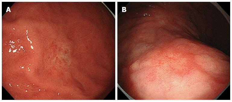
High definition white light endoscopy view. A: A depressed lesion with mucosal discoloration due to early gastric cancer; B: High definition white light endoscopy view of early gastric cancer.
Very few studies have directly compared HD-endoscopy with SD-endoscopy. For example in colon, these HD endoscopes showed marginal benefit in a meta-analysis with a number needed to treat of 25 to identify one additional polyp or adenoma[5]. Such objective data are not available for the upper GI tract but the expectation is similar.
WLE WITH MAGNIFICATION
WLE with magnification has potential to provide detailed mucosal views. In 1978, Sakaki et al[6] described classification of the stomach mucosa according to the “minute gastric mucosal patterns”. Since 1999, true magnifying endoscopes (for example, GIF-Q240Z by Olympus Corporation or EG-450ZH by Fujinon) were introduced which could optically zoom the image by up to 80 times. Such spatially resolved and magnified images revealed surface and microvascular patterns of stomach mucosa in great details. These studies are summarized in Table 1.
Table 1.
Summary of studies using magnification with white light
| Use in stomach | Type of evidence | Description | Remarks |
| Identification of normal gastric mucosa | Descriptive study[7]; Cross-sectional study with comparison to histology[8] | Normal corpus: Regular honeycomb pattern Normal antrum: Coil-shaped network with rare collecting venules | Different descriptive classifications have been used, but all emphasize on regular and uninterrupted mucosal and vascular patterns |
| Diagnosis of H. pylori gastritis | Six prospective studies with histology as the comparator[8-13] | High sensitivities and specificities for diagnosis of H. pylori | Multiple and varied pattern classifications with different endoscopes. Inherent subjectivity in classifications is an issue |
| Characterization of EGC | Six prospective studies with histology as the comparator[15-20] | Better results as compared to the traditional white light endoscopy | Multiple classifications bring inherent subjectivity; the most prevalent classification is the “VS” classification[17] which describes: Differentiated EGC: Irregular microvessels with a demarcation line Undifferentiated EGC: Absent demarcation line and absent sub-epithelial capillary networks |
H. pylori: Helicobacter pylori; EGC: Early gastric cancer.
Appearance of normal gastric mucosa with “only ME”
Magnified views of normal gastric mucosa have been classified and named differently by various authors. In a preliminary study in 2001, Yao et al[7] described the magnifying views of antrum as coil-shaped network with rare collecting venules, and of corpus as honeycomb pattern with interspersed collecting venules. Absence of sub-epithelial capillary network (SECN) along with proliferation of irregular microvessels was observed in differentiated early gastric cancer (EGC). Similarly, Yagi et al[8] studied the mucosal patterns in normal stomach without Helicobacter pylori (H. pylori) infection. The study concluded that the presence of regular arrangement of collecting venules (RAC) could predict the absence of H. pylori gastritis with 95.5% accuracy. Moreover, the presence of well-defined ridge pattern (wDRP) in antrum had 100% specificity for the absence of H. pylori gastritis, although its sensitivity was relatively low at 54.5%. In another study, the ME views of corpus were grouped into four types (Z-0 to Z-3) and almost all patients without H. pylori gastritis had Z-0 pattern[9].
H. pylori assessment with “only ME”
Six prospective studies have reported the use of “only ME” in predicting the gastritis (especially H. pylori gastritis)[8-13]. However, a variety of different mucosal classifications were used and proposed to correlate with H. pylori status. As mentioned in the earlier section, Yagi et al[8,9] correlated H. pylori status with RAC in corpus, wDRP in antrum and Z0-Z4 classification in corpus with good success. Based on the same Z0-Z4 classification, a group from Turkey showed superior results with H. pylori detection when compared with standard endoscopy[10]. In another study, Anagnostopoulos et al[11] grouped ME views of corpus (GIF Q240-Z, 115 × magnifications) into four types with high inter-observer agreement. Their classification identified H. pylori gastritis with sensitivity and specificity of 100% and 92.7% respectively. Similarly, other authors have reported excellent results with other classifications.
Worldwide, H. pylori gastric infection is considered the primary carcinogen for development of gastric adenocarcinoma[14]. Real-time diagnosis of H. pylori and other types of gastritis with ME may be beneficial in a sense that detection of such types of abnormal mucosal patterns may make an endoscopist more vigilant to the possibility of dysplastic gastric lesions. However it should be emphasized that there are inherent difficulties in interpretation of these subjective classifications and all studies have been done by experts. Moreover, given the widespread availability of relatively cheap, objective and sensitive tests to detect H. pylori gastritis (e.g., rapid urease test), routine utilization of ME alone for diagnosis of H. pylori should be undertaken with caution and biopsy based tests remain the standard for diagnosis.
Characterization of EGC with “only ME”
As the area of mucosal view is small with ME, its role for screening or identification of pre or early malignant lesions in stomach is limited. However, ME has a role in characterization of subtle gastric lesions which are detected by screening WLE. A variety of patterns have been described to differentiate EGC from benign gastric mucosa. A variety of patterns have also been described for characterization of differentiated EGC from undifferentiated EGC. There have been six prospective studies using ME-alone for characterization of EGC[15-20]. In 2001, Tobita[15] from Japan first described ME findings in 103 depressed gastric lesions (including 63 malignant lesions) using Fujinon, EG-410CR at × 60 magnification. It was concluded that the findings of “irregular protrusion” and “minute vessels in amorphia” were specific for malignancy. However, the classification had inherent subjectivity as it was assessed by one expert and was not been validated externally. In another prospective study in 2002, Tajiri et al[16] compared WLE-examination with ME (Olympus, GIF-Q240Z) in 211 consecutive gastric lesions. The authors found 89 EGC (58 depressed-type, 31 elevated-type). Using their classification, the accuracy of ME-examination was significantly superior to WLE-examination for any type of small (≤ 1 cm) EGC. In the same year, Yao et al[17] proposed a classification of magnified views of gastric mucosa which subsequently became the most widely utilized classification in studies of ME with IEE techniques. The mucosa was classified based on “microvascular” and “microsurface” patterns, later known as the “VS classification”. Non-cancerous mucosa were found to have regular appearances of SECN, all differentiated EGC had a demarcation line and irregular microvessels, while all undifferentiated EGC had absent demarcation line with absent or reduced SECN. In 2007, Yao et al[18] validated their classification on a larger sample. For characterization of EGC with ME, other authors have used varied classification with good results[19,20].
Overall, the efficacy of ME-alone in the stomach has been studied by a few authors, mainly from Japan. The various uses of ME-alone in stomach are summarized in Table 1. However as will be discussed later, the majority of studies of ME in stomach have been performed in combination with other IEE techniques.
CONVENTIONAL CHROMOENDOSCOPY
Introduced in the 1990s, CE refers to spraying of harmless dyes to stain the mucosal surface. This is usually done after a preliminary inspection with WLE. The staining of surface makes subtle mucosal patterns more obvious. A variety of stains have been used in the GI tract and these are classified into three types based on their actions: Absorptive (or vital) stains, contrast stains, and reactive stains. Absorptive stains (e.g., Lugol’s iodine, methylene blue) have property of differential absorption into different cell types, thus highlighting one type of tissue over other. For example, methylene blue is absorbed by cells of small intestine and colon, and therefore a stained focus in the stomach theoretically indicates IM. On the contrary, the contrast stains (e.g., indigo carmine) do not react with the cells, but accumulate in the pits and crevices of a mucosal lesion thus accentuating the surface pattern (or the topography), mucosal irregularities and boundaries of a lesion (Figure 2). The last category, the reactive stains (e.g., acetic acid, phenol red) change color by coming in contact with a particular protein on the surface. For example, Phenol red and Congo red are reactive stains which turn red in an alkaline gastric environment signalling infection with H. pylori.
Figure 2.
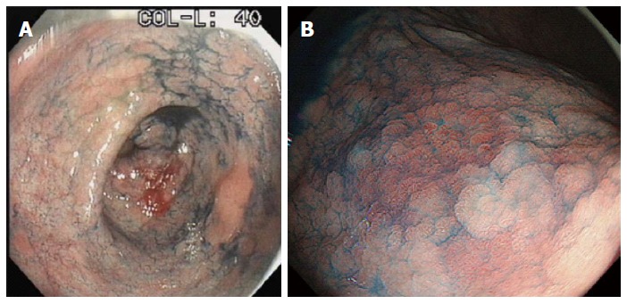
Mucosal irregularities and boundaries of a lesion. A: Gastric adenoma accentuated by indigo carmine; B: Early gastric cancer accentuated by indigo carmine.
CE can be performed in two ways: (1) pan-CE, which involves spraying the dye blindly and voluminously to screen for any abnormal areas; or (2) targeted staining, where a dye is sprayed over a lesion of interest to further characterize it. Several studies have attempted a variety of stains in the stomach, either alone or in combination, to identify, characterize and outline focal lesions (e.g., IM or EGC). CE is technically easy to perform and has shown significant advantages in detecting flat colorectal neoplasia and colitis-associated neoplasia[21].
Use of acetic acid in stomach
Acetic acid causes a reversible and transient alteration in the tertiary structure of the cellular proteins which leads to temporary opacification of mucosal surface. This produces a vivid mucosal image with crypts turning brown while the intervening epithelial surface appearing white. While the non-cancerous mucosa changes into white, the dysplastic and cancerous cells remain unstained, producing a good contrast. After spray of acetic acid, the mucosal details are visualized with magnification. This combination of acetic acid instillation and ME is often termed as “Enhanced-magnification endoscopy (EME)”, which allows visualization of villi and crypts. In 2005, Yagi et al[22] studied the value of EME in stomach in 45 patients. The mean duration of whitening differed with each histologic type: Low-grade adenoma, 94 s; high-grade adenoma, 24.3 s; non-invasive carcinoma, 20.1 s; invasive intramucosal carcinoma, 3.5 s; and submucosal carcinoma or beyond, 2.5 s. Therefore, the acetic-acid with ME was useful in differentiating between neoplasia and non-neoplasia based on duration of whitening.
One year later in 2006, Tanaka et al[23] proposed a classification of EME findings in stomach based on forty seven consecutive patients, into five categories: Type I, small round pits; type II, slit-like pits; type III, gyrus and villous patterns; type IV, irregular arrangements and sizes; type V, destructive patterns. Elevated gastric carcinomas showed type III or IV patterns; while depressed carcinomas showed type IV or V patterns. Later the same group, in a separate observational study, found that the surface patterns were evident in 100% of lesions by EME as compared to only 66.4% with conventional or magnification endoscopies[24]. The type IV-V lesions were strongly associated with gastric cancer with a sensitivity of 100% and specificity of 89.7%.
Use of congo-red and phenol-red in stomach
Utilizing its tendency to turn red in an alkaline environment, this Congo-red has been used for detection of AG, H. pylori infection and IM. Data are limited. Phenol-red has been used in old studies to map the gastric mucosa for H. pylori infection. With phenol red spraying endoscopy, Kohli et al[25] identified H. pylori infection with sensitivity and specificity of 100% and 84.6% respectively.
Use of methylene-blue in stomach
Methylene blue is absorbed by intestinal cells and thus highlights gastric IM (Figure 3). Dinis-Ribeiro et al[26] examined and proposed a classification in 136 patients using ME after methylene blue (1%) spray and could identify IM and dysplasia with 84% and 83% accuracy respectively. The findings were externally validated at another centre in Portugal in forty two patients with AG with or without IM, and the results showed excellent reproducibility for the classification[27]. In a tandem study with only thirty-three patients, Taghavi et al[28] compared conventional endoscopy against CE with methylene blue. The CE group yielded more IM lesions compared to conventional endoscopy.
Figure 3.
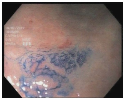
Gastric intestinal metaplasia highlighted by methylene blue.
Use of haematoxylin in stomach
Haematoxylin is a common stain used in histological assessment since it stains the nuclei of cells. To date, there is only a single study utilizing haematoxylin as CE on a heterogeneous sample of gastric abnormalities[29]. Although high sensitivity (92.9%) and specificity (89.3%) for diagnosing gastric neoplasia were reported, there were only three cases of cancer.
Use of “acetic acid plus indigo carmine” in stomach
Acetic acid whitens the non-cancerous gastric mucosal epithelial cell while the cancerous cells remain unstained. As detailed above, multiple studies have utilized acetic acid with ME in technique known EME. However, some authors believe that the use of ME may be cumbersome for neoplastic lesions (especially if lesion is large or located at a difficult position). Therefore, CE using a sequential combination of acetic acid spray followed by another spray with indigo carmine (AI) has been proposed for examination of a mucosal neoplastic lesion. Multiple studies have shown the efficacy of AI for delineating the margins of EGC before endoscopic resection. In the first published study on AI use with 114 patients, AI was much more effective in delineating the lateral spread of cancers as compared to indigo carmine alone[30]. In another prospective comparative study, Sakai et al[31] used AI in 53 consecutive gastric lesions before endoscopic submucosal dissection and good interobserver agreement was reported between the two endoscopists (kappa = 0.764). The diagnostic performance of AI was significantly better than either indigo carmine or acetic acid alone. In another prospective study on 108 EGC lesions, Kawahara et al[32] compared WLE, indigo-carmine and AI for delineation of margins before ESD. All endoscopic examinations were performed by one endoscopist. When correlated with pathological specimens, the diagnostic accuracy of AI was higher when compared to WLE or indigo-carmine (90.7% vs 50.0% vs 75.9%, respectively).
Overall, the studies with AI have shown an excellent efficacy in demarcation of EGC before endoscopic resection. The technique does not require additional equipment (e.g., magnification endoscope).
NARROW BAND IMAGING
NBI is a proprietary optical image-enhancement technology launched by the Olympus Corporation (Tokyo, Japan) in year 2005. NBI is the most widely utilized electronic IEE technique with demonstrated scientific evidence for its efficacy in GI diseases. Normally, the wavelengths of white light range from 400 to 700 nm. During conventional WLE, the illuminating white light travels from the xenon lamp via a rotating red-green-blue (RGB) rotatory filter. In NBI, an additional filter is placed between the xenon lamp and the RGB filter[33]. This whole NBI system is simply activated by a push of button on the control handle of the endoscope without interrupting the views on monitor. By this additional NBI filter, the light is converted from a broad RGB into narrow bands of blue and green at 415 (± 15) nm and 540 (± 15) nm wavelengths respectively. The narrow wavelengths of illuminating light increase the saturation. Moreover, biological tissues behave differently at different wavelengths of light due to their characteristic patterns of absorption and scattering of light. Since haemoglobin molecule has two absorption peaks at 415 nm and 540 nm, the mucosal microvascular patterns are highlighted in extensive detail with NBI[34].
NBI can diagnose the subtle and flat mucosal GI lesions which are often missed or remain uncharacterized on WLE. Since the sub-epithelial capillaries of stomach have minimum diameter of 8 μm[17], combining ME with NBI has been studied for detailed examination of capillary patterns in stomach. As described below, most of the published studies have utilized a simultaneous combination of ME and NBI. It must be recognized that there are two NBI systems in use, the EVIS EXERA and the EVIS LUCERA systems. For the EXERA system, the magnification achieved is by digital magnification and a specific technique called “Dual Focus” which allows near mode imaging; in contrast, the LUCERA system allows optical magnification and this is the main system used in prior studies of magnifying NBI. An overview of NBI studies are provided in Table 2.
Table 2.
Summary of studies using narrow band imaging in stomach
| Use in stomach | Type of evidence | Description | Remarks |
| Screening of focal lesions in stomach | Five prospective studies[35-39] studied screening with WLE followed by characterization of detected lesions with NBI Single randomized prospective study with bright-NBI[40] | WLE followed by characterization with NBI seems to increase confidence in taking targeted biopsies New generation “bright-NBI” appears promising to increase yield of FGL as single step examination in stomach | Majority of the detected FGLs are intestinal metaplasia Due to small sample sizes in these studies, it is unclear whether such strategy will improve detection of subtle malignant gastric lesions |
| Diagnosis of H. pylori gastritis | Two prospective trials[41,42] using M-NBI with histology as comparator | Subjective classifications of mucosal microvascular patterns showed high sensitivity and specificity for real-time diagnosis of H. pylori gastritis | Inherent subjectivity in the classification is an issue |
| Diagnosis of IM | Multiple prospective studies and one recent meta-analysis[44] using M-NBI for diagnosis of IM | Multiple patterns have been assigned for diagnosis of IM. The most prevalent is the “LBC” sign The pooled sensitivity and specificity of LBC for diagnosis of IM are 84% and 93% respectively | LBC sign with M-NBI appears easy to learn and reliable for real-time diagnosis of IM |
| Characterization of an EGC | Multiple prospective studies including two recent meta-analyses[52,53] using M-NBI for characterization of an EGC | Various pattern-classification systems with M-NBI have been used in different studies to characterize a lesion as EGC. The pooled sensitivity: 0.83-0.85 The pooled specificity: 0.96 | Inherent subjectivities in a variety of classifications remain an issue Significant heterogeneity were observed in both meta-analyses |
| Prediction of histological differentiation of an EGC | At least two prospective studies[54,55] | Subjective pattern assignments were given; Only moderate sensitivities and specificities to determine histological differentiation of an EGC | Inherent subjectivities in the classification system. Currently, histology is still required to determine histological differentiation of an EGC |
| Determination of horizontal extent of an EGC | Few studies with small sample sizes | One study[58] showed better accuracy than indigo carmine chromoendoscopy | Real-time estimation of an EGC is useful before endoscopic resection. However, the histology still remains the gold-standard |
| Determination of depth of an EGC | Two prospective[61,62] studies | Subjective classifications but with excellent accuracy | Inherent subjectivities in the classification system. Currently, histology is still required to determine depth of an EGC |
FGL: Focal gastric lesion; H. pylori: Helicobacter pylori; EGC: Early gastric cancer; M-NBI: Magnifying narrow band imaging; IM: Intestinal metaplasia; LBC: Light blue-crest; WLE: White light endoscopy.
NBI for screening of gastric pathologies
At narrow wavelengths of light with NBI, the intensity of illumination is compromised resulting in darker images when compared to images during WLE. This is especially relevant while examining the stomach, a capacious organ, where dark views result in NBI being not so useful for screening of focal gastric lesions. However, NBI can be utilized as a second-look method to focus on lesions detected upon screening with WLE. This technique appears to increase detection rate of gastric focal lesions. At least five prospective studies have studied NBI using this approach[35-39]. In a prospective study on an unselected population, our group screened for focal gastric lesions using WLE followed by characterization of detected lesions by magnified NBI (M-NBI)[34]. Additional 15% lesions (mostly IM) were detected with M-NBI (Figure 4). In another multicentre prospective study using the similar sequence (WLE followed by M-NBI), the accuracy of M-NBI for high confidence diagnoses of gastric lesions was 98%[35]. In a recent prospective study with more than three thousands non-selected patients, gastric examinations were performed with high-definition white light (HD-WLE) followed by ME and then with M-NBI[36]. Using such strategy to detect EGC, ME and M-NBI had significantly higher sensitivities when compared to HD-WLE. However, there were no differences among specificities of the techniques.
Figure 4.
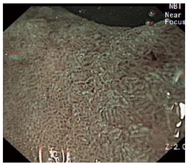
Gastric intestinal metaplasia highlighted by narrow band imaging using the EXERA III system with dual focus.
The new generation NBI system introduced in 2012 (e.g., the “EVIS EXERA III” or “EVIS LUCERA” from Olympus Corporation) attempt to overcome the drawback of dark views by having an upgraded xenon light source. Besides this, the new systems also have two filters for blue light and one filter for green light in contrast with previous NBI system where only one filter each is used for blue and green. Thus, this new generation NBI system (sometimes known as bright-NBI) produces brighter NBI images even from a distance. In a recent multicentre prospective randomized study, our group compared the HD-WLE with the new generation bright-NBI system (either 190-NBI or 290-NBI) for screening of focal gastric lesions (FGL)[40]. The detection rate of FGL was higher with bright-NBI than with HD-WLE (41% vs 29%; P = 0.003).
Magnifying-NBI for diagnosis of H. pylori gastritis
Since H. pylori infection produces alterations in the microsurface structures and microvascular patterns of gastric mucosa, it is postulated that the magnified NBI views may be helpful for real-time diagnosis of H. pylori gastritis. For this pathology, there has been considerable interest by researchers for two reasons. First, the high-confidence real-time diagnosis of H. pylori may stimulate an endoscopist to be more vigilant for concomitant pre-malignant and malignant lesions of stomach. Also, the real-time gastric mucosal pattern analyses may theoretically help in obtaining targeted biopsies instead of routine practice of blind-biopsies for H. pylori check. In a prospective study in 2009, Tahara et al[41] attempted to correlate gastric mucosal patterns on magnifying-NBI (M-NBI) with H. pylori gastritis. At M-NBI, the normal gastric corpus pattern was identified as small, round pits with regular subepithelial capillary networks (SECN). The abnormal patterns were classified into three (type 1 to 3) categories. The sensitivity and specificity of abnormal patterns (type 1 + 2 + 3) for H. pylori gastritis were 95.2% and 82.2%, respectively. In a comparative trial in 2014, Yagi et al[42] compared conventional WLE with M-NBI for detection of H. pylori gastritis in patients diagnosed with EGC. For diagnosis of H. pylori gastritis, the sensitivity and specificity in M-NBI group were higher than in WLE group.
M-NBI for diagnosis of AG and IM
AG and IM represent significant milestones in the sequence of gastric carcinogenesis[14]. On conventional WLE, corpus AG is suspected based on a paucity of gastric rugae with more marked appearances of submucosal vessels; while IM appears as patchy, white, raised or flat spots. However, WLE is considered insensitive for diagnosis of AG and IM. On NBI, AG is characterized by a complete loss of pit-pattern and SECN, with presence of only collecting venules. In a randomized, prospective and crossover study by Dutta et al[38] NBI was superior to WLE for detection of AG. With NBI various appearances have been proposed for characterization of IM. The most important of these is the identification of light blue crests (LBC).
In 2006, Uedo et al[43] first described LBC as fine blue-white lines on the crests of the epithelial surface/gyri, similar to a light reflected from mirror. In this seminal study, the sensitivity and specificity of LBC for diagnosis of IM were 89% and 93% respectively. Similarly, high diagnostic values of LBC for characterization of IM have been shown by other authors (Figure 5A). A recent meta-analysis by Wang et al[44], which included four prospective studies without significant heterogeneity, documented that the pooled sensitivity and specificity of LBC for diagnosis of IM were 84% and 93% respectively. In a pilot feasibility trial, Bansal et al[45] found sensitivity and specificity of “ridge/villous pattern” for IM to be 80% and 100% respectively.
Figure 5.

Best visualized with optical magnification. A: Magnifying narrow band image of gastric intestinal metaplasia showing, the light blue crest sign, surrounding central area of early gastric cancer; B: Magnifying narrow band image of early gastric cancer; C: Magnifying narrow band image of early gastric cancer.
M-NBI for characterization of EGC
Perhaps the most important and most extensively investigated use of NBI in the stomach is for the characterization of EGC. The accurate identification of EGC is important since endoscopic resection for such early cancers achieves > 90% five-year survival. Morphologically EGC are categorized mainly into three types by the Paris classification[46]: Superficial elevated (0-IIa), superficial flat (0-IIb), and superficial depressed (0-IIc). EGCs often produce subtle mucosal changes (sometimes known as “gastritis-like cancers”) and can be easily missed on conventional WLE. The major contribution of M-NBI lies in accurate differentiation of such lesions from normal or inflamed gastric mucosa.
The most widely studied and utilized classification is the “VS classification” by Yao et al[47] where “V” stands for microvascular patterns while “S” stands for surface microstructures. In this classification, an EGC is accurately identified based on two features: (1) a demarcation line (DL) with loss of SECN; and (2) an irregular microvascular pattern (IMVP) or an irregular microstructural pattern. These features are best visualized with optical magnification (Figure 5). Digital magnification combined with dual focus imaging may provide an adequate view of the demarcation line and microstructural pattern, but will not be able to clearly visualize the microvascular pattern (Figure 6).
Figure 6.
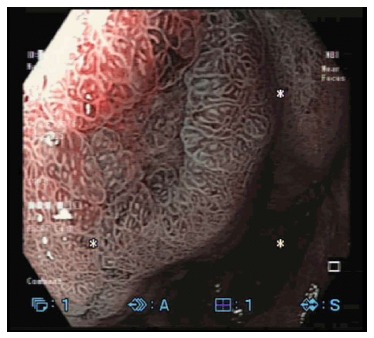
Image of early gastric cancer visualized using narrow band imaging with digital magnification and dual focus imaging.
In 2010, Ezoe et al[48] prospectively compared diagnostic efficacies of ME with M-NBI using VS classification in a sample of 57 depressed gastric lesions (including 27 malignant). For accurate diagnosis of EGC, the diagnostic accuracy and sensitivity were significantly higher for M-NBI as compared to ME. Subsequently, the same comparison was studied in much larger sample in a randomized and multicentre trial[49]. Again for diagnosis of depressed-type EGC, the M-NBI was superior to ME. The sensitivity and specificity of M-NBI were 95.0% and 96.8% respectively.
In the recent post-hoc analysis of this study, a sequential strategy for diagnosing a cancerous gastric lesion was proposed[50]. For a depressed gastric lesion on white-light examination, M-NBI was suggested to look for a DL first since presence of DL had high sensitivity and high negative predictive value for a malignant lesion. In lesions with DL, an absence of IMVP was proposed to rule out malignant lesions because of high specificity of IMVP. In another prospective study, Kato et al[51] surveyed 111 high-risk patients for EGC based on a triad of findings on M-NBI: (1) disappearance of mucosal pattern; (2) microvascular dilatation; and (3) heterogeneity. Although only 14 gastric cancers were detected, the sensitivity and specificity of M-NBI (92.9% and 94.7% respectively) were superior to WLE (42.9% and 61.0% respectively).
In a recent meta-analysis by Zhang et al[52] the pooled sensitivity and specificity of M-NBI for diagnosis of EGC were 0.83 and 0.96 respectively. However, there were significant heterogeneity among the studies and also, a combination of retrospective and prospective studies were pooled together. Another recent meta-analysis, which only included six prospective studies, also showed high pooled sensitivity (0.85) and specificity (0.96) for M-NBI diagnosis of EGC[53]. A significant heterogeneity among studies was also observed in this meta-analysis.
M-NBI for histological differentiation of EGC
Besides differentiating between cancerous and non-cancerous lesions, several studies have attempted to use M-NBI for prediction of histologic differentiation of EGC. During the early years of NBI technique, Nakayoshi et al[54] studied 165 depressed-type of EGC with M-NBI. The microvascular patterns were divided into three patterns: Fine network, corkscrew and unclassified. The fine network patterns were seen more commonly in differentiated EGC as compared to the undifferentiated type (66.1% vs 3.7%), whereas corkscrew patterns were seen more commonly in undifferentiated-type (85.7% vs 3.6%; P = 0.0011). However, the conclusion was that the real-time optical diagnosis with M-NBI, although beneficial, was still not sufficient to replace histopathological confirmation.
Similarly, Yokoyama et al[55] studied 257 consecutive EGC with M-NBI and divided the microvascular patterns into four categories: Fine-network, corkscrew, intra-lobular loop-1, and intra-lobular loop-2. When correlated with histopathology, differentiated-type EGC mostly had fine-network pattern or intra-lobular loop patterns. On the contrary, the undifferentiated-type of EGC had intra-lobular loop-2 pattern and corkscrew pattern in almost all patients (41.2% and 58.2% respectively).
M-NBI to determine the horizontal extent of EGC
Delineating the horizontal extent of EGC is important for margin-free endoscopic resection. Traditionally, CE with indigo-carmine has been used to highlight abnormal mucosal patterns before endoscopic resection. In 2002, Yao et al[56] published a case report where demarcation of a well-differentiated EGC was made with an image-enhanced technology using “hemoglobin index”.
Subsequently in a sample of twelve EGC, Sumiyama et al[57] used a combination of M-NBI and a multibending endoscope for en bloc endoscopic mucosal resection. Using this combination, 91.7% (11/12) en bloc resections were made feasible as compared to 35% in conventional endoscopy group. However, authors stated that it was unclear as to which of these factors (i.e., either NBI or multibending endoscope or both) led to this satisfactory outcome.
Kiyotoki et al[58] compared M-NBI with indigo-carmine based CE in 118 EGC. For delineating margins of EGC, the accuracy was higher in M-NBI group as compared to the indigo-carmine (97.4% and 77.8% respectively; P = 0.009). Another prospective study by Nagahama et al[59] documented 72.6% accuracy for identifying margins of EGC which could not be delineated with acetic acid CE.
Overall, studies have shown beneficial results with M-NBI for delineation of margins only in the differentiated-type of EGC. Demarcation of undifferentiated-type of EGC is considered difficult since the malignant growth seems to creep more into the lamina propria which may not always produce endoscopically visible mucosal changes. For example in the study by Nagahama et al[59] the endoscopic delineation remained difficult for undifferentiated lesions. However one prospective study also showed high accuracy (81.6%) using M-NBI for demarcation of undifferentiated-type of EGC[60].
It can be concluded that there is good evidence with prospective trials in support of M-NBI for demarcation of differentiated EGC before performing endoscopic resection. However it should also be noted that the trials have generally included a small number of patients in whom experts have performed endoscopic examination while utilizing various types of classification for pattern categorization. Although histopathology is still considered the gold-standard for retrospective confirmation of clearance of margins, the real-time estimation of horizontal margins with M-NBI is helpful as a prospective guide for accurate en-bloc resection.
M-NBI to determine the depth of EGC
According to the Japanese Gastric Cancer Handling Codes, the submucosal EGC are divided into three types (SM1 to SM3) based on the depth of cancer invasion. The differentiated-type of SM1 (i.e., vertical depth up to 500 μm) EGC can be considered as an expanded indication for endoscopic resection. But, surgical resection should be considered for SM2 and SM3 cancers as the probability of lymph node metastatic disease is high. Prospective estimation of the depth of invasion is difficult and therefore endoscopic resection is considered complete only after histopathological assessment of the resected specimen. Knowledge of deep invasion will avoid unwarranted endoscopic resection. Presence of ulceration on standard WLE suggests deep invasion. But, it may be especially difficult to estimate depth of invasion in flat (Paris 0-IIa, 0-IIb and 0-IIc) EGC.
In 2008, Yagi et al[61] correlated the M-NBI patterns of 72 differentiated-type EGC (10 elevated, 27 flat, and 35 depressed-type) with histopathology. All endoscopic examinations were performed by one expert and the patterns were classified into three types: Mesh, loop and interrupted. The mesh or loop pattern were seen in 94.9% of mucosal EGC while 92.3% of submucosal EGC had interrupted patterns. In another prospective study from China, Li et al[62] reported findings of M-NBI in 164 suspected gastric lesions. The patterns of M-NBI were categorized into three groups (A, B and C) based on both surface pattern and microvascular architecture. Besides excellent diagnostic values for characterization and differentiation of EGC, M-NBI classification was able to accurately predict the depth of invasion in 37 out of 39 differentiated adenocarcinomas (95%).
Two retrospective studies have also attempted correlation of M-NBI images (taken before the resection) with histopathology of resected specimens. In the first study by Kobara et al[63] it was concluded that the presence of non-structure, scattery vessels and multi-caliber vessels can possibly serve as indicators of SM2 invasion in differentiated-type of EGC. In the second study by Kikuchi et al[64] M-NBI images were examined for dilated vessels (D-vessels) which were defined as vessels with diameter 3 times larger than that of the irregular microvessels. The sensitivity and specificity of D-vessels for SM2 invasion were 37.5% and 88.3% respectively.
FICE
This technology is also known as “optimal band imaging” or “multi-band imaging”. FICE was introduced by Fujinon (Tokyo, Japan) in year 2005 and is currently its proprietary technology. In FICE, the ordinary white-light images are captured by the CCD and are mathematically processed in the processor by assigning specific ranges of wavelengths. Thereafter, electronically enhanced and reconstructed color images are displayed on the monitor[65]. This is in contrast with the NBI where raw and enhanced images are captured by putting an optical filter in the path of illuminating light. Since FICE processes well-illuminated white-light images, this means that the FICE can provide enhanced images without compromising on brightness. At present, there are ten pre-settings of FICE which can be instantaneously activated by pushing a button on an accompanying keyboard. A total of three presets can also be assigned to the buttons on the control handle of the endoscope to ease the switching of the different FICE images. In recent years, several studies have claimed superior efficacy of FICE as compared to WLE for various pathologies of esophagus, colon and stomach. Only a few studies have studied FICE in stomach, with most performed for delineation of margins of EGCs. However, a learning curve for pattern recognition, a requirement for separate endoscopic equipment and lack of consensus for objective diagnostic criteria has restricted use of FICE.
Use of FICE in EGC
In the management of EGC, the use of FICE has been limited to demarcation of already identified lesions. FICE has not been objectively studied as a screening tool to pick up early malignant lesion. In 2008, Osawa et al[66] published the first clinical study in stomach. In this real-time prospective study with a small sample of twenty-seven patients, four endoscopists, in real-time demarcated the depressed type of EGC with accuracy of 96%. In the same year, another study by the same group claimed efficacy in demarcation of elevated and depressed type of EGC[67]. However in this study, the objective results of efficacy of this method were not reported. In another study from the same group, it enabled delineation of elevated-type of ECG in the background of AG[68].
Since a variety of wavelengths were being used for gastric examination with FICE, the most effective wavelength was studied in a retrospective fashion by another group in Japan[69]. Previously captured white-light images of EGC were processed by the FICE processor and analysed. It was noted that the wavelength of 530 nm generated maximal difference in spectral reflectance between EGC and normal mucosa and there was significant improvement from the WLE images to the FICE images. FICE is proposed as a potential alternative to conventional CE because it provides contrast enhancement of tissue surface structures. However at present, the evidence for its support has come from a few studies with small sample. Since the technology requires a new set of equipment, further external and large-scale validation will be required before its widespread use.
Use of FICE for other gastric pathologies
In one study, FICE was studied for differentiation of non-neoplastic lesions, adenomas and cancers in the stomach[70]. A total of 171 gastric lesions were examined and FICE performed better than magnifying-WLE. Another study examined the role of FICE for diagnosis of gastric intestinal metaplasia[71]. FICE had sensitivity and specificity of IM diagnosis of 60% and 87% based on histological confirmation. Although this study might have diagnosed IM based on LBC, there is still no consensus on diagnostic criteria for IM on FICE.
Small-caliber gastroscope with FICE
Small-caliber endoscope has lower resolution but is more comfortable for the patients, especially if used as a screening tool for gastric pathologies. Theoretically, FICE combined with small-caliber gastroscope can enhance the color contrast of gastric pathologies. To date, there are two studies, from Japan, which have evaluated this hypothesis. Tanioka et al[72] retrospectively examined 50 gastric lesions which were previously identified on screening endoscopy with Ultraslim endoscope (Fujinon EG-530N2). After conversion into FICE images, superior visibility was seen in 54.7% upper GI lesions as compared to conventional images. In another study by Osawa et al[73] 82 depressed-type EGC (which were already diagnosed with conventional normal-caliber endoscopy) were examined with small-caliber (Fujinon EG-530N2) endoscopy and FICE by endoscopists who were blinded of the locations of the lesions. Most EGC could be detected as reddish lesions on FICE with clear demarcation.
FICE combined with indigo carmine in stomach
A single study from Japan has evaluated the usefulness of adding indigo-carmine to FICE examination (I-FICE)[74]. In a small sample of 29 well-differentiated EGC, I-FICE was superior for demarcation of the lesion when compared to WLE, FICE and indigo-carmine CE.
I-SCAN
Conceptually similar to FICE, I-SCAN is a post-processing image-enhancement technology introduced by Pentax Corporation (Tokyo, Japan) in year 2007, allowing detailed views of mucosal vascular patterns[75]. Special processors (EPKi high-definition, Pentax) are required for I-SCAN. The white-light images are processed arithmetically into three types of enhancements: Surface enhancement (SE), contrast enhancement (CE), and tone enhancement (TE). The SE and CE are suggested useful for screening of early GI lesions, while TE is proposed for further characterization of identified lesions. The modes can be switched back and forth by pressing a button on the endoscope, and two modes can be displayed simultaneously. A small number of studies have explored this technique for colonic and esophageal pathologies, where equivocal benefits have been seen. Till date, there is only one study of I-SCAN’s use in stomach. Using magnified I-SCAN in a small sample of 43 patients, Li et al[76] performed a feasibility trial. Magnified I-SCAN was attempted to characterize a heterogeneous variety of small superficial gastric lesions. With histology as gold-standard, the specificity of real-time magnified I-SCAN was only 77% for neoplastic lesions. Therefore at present, the data for gastric use of I-SCAN is almost non-existent and further work will be required before routine use in stomach.
BLUE-LASER IMAGING
Introduced in year 2012 by Fujifilm Corporation (Tokyo, Japan), this is the latest addition to the field of IEE. Contrary to NBI which, in the process of producing a narrow bandwidth, darkens the image, blue-laser imaging (BLI) generates brighter and sharper images. BLI utilizes two laser sources: One at 410 nm for illumination of superficial mucosa and another at 450 nm to visualize deep vascular mucosal image. Therefore, BLI produces more vivid mucosal and microvascular architectural details. In 2014, first in-human clinical trial with BLI in stomach was reported by Kaneko et al[77] from Japan. Out of a variety of GI lesions, 14 patients had EGC. BLI-bright produced better far-field view as compared to the first generation NBI. Since the BLI technology is new, more extensive work is warranted before any conclusion can be drawn. However, it would appear to be very promising, and be similar to NBI in terms of performance characteristics (Figure 7).
Figure 7.
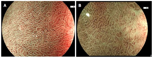
Performance characteristics. A: Image of gastric intestinal metaplasia visualized by blue laser imaging with optical magnification; B: Image of early gastric cancer visualized by blue laser imaging with optical magnification.
CONCLUSION
Especially in last 15 years, there has been a proliferation of research for use of IEE in detection and characterization of gastric pathologies. The role of IEE in screening is still limited and WLE should still be used as first line method of examination. However, there is large amount of data in support of M-NBI for diagnosis, delineation and depth-estimation of EGC. M-NBI also has excellent diagnostic characteristics for IM. It is important to emphasize that almost all studies have been done by experts in IEE highlighting the importance of a proper training in pattern recognition before general use. The published data for other IEE techniques are more limited. Since the principle of BLI is the same as NBI, it would be expected to provide similar efficacy as NBI, especially with optical magnification. In contrast, it is probably not possible to extrapolate the NBI data to FICE and I-SCAN, since these are processed images without optical magnification (Table 3).
Table 3.
Summary of image-enhanced endoscopy in stomach
| Technique | Use | Evidence | Remarks |
| High-definition WLE | Standard of care for initial examination of gastric mucosa | Not available | |
| WLE with magnification | Helpful in describing normal mucosal patterns in corpus and antrum. Appears useful in predicting real-time diagnosis of H. pylori infection. Better than WLE for characterization of EGCs | Multiple prospective comparative studies for identifying H. pylori infection and for characterization of EGCs | A variety of classifications in describing the normal and abnormal mucosal pattern makes interpretation difficult for widespread use |
| Dye-based chromoendoscopy | Traditionally used for demarcation of EGC before resection | Few prospective studies are available, and more data will be needed | There are heterogeneity in the types of stain, technique of staining, classification in defining mucosal patterns |
| NBI | Good for characterization of a focal lesion detected on WLE May be useful for real-time diagnosis of H. pylori Appears reliable for diagnosis of intestinal metaplasia High specificity for characterization of EGCs May be useful for prediction of histological differentiation, prediction of depth of invasion, and in determination of horizontal extent of EGCs | Multiple prospective comparative study show good evidence in support of NBI for diagnosis of intestinal metaplasia and characterization of EGCs More evidence will be needed for other indications | Identifying intestinal metaplasia appears straightforward A variety of classifications for different mucosal pattern bring difficulty in generalization of NBI |
| FICE | May be useful for diagnosis of focal gastric lesions | Not much comparative prospective data is available | |
| I-SCAN | No comparative data for use of I-SCAN in stomach | ||
| Blue-laser imaging | Is expected to be used in similar manner as NBI | Data mainly based on case series rather than comparative studies | Based on anecdotal experience it is similar to NBI and therefore would be expected to provide similar outcomes |
WLE: White light endoscopy; EGC: Early gastric cancer; NBI: Narrow-band imaging; FICE: Flexible spectral imaging color enhancement; BNI: Narrow band imaging; H. pylori: Helicobacter pylori.
ACKNOWLEDGMENTS
We would like to acknowledge Dr Noriya Uedo (Osaka, Japan) and FUJIFILM Asia Pacific Pte. Ltd., for contribution of images used in this paper.
Footnotes
Conflict-of-interest statement: Authors declare no conflict of interests for this article.
Manuscript source: Invited manuscript
Specialty type: Gastroenterology and hepatology
Country of origin: Singapore
Peer-review report classification
Grade A (Excellent): 0
Grade B (Very good): 0
Grade C (Good): C
Grade D (Fair): 0
Grade E (Poor): 0
Peer-review started: April 1, 2016
First decision: June 12, 2016
Article in press: September 22, 2016
P- Reviewer: Tham TCK S- Editor: Qiu S L- Editor: A E- Editor: Wu HL
References
- 1.Subramanian V, Ragunath K. Advanced endoscopic imaging: a review of commercially available technologies. Clin Gastroenterol Hepatol. 2014;12:368–376.e1. doi: 10.1016/j.cgh.2013.06.015. [DOI] [PubMed] [Google Scholar]
- 2.Miyahara R, Niwa Y, Matsuura T, Maeda O, Ando T, Ohmiya N, Itoh A, Hirooka Y, Goto H. Prevalence and prognosis of gastric cancer detected by screening in a large Japanese population: data from a single institute over 30 years. J Gastroenterol Hepatol. 2007;22:1435–1442. doi: 10.1111/j.1440-1746.2007.04991.x. [DOI] [PubMed] [Google Scholar]
- 3.Tajiri H, Niwa H. Proposal for a consensus terminology in endoscopy: how should different endoscopic imaging techniques be grouped and defined? Endoscopy. 2008;40:775–778. doi: 10.1055/s-2008-1077507. [DOI] [PubMed] [Google Scholar]
- 4.Kwon RS, Adler DG, Chand B, Conway JD, Diehl DL, Kantsevoy SV, Mamula P, Rodriguez SA, Shah RJ, Wong Kee Song LM, et al. High-resolution and high-magnification endoscopes. Gastrointest Endosc. 2009;69:399–407. doi: 10.1016/j.gie.2008.12.049. [DOI] [PubMed] [Google Scholar]
- 5.Subramanian V, Mannath J, Hawkey CJ, Ragunath K. High definition colonoscopy vs. standard video endoscopy for the detection of colonic polyps: a meta-analysis. Endoscopy. 2011;43:499–505. doi: 10.1055/s-0030-1256207. [DOI] [PubMed] [Google Scholar]
- 6.Sakaki N, Iida Y, Okazaki Y, Kawamura S, Takemoto T. Magnifying endoscopic observation of the gastric mucosa, particularly in patients with atrophic gastritis. Endoscopy. 1978;10:269–274. doi: 10.1055/s-0028-1098307. [DOI] [PubMed] [Google Scholar]
- 7.Yao K, Oishi T. Microgastroscopic findings of mucosal microvascular architecture as visualized by magnifying endoscopy. Dig Endosc. 2001;13:S27–S33. [Google Scholar]
- 8.Yagi K, Nakamura A, Sekine A. Characteristic endoscopic and magnified endoscopic findings in the normal stomach without Helicobacter pylori infection. J Gastroenterol Hepatol. 2002;17:39–45. doi: 10.1046/j.1440-1746.2002.02665.x. [DOI] [PubMed] [Google Scholar]
- 9.Yagi K, Nakamura A, Sekine A. Comparison between magnifying endoscopy and histological, culture and urease test findings from the gastric mucosa of the corpus. Endoscopy. 2002;34:376–381. doi: 10.1055/s-2002-25281. [DOI] [PubMed] [Google Scholar]
- 10.Gonen C, Simsek I, Sarioglu S, Akpinar H. Comparison of high resolution magnifying endoscopy and standard videoendoscopy for the diagnosis of Helicobacter pylori gastritis in routine clinical practice: a prospective study. Helicobacter. 2009;14:12–21. doi: 10.1111/j.1523-5378.2009.00650.x. [DOI] [PubMed] [Google Scholar]
- 11.Anagnostopoulos GK, Yao K, Kaye P, Fogden E, Fortun P, Shonde A, Foley S, Sunil S, Atherton JJ, Hawkey C, et al. High-resolution magnification endoscopy can reliably identify normal gastric mucosa, Helicobacter pylori-associated gastritis, and gastric atrophy. Endoscopy. 2007;39:202–207. doi: 10.1055/s-2006-945056. [DOI] [PubMed] [Google Scholar]
- 12.Yang JM, Chen L, Fan YL, Li XH, Yu X, Fang DC. Endoscopic patterns of gastric mucosa and its clinicopathological significance. World J Gastroenterol. 2003;9:2552–2556. doi: 10.3748/wjg.v9.i11.2552. [DOI] [PMC free article] [PubMed] [Google Scholar]
- 13.Nakagawa S, Kato M, Shimizu Y, Nakagawa M, Yamamoto J, Luis PA, Kodaira J, Kawarasaki M, Takeda H, Sugiyama T, et al. Relationship between histopathologic gastritis and mucosal microvascularity: observations with magnifying endoscopy. Gastrointest Endosc. 2003;58:71–75. doi: 10.1067/mge.2003.316. [DOI] [PubMed] [Google Scholar]
- 14.Correa P. Human gastric carcinogenesis: a multistep and multifactorial process--First American Cancer Society Award Lecture on Cancer Epidemiology and Prevention. Cancer Res. 1992;52:6735–6740. [PubMed] [Google Scholar]
- 15.Tobita K. Study on minute surface structures of the depressed-type early gastric cancer with magnifying endoscopy. Dig Endosc. 2001;13:121–126. [Google Scholar]
- 16.Tajiri H, Doi T, Endo H, Nishina T, Terao T, Hyodo I, Matsuda K, Yagi K. Routine endoscopy using a magnifying endoscope for gastric cancer diagnosis. Endoscopy. 2002;34:772–777. doi: 10.1055/s-2002-34267. [DOI] [PubMed] [Google Scholar]
- 17.Yao K, Oishi T, Matsui T, Yao T, Iwashita A. Novel magnified endoscopic findings of microvascular architecture in intramucosal gastric cancer. Gastrointest Endosc. 2002;56:279–284. doi: 10.1016/s0016-5107(02)70194-6. [DOI] [PubMed] [Google Scholar]
- 18.Yao K, Iwashita A, Tanabe H, Nagahama T, Matsui T, Ueki T, Sou S, Kikuchi Y, Yorioka M. Novel zoom endoscopy technique for diagnosis of small flat gastric cancer: a prospective, blind study. Clin Gastroenterol Hepatol. 2007;5:869–878. doi: 10.1016/j.cgh.2007.02.034. [DOI] [PubMed] [Google Scholar]
- 19.Otsuka Y, Niwa Y, Ohmiya N, Ando N, Ohashi A, Hirooka Y, Goto H. Usefulness of magnifying endoscopy in the diagnosis of early gastric cancer. Endoscopy. 2004;36:165–169. doi: 10.1055/s-2004-814184. [DOI] [PubMed] [Google Scholar]
- 20.Yoshida T, Kawachi H, Sasajima K, Shiokawa A, Kudo SE. The clinical meaning of a nonstructural pattern in early gastric cancer on magnifying endoscopy. Gastrointest Endosc. 2005;62:48–54. doi: 10.1016/s0016-5107(05)00373-1. [DOI] [PubMed] [Google Scholar]
- 21.Kiesslich R, Fritsch J, Holtmann M, Koehler HH, Stolte M, Kanzler S, Nafe B, Jung M, Galle PR, Neurath MF. Methylene blue-aided chromoendoscopy for the detection of intraepithelial neoplasia and colon cancer in ulcerative colitis. Gastroenterology. 2003;124:880–888. doi: 10.1053/gast.2003.50146. [DOI] [PubMed] [Google Scholar]
- 22.Yagi K, Aruga Y, Nakamura A, Sekine A, Umezu H. The study of dynamic chemical magnifying endoscopy in gastric neoplasia. Gastrointest Endosc. 2005;62:963–969. doi: 10.1016/j.gie.2005.08.050. [DOI] [PubMed] [Google Scholar]
- 23.Tanaka K, Toyoda H, Kadowaki S, Kosaka R, Shiraishi T, Imoto I, Shiku H, Adachi Y. Features of early gastric cancer and gastric adenoma by enhanced-magnification endoscopy. J Gastroenterol. 2006;41:332–338. doi: 10.1007/s00535-005-1760-3. [DOI] [PubMed] [Google Scholar]
- 24.Tanaka K, Toyoda H, Kadowaki S, Hamada Y, Kosaka R, Matsuzaki S, Shiraishi T, Imoto I, Takei Y. Surface pattern classification by enhanced-magnification endoscopy for identifying early gastric cancers. Gastrointest Endosc. 2008;67:430–437. doi: 10.1016/j.gie.2007.10.042. [DOI] [PubMed] [Google Scholar]
- 25.Kohli Y, Kato T, Ito S. Helicobacter pylori an chronic atrophic gastritis. J Gastroenterol. 1994;29 Suppl 7:105–109. [PubMed] [Google Scholar]
- 26.Dinis-Ribeiro M, da Costa-Pereira A, Lopes C, Lara-Santos L, Guilherme M, Moreira-Dias L, Lomba-Viana H, Ribeiro A, Santos C, Soares J, et al. Magnification chromoendoscopy for the diagnosis of gastric intestinal metaplasia and dysplasia. Gastrointest Endosc. 2003;57:498–504. doi: 10.1067/mge.2003.145. [DOI] [PubMed] [Google Scholar]
- 27.Areia M, Amaro P, Dinis-Ribeiro M, Cipriano MA, Marinho C, Costa-Pereira A, Lopes C, Moreira-Dias L, Romãozinho JM, Gouveia H, et al. External validation of a classification for methylene blue magnification chromoendoscopy in premalignant gastric lesions. Gastrointest Endosc. 2008;67:1011–1018. doi: 10.1016/j.gie.2007.08.044. [DOI] [PubMed] [Google Scholar]
- 28.Taghavi SA, Membari ME, Eshraghian A, Dehghani SM, Hamidpour L, Khademalhoseini F. Comparison of chromoendoscopy and conventional endoscopy in the detection of premalignant gastric lesions. Can J Gastroenterol. 2009;23:105–108. doi: 10.1155/2009/594983. [DOI] [PMC free article] [PubMed] [Google Scholar]
- 29.Mouzyka S, Fedoseeva A. Chromoendoscopy with hematoxylin in the classification of gastric lesions. Gastric Cancer. 2008;11:15–21; discussion 21-22. doi: 10.1007/s10120-007-0445-4. [DOI] [PubMed] [Google Scholar]
- 30.Iizuka T, Kikuchi D, Hoteya S, Yahagi N. The acetic acid + indigocarmine method in the delineation of gastric cancer. J Gastroenterol Hepatol. 2008;23:1358–1361. doi: 10.1111/j.1440-1746.2008.05528.x. [DOI] [PubMed] [Google Scholar]
- 31.Sakai Y, Eto R, Kasanuki J, Kondo F, Kato K, Arai M, Suzuki T, Kobayashi M, Matsumura T, Bekku D, et al. Chromoendoscopy with indigo carmine dye added to acetic acid in the diagnosis of gastric neoplasia: a prospective comparative study. Gastrointest Endosc. 2008;68:635–641. doi: 10.1016/j.gie.2008.03.1065. [DOI] [PubMed] [Google Scholar]
- 32.Kawahara Y, Takenaka R, Okada H, Kawano S, Inoue M, Tsuzuki T, Tanioka D, Hori K, Yamamoto K. Novel chromoendoscopic method using an acetic acid-indigocarmine mixture for diagnostic accuracy in delineating the margin of early gastric cancers. Dig Endosc. 2009;21:14–19. doi: 10.1111/j.1443-1661.2008.00824.x. [DOI] [PubMed] [Google Scholar]
- 33.Gono K, Obi T, Yamaguchi M, Ohyama N, Machida H, Sano Y, Yoshida S, Hamamoto Y, Endo T. Appearance of enhanced tissue features in narrow-band endoscopic imaging. J Biomed Opt. 2004;9:568–577. doi: 10.1117/1.1695563. [DOI] [PubMed] [Google Scholar]
- 34.Gono K. Narrow Band Imaging: Technology Basis and Research and Development History. Clin Endosc. 2015;48:476–480. doi: 10.5946/ce.2015.48.6.476. [DOI] [PMC free article] [PubMed] [Google Scholar]
- 35.Ang TL, Fock KM, Teo EK, Tan J, Poh CH, Ong J, Ang D. The diagnostic utility of narrow band imaging magnifying endoscopy in clinical practice in a population with intermediate gastric cancer risk. Eur J Gastroenterol Hepatol. 2012;24:362–367. doi: 10.1097/MEG.0b013e3283500968. [DOI] [PubMed] [Google Scholar]
- 36.Yao K, Doyama H, Gotoda T, Ishikawa H, Nagahama T, Yokoi C, Oda I, Machida H, Uchita K, Tabuchi M. Diagnostic performance and limitations of magnifying narrow-band imaging in screening endoscopy of early gastric cancer: a prospective multicenter feasibility study. Gastric Cancer. 2014;17:669–679. doi: 10.1007/s10120-013-0332-0. [DOI] [PubMed] [Google Scholar]
- 37.Yu H, Yang AM, Lu XH, Zhou WX, Yao F, Fei GJ, Guo T, Yao LQ, He LP, Wang BM. Magnifying narrow-band imaging endoscopy is superior in diagnosis of early gastric cancer. World J Gastroenterol. 2015;21:9156–9162. doi: 10.3748/wjg.v21.i30.9156. [DOI] [PMC free article] [PubMed] [Google Scholar]
- 38.Dutta AK, Sajith KG, Pulimood AB, Chacko A. Narrow band imaging versus white light gastroscopy in detecting potentially premalignant gastric lesions: a randomized prospective crossover study. Indian J Gastroenterol. 2013;32:37–42. doi: 10.1007/s12664-012-0246-5. [DOI] [PubMed] [Google Scholar]
- 39.Xirouchakis E, Laoudi F, Tsartsali L, Spiliadi C, Georgopoulos SD. Screening for gastric premalignant lesions with narrow band imaging, white light and updated Sydney protocol or both? Dig Dis Sci. 2013;58:1084–1090. doi: 10.1007/s10620-012-2431-x. [DOI] [PubMed] [Google Scholar]
- 40.Ang TL, Pittayanon R, Lau JY, Rerknimitr R, Ho SH, Singh R, Kwek AB, Ang DS, Chiu PW, Luk S, et al. A multicenter randomized comparison between high-definition white light endoscopy and narrow band imaging for detection of gastric lesions. Eur J Gastroenterol Hepatol. 2015;27:1473–1478. doi: 10.1097/MEG.0000000000000478. [DOI] [PubMed] [Google Scholar]
- 41.Tahara T, Shibata T, Nakamura M, Yoshioka D, Okubo M, Arisawa T, Hirata I. Gastric mucosal pattern by using magnifying narrow-band imaging endoscopy clearly distinguishes histological and serological severity of chronic gastritis. Gastrointest Endosc. 2009;70:246–253. doi: 10.1016/j.gie.2008.11.046. [DOI] [PubMed] [Google Scholar]
- 42.Yagi K, Saka A, Nozawa Y, Nakamura A. Prediction of Helicobacter pylori status by conventional endoscopy, narrow-band imaging magnifying endoscopy in stomach after endoscopic resection of gastric cancer. Helicobacter. 2014;19:111–115. doi: 10.1111/hel.12104. [DOI] [PubMed] [Google Scholar]
- 43.Uedo N, Ishihara R, Iishi H, Yamamoto S, Yamamoto S, Yamada T, Imanaka K, Takeuchi Y, Higashino K, Ishiguro S, et al. A new method of diagnosing gastric intestinal metaplasia: narrow-band imaging with magnifying endoscopy. Endoscopy. 2006;38:819–824. doi: 10.1055/s-2006-944632. [DOI] [PubMed] [Google Scholar]
- 44.Wang L, Huang W, Du J, Chen Y, Yang J. Diagnostic yield of the light blue crest sign in gastric intestinal metaplasia: a meta-analysis. PLoS One. 2014;9:e92874. doi: 10.1371/journal.pone.0092874. [DOI] [PMC free article] [PubMed] [Google Scholar]
- 45.Bansal A, Ulusarac O, Mathur S, Sharma P. Correlation between narrow band imaging and nonneoplastic gastric pathology: a pilot feasibility trial. Gastrointest Endosc. 2008;67:210–216. doi: 10.1016/j.gie.2007.06.009. [DOI] [PubMed] [Google Scholar]
- 46.The Paris endoscopic classification of superficial neoplastic lesions: esophagus, stomach, and colon: November 30 to December 1, 2002. Gastrointest Endosc. 2003;58:S3–43. doi: 10.1016/s0016-5107(03)02159-x. [DOI] [PubMed] [Google Scholar]
- 47.Yao K, Takaki Y, Matsui T, Iwashita A, Anagnostopoulos GK, Kaye P, Ragunath K. Clinical application of magnification endoscopy and narrow-band imaging in the upper gastrointestinal tract: new imaging techniques for detecting and characterizing gastrointestinal neoplasia. Gastrointest Endosc Clin N Am. 2008;18:415–433, vii-viii. doi: 10.1016/j.giec.2008.05.011. [DOI] [PubMed] [Google Scholar]
- 48.Ezoe Y, Muto M, Horimatsu T, Minashi K, Yano T, Sano Y, Chiba T, Ohtsu A. Magnifying narrow-band imaging versus magnifying white-light imaging for the differential diagnosis of gastric small depressive lesions: a prospective study. Gastrointest Endosc. 2010;71:477–484. doi: 10.1016/j.gie.2009.10.036. [DOI] [PubMed] [Google Scholar]
- 49.Ezoe Y, Muto M, Uedo N, Doyama H, Yao K, Oda I, Kaneko K, Kawahara Y, Yokoi C, Sugiura Y, et al. Magnifying narrowband imaging is more accurate than conventional white-light imaging in diagnosis of gastric mucosal cancer. Gastroenterology. 2011;141:2017–2025.e3. doi: 10.1053/j.gastro.2011.08.007. [DOI] [PubMed] [Google Scholar]
- 50.Yamada S, Doyama H, Yao K, Uedo N, Ezoe Y, Oda I, Kaneko K, Kawahara Y, Yokoi C, Sugiura Y, et al. An efficient diagnostic strategy for small, depressed early gastric cancer with magnifying narrow-band imaging: a post-hoc analysis of a prospective randomized controlled trial. Gastrointest Endosc. 2014;79:55–63. doi: 10.1016/j.gie.2013.07.008. [DOI] [PubMed] [Google Scholar]
- 51.Kato M, Kaise M, Yonezawa J, Toyoizumi H, Yoshimura N, Yoshida Y, Kawamura M, Tajiri H. Magnifying endoscopy with narrow-band imaging achieves superior accuracy in the differential diagnosis of superficial gastric lesions identified with white-light endoscopy: a prospective study. Gastrointest Endosc. 2010;72:523–529. doi: 10.1016/j.gie.2010.04.041. [DOI] [PubMed] [Google Scholar]
- 52.Zhang Q, Wang F, Chen ZY, Wang Z, Zhi FC, Liu SD, Bai Y. Comparison of the diagnostic efficacy of white light endoscopy and magnifying endoscopy with narrow band imaging for early gastric cancer: a meta-analysis. Gastric Cancer. 2016;19:543–552. doi: 10.1007/s10120-015-0500-5. [DOI] [PubMed] [Google Scholar]
- 53.Lv X, Wang C, Xie Y, Yan Z. Diagnostic efficacy of magnifying endoscopy with narrow-band imaging for gastric neoplasms: a meta-analysis. PLoS One. 2015;10:e0123832. doi: 10.1371/journal.pone.0123832. [DOI] [PMC free article] [PubMed] [Google Scholar]
- 54.Nakayoshi T, Tajiri H, Matsuda K, Kaise M, Ikegami M, Sasaki H. Magnifying endoscopy combined with narrow band imaging system for early gastric cancer: correlation of vascular pattern with histopathology (including video) Endoscopy. 2004;36:1080–1084. doi: 10.1055/s-2004-825961. [DOI] [PubMed] [Google Scholar]
- 55.Yokoyama A, Inoue H, Minami H, Wada Y, Sato Y, Satodate H, Hamatani S, Kudo SE. Novel narrow-band imaging magnifying endoscopic classification for early gastric cancer. Dig Liver Dis. 2010;42:704–708. doi: 10.1016/j.dld.2010.03.013. [DOI] [PubMed] [Google Scholar]
- 56.Yao K, Yao T, Iwashita A. Determining the horizontal extent of early gastric carcinoma: two modern techniques based on differences in the mucosal microvascular architecture and density between carcinomatous and non-carcinomatous mucosal. Dig Endosc. 2002;14:S83–S87. [Google Scholar]
- 57.Sumiyama K, Kaise M, Nakayoshi T, Kato M, Mashiko T, Uchiyama Y, Goda K, Hino S, Nakamura Y, Matsuda K, et al. Combined use of a magnifying endoscope with a narrow band imaging system and a multibending endoscope for en bloc EMR of early stage gastric cancer. Gastrointest Endosc. 2004;60:79–84. doi: 10.1016/s0016-5107(04)01285-4. [DOI] [PubMed] [Google Scholar]
- 58.Kiyotoki S, Nishikawa J, Satake M, Fukagawa Y, Shirai Y, Hamabe K, Saito M, Okamoto T, Sakaida I. Usefulness of magnifying endoscopy with narrow-band imaging for determining gastric tumor margin. J Gastroenterol Hepatol. 2010;25:1636–1641. doi: 10.1111/j.1440-1746.2010.06379.x. [DOI] [PubMed] [Google Scholar]
- 59.Nagahama T, Yao K, Maki S, Yasaka M, Takaki Y, Matsui T, Tanabe H, Iwashita A, Ota A. Usefulness of magnifying endoscopy with narrow-band imaging for determining the horizontal extent of early gastric cancer when there is an unclear margin by chromoendoscopy (with video) Gastrointest Endosc. 2011;74:1259–1267. doi: 10.1016/j.gie.2011.09.005. [DOI] [PubMed] [Google Scholar]
- 60.Horiuchi Y, Fujisaki J, Yamamoto N, Shimizu T, Miyamoto Y, Tomida H, Omae M, Ishiyama A, Yoshio T, Hirasawa T, et al. Accuracy of diagnostic demarcation of undifferentiated-type early gastric cancers for magnifying endoscopy with narrow-band imaging: endoscopic submucosal dissection cases. Gastric Cancer. 2016;19:515–523. doi: 10.1007/s10120-015-0488-x. [DOI] [PubMed] [Google Scholar]
- 61.Yagi K, Nakamura A, Sekine A, Umezu H. Magnifying endoscopy with narrow band imaging for early differentiated gastric adenocarcinoma. Dig Endosc. 2008;20:115–122. [Google Scholar]
- 62.Li HY, Dai J, Xue HB, Zhao YJ, Chen XY, Gao YJ, Song Y, Ge ZZ, Li XB. Application of magnifying endoscopy with narrow-band imaging in diagnosing gastric lesions: a prospective study. Gastrointest Endosc. 2012;76:1124–1132. doi: 10.1016/j.gie.2012.08.015. [DOI] [PubMed] [Google Scholar]
- 63.Kobara H, Mori H, Fujihara S, Kobayashi M, Nishiyama N, Nomura T, Kato K, Ishihara S, Morito T, Mizobuchi K, et al. Prediction of invasion depth for submucosal differentiated gastric cancer by magnifying endoscopy with narrow-band imaging. Oncol Rep. 2012;28:841–847. doi: 10.3892/or.2012.1889. [DOI] [PubMed] [Google Scholar]
- 64.Kikuchi D, Iizuka T, Hoteya S, Yamada A, Furuhata T, Yamashita S, Domon K, Nakamura M, Matsui A, Mitani T, et al. Usefulness of magnifying endoscopy with narrow-band imaging for determining tumor invasion depth in early gastric cancer. Gastroenterol Res Pract. 2013;2013:217695. doi: 10.1155/2013/217695. [DOI] [PMC free article] [PubMed] [Google Scholar]
- 65.Cho JH. Advanced Imaging Technology Other than Narrow Band Imaging. Clin Endosc. 2015;48:503–510. doi: 10.5946/ce.2015.48.6.503. [DOI] [PMC free article] [PubMed] [Google Scholar]
- 66.Osawa H, Yoshizawa M, Yamamoto H, Kita H, Satoh K, Ohnishi H, Nakano H, Wada M, Arashiro M, Tsukui M, et al. Optimal band imaging system can facilitate detection of changes in depressed-type early gastric cancer. Gastrointest Endosc. 2008;67:226–234. doi: 10.1016/j.gie.2007.06.067. [DOI] [PubMed] [Google Scholar]
- 67.Yoshizawa M, Osawa H, Yamamoto H, Satoh K, Nakano H, Tsukui M, Sugano K. Newly developed optimal band imaging system for the diagnosis of early gastric cancer. Dig Endosc. 2008;20:194–197. [Google Scholar]
- 68.Yoshizawa M, Osawa H, Yamamoto H, Kita H, Nakano H, Satoh K, Shigemori M, Tsukui M, Sugano K. Diagnosis of elevated-type early gastric cancers by the optimal band imaging system. Gastrointest Endosc. 2009;69:19–28. doi: 10.1016/j.gie.2008.09.007. [DOI] [PubMed] [Google Scholar]
- 69.Mouri R, Yoshida S, Tanaka S, Oka S, Yoshihara M, Chayama K. Evaluation and validation of computed virtual chromoendoscopy in early gastric cancer. Gastrointest Endosc. 2009;69:1052–1058. doi: 10.1016/j.gie.2008.08.032. [DOI] [PubMed] [Google Scholar]
- 70.Jung SW, Lim KS, Lim JU, Jeon JW, Shin HP, Kim SH, Lee EK, Park JJ, Cha JM, Joo KR, et al. Flexible spectral imaging color enhancement (FICE) is useful to discriminate among non-neoplastic lesion, adenoma, and cancer of stomach. Dig Dis Sci. 2011;56:2879–2886. doi: 10.1007/s10620-011-1831-7. [DOI] [PubMed] [Google Scholar]
- 71.Kikuste I, Stirna D, Liepniece-Karele I, Leja M, Dinis-Ribeiro M. The accuracy of flexible spectral imaging colour enhancement for the diagnosis of gastric intestinal metaplasia: do we still need histology to select individuals at risk for adenocarcinoma? Eur J Gastroenterol Hepatol. 2014;26:704–709. doi: 10.1097/MEG.0000000000000108. [DOI] [PubMed] [Google Scholar]
- 72.Tanioka Y, Yanai H, Sakaguchi E. Ultraslim endoscopy with flexible spectral imaging color enhancement for upper gastrointestinal neoplasms. World J Gastrointest Endosc. 2011;3:11–15. doi: 10.4253/wjge.v3.i1.11. [DOI] [PMC free article] [PubMed] [Google Scholar]
- 73.Osawa H, Yamamoto H, Miura Y, Ajibe H, Shinhata H, Yoshizawa M, Sunada K, Toma S, Satoh K, Sugano K. Diagnosis of depressed-type early gastric cancer using small-caliber endoscopy with flexible spectral imaging color enhancement. Dig Endosc. 2012;24:231–236. doi: 10.1111/j.1443-1661.2011.01224.x. [DOI] [PubMed] [Google Scholar]
- 74.Dohi O, Yagi N, Wada T, Yamada N, Bito N, Yamada S, Gen Y, Yoshida N, Uchiyama K, Ishikawa T, et al. Recognition of endoscopic diagnosis in differentiated-type early gastric cancer by flexible spectral imaging color enhancement with indigo carmine. Digestion. 2012;86:161–170. doi: 10.1159/000339878. [DOI] [PubMed] [Google Scholar]
- 75.Kodashima S, Fujishiro M. Novel image-enhanced endoscopy with i-scan technology. World J Gastroenterol. 2010;16:1043–1049. doi: 10.3748/wjg.v16.i9.1043. [DOI] [PMC free article] [PubMed] [Google Scholar]
- 76.Li CQ, Li Y, Zuo XL, Ji R, Li Z, Gu XM, Yu T, Qi QQ, Zhou CJ, Li YQ. Magnified and enhanced computed virtual chromoendoscopy in gastric neoplasia: a feasibility study. World J Gastroenterol. 2013;19:4221–4227. doi: 10.3748/wjg.v19.i26.4221. [DOI] [PMC free article] [PubMed] [Google Scholar]
- 77.Kaneko K, Oono Y, Yano T, Ikematsu H, Odagaki T, Yoda Y, Yagishita A, Sato A, Nomura S. Effect of novel bright image enhanced endoscopy using blue laser imaging (BLI) Endosc Int Open. 2014;2:E212–E219. doi: 10.1055/s-0034-1390707. [DOI] [PMC free article] [PubMed] [Google Scholar]


