
Figure 5.
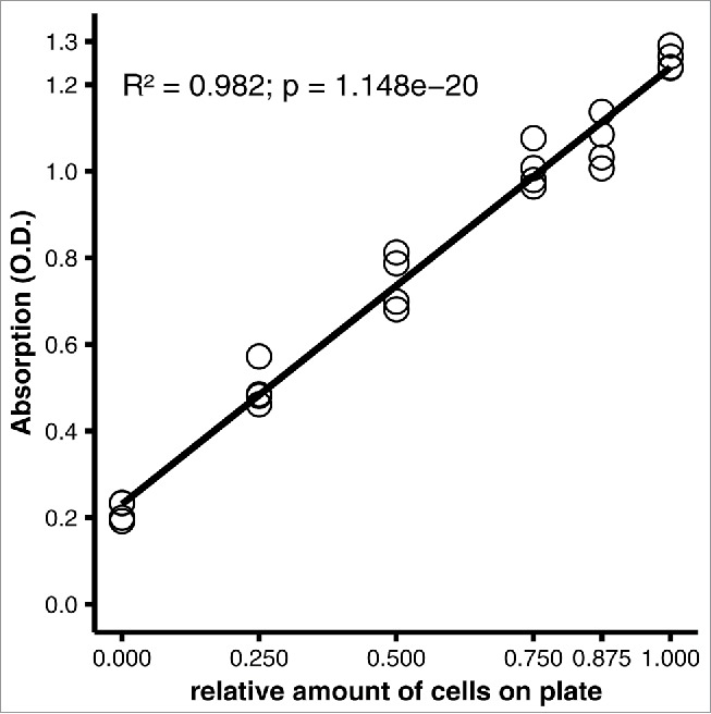
Applicability of the staining protocol to a different culture plate format. 3T3-L1 adipocytes were differentiated on 12-well plates, then stained using the optimized protocol (see Materials and Methods). Linear regression analysis of quantitative staining of varying amounts of cells in a well. All wells were initially covered with a confluent monolayer of preadipocytes; the amount of cells was varied experimentally by scraping off the cells. Efficacy of scraping was verified under the microscope.